

Laser Maculopathy
<< Back to Cases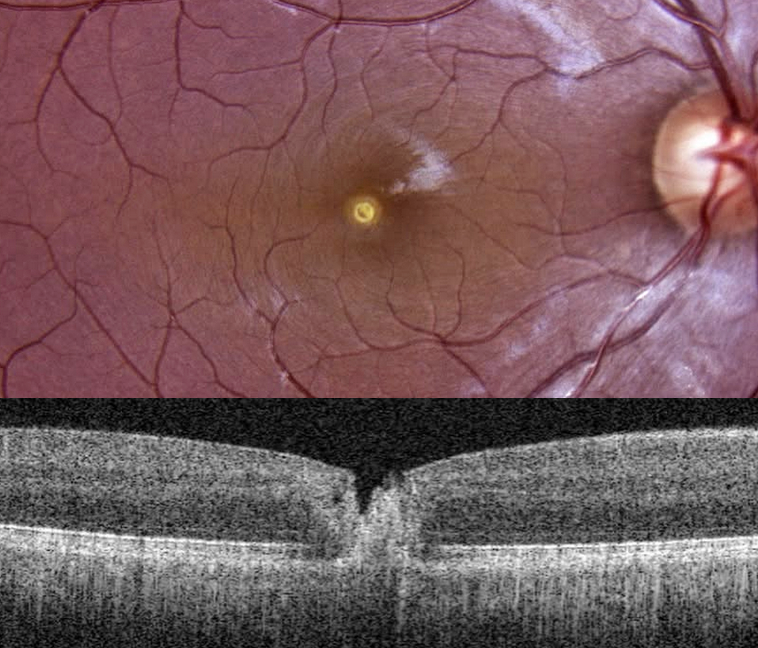
LASER MACULOPATHY
The European VitreoRetinal Society (EVRS) and Aniruddh Soni
Originally posted on @retina.rocks June 12, 2026
This 26YO male DJ noticed immediate blurred vision in his right eye after looking at a laser disco light during an event 5 days earlier. Vision was 20/120 OD.
Color photography shows a small, round, yellow lesion involving or quite close to the foveal center. OCT scanning shows full-thickness foveal disruption and hyperreflectivity.
Learning Points:
Most cases of laser maculopathy in the literature are caused by purposeful or inadvertent exposure to laser pointers (Bhavsar et al, Survey Ophthalmology 2021;66:231-260). However, more recently, there have been reports of macular injury from exposure to high-power lasers during concerts or from DJ laser machines (Perz-Montano et al, Doc Ophthalmol 2019;138:71-76). Most eyes recover fairly good vision with conservative management.
LASER MACULOPATHY
Vaibhav Sethi
Originally posted on @retina.rocks January 20, 2026
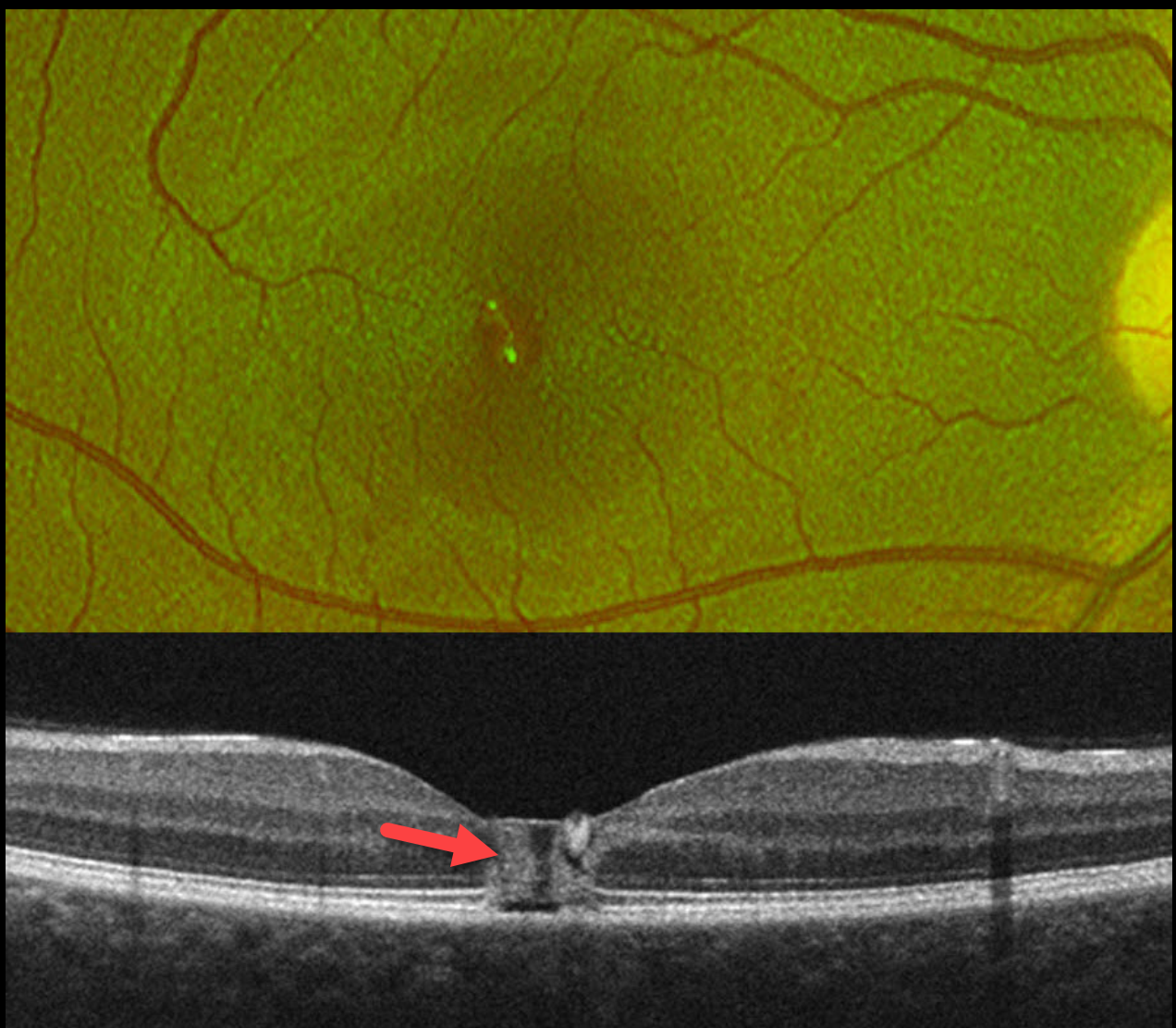
This healthy 19YO female noticed immediate blurred vision in her right eye after looking at a laser during a party. Her vision in this eye was 20/60 when she presented to us the following day.
Optos color RG imaging shows a foveal pseudohole. OCT scanning shows a central foveal column of full-thickness hyperreflectivity. There is loss of the outer segment bands overlying the RPE.
Learning Points:
Most cases of laser maculopathy reported in the literature are caused by intentional or inadvertent exposure to laser pointers (Bhavsar et al, Survey Ophthalmology 2021;66:231-260). However, more recently, there have been reports of macular injury from exposure to high-power lasers during concerts or from DJ laser machines (Perz-Montano et al, Doc Ophthalmol 2019;138:71-76). Most eyes recover fairly good vision with conservative management.
Our patient was immediately lost to follow-up.
LASER MACULOPATHY
The European VitreoRetina Society (EVRS) and Shraddha Chandorkar
Originally posted on @retina.rocks October 17, 2025
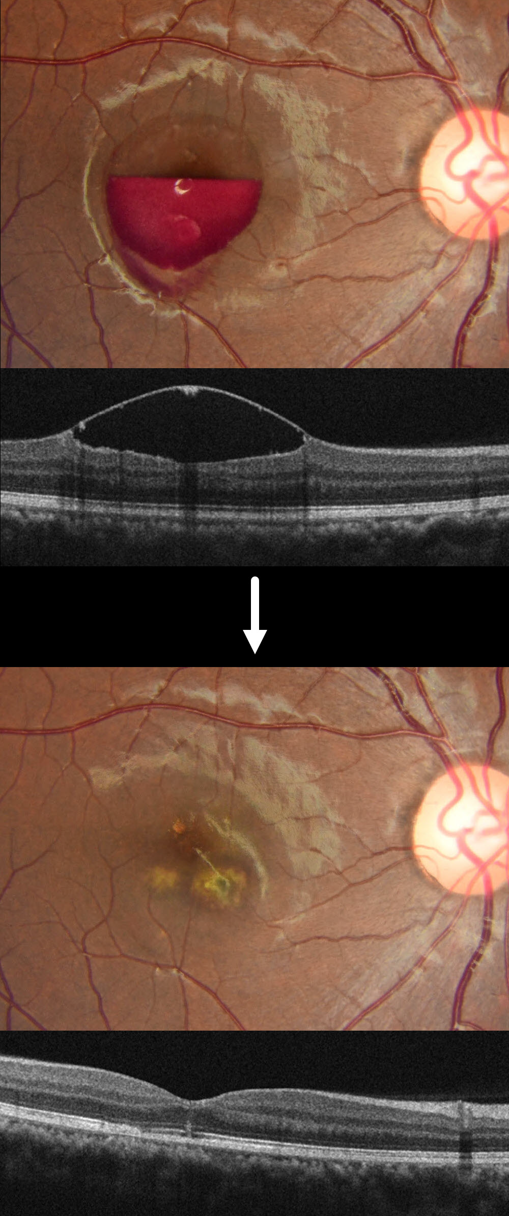
This healthy 19YO female noted sudden vision loss in her right eye immediately after looking directly at the light while attending a DJ laser party. When she presented to us 2 days later, her vision was hand motion OD and 20/20 in her normal OS.
Fundus photography and OCT show a partially layered central macular sub-internal limiting membrane (ILM) hemorrhage.
When examined 1 week later, the blood was drained via a YAG laser due to no improvement in her vision or sub-ILM blood. Two months later, vision improved to 20/20 despite multiple paracentral scars.
Learning Points:
DJ laser machines typically fall into Class 3R or Class 4, depending on their power output. These lasers emit visible light in the red (635-650 nm), green (520-532 nm), and blue (445-460 nm) wavelengths, often combined for full RGB effects. While visually stunning, high-powered lasers, especially Class 4, can pose serious risks, including retinal burns, if beams enter the audience area or are viewed directly. Blue wavelengths are particularly hazardous due to their shorter wavelength and higher energy density. A strikingly similar case to our patient was reported by Perz-Montano et al (Doc Ophthalmol 2019;138:71-76).
To protect audiences, professional setups use a combination of engineering controls and regulatory compliance. This includes fail-safe scanning systems, beam termination zones, and power-limiting protocols to remain below the Maximum Permissible Exposure. In the US, the FDA’s Center for Devices and Radiological Health oversees these devices, requiring manufacturers to submit safety documentation and operators to file a Laser Light Show Variance for public performances. Proper mounting, trained personnel, and adherence to ANSI and IEC standards are essential to ensure safe and compliant laser shows.
LASER MACULOPATHY
Originally posted on @retina.rocks October 3, 2024
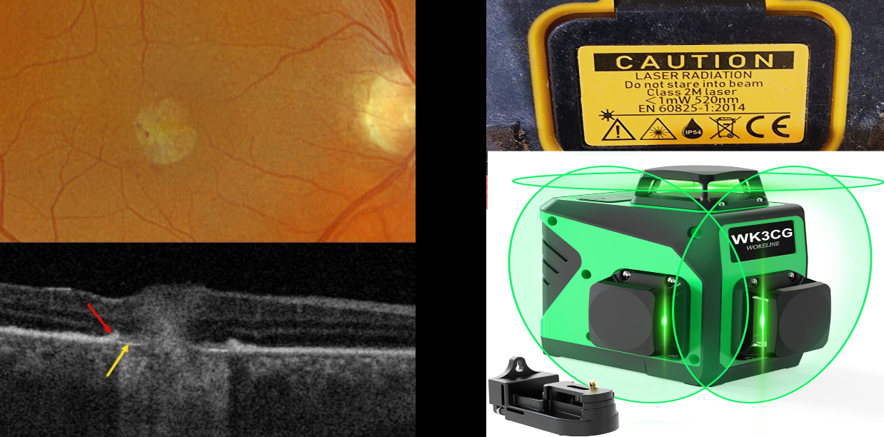
This 46YO male presented with a several-week history of unilateral vision loss. Vision was 20/20 in his normal left eye and counting fingers in his right eye.
Optos color RGB imaging shows a mostly round foveal scar with a few temporal blood dots. Triton swept-source OCT scanning shows the normal foveal architecture replaced by full-thickness, hyperreflective, disorganized tissue. The RPE is thickened and somewhat elevated along the lesion’s edge, with bare Bruch’s membrane more centrally.
Upon further questioning, we found that the patient works as a contractor and lost vision immediately following the use of a laser leveler. He didn’t recall looking directly at the laser beam. He showed us the device, a WOKELINE WGR12L, which is used as a full-room-level laser device. A label on the back of the device defines it as a 520nm Class 2M laser with an output of less than 1 milliwatt. We found an image online of a similar device showing how this device projects a 3D green laser light in 2 vertical planes and a single horizontal plane to cover the floor, ceiling, and all walls simultaneously.
Learning Points:
A Class 2 laser device emits light in the visible spectrum, 400-700 nm. It is presumed that the natural aversion response to the very bright light will be sufficient to prevent damaging exposure, although prolonged viewing may be dangerous. Although laser pointers, Class 2 or 3A devices, are supposed to have an output of under 5 milliwatts, real-world findings reveal that the power output of many of these pointers often far exceeds what is displayed in their safety information. Laser pointer maculopathy is well reported in the literature (Bhavasar et al., Surv Ophthalmology 2021;66:231-360), although we are not aware of a prior case involving a laser-level device, as in our patient.
LASER-INDUCED MACULAR HOLE
Tareq Alsulami and Naser Alsaedi
Originally posted on @retina.rocks June 17, 2024
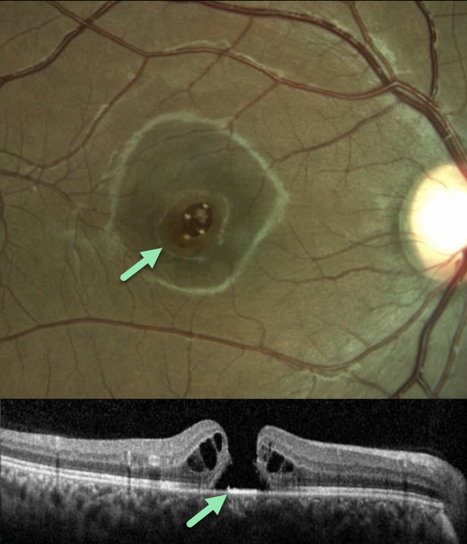
This 14YO boy presented with vision loss in his right eye following self-directed exposure to a blue wavelength laser pointer. Vision was 20/200 in his right eye and 20/20 in his normal left eye.
Color imaging and OCT scanning show an irregular full-thickness macula hole. The hole closed following vitrectomy surgery, but vision failed to improve.
Learning Points:
Laser pointers can cause significant macular injury, including solar maculopathy-like outer foveal microdefects, hemorrhage, pseudo macular dystrophy, vitelliform lesions, and macular scarring (Bhavsar et al, Surv Ophthalmology 2021;66:231-260). There are also numerous reports of a secondary full-thickness macular hole, as developed in our patient.
LASER MACULOPATHY
Mattie Adams
Originally posted on @retina.rocks April 22, 2024
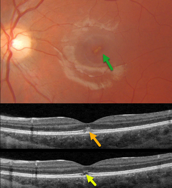
This 13YO girl presented with immediate vision loss in her left eye after looking at a laser pointer one week earlier. Vision was 20/20 in her normal right eye and 20/70 in her left eye.
Triton color photography shows an oblique, slightly linear area of foveal pigment loss. Swept-source OCT scans through this lesion show focal hyperreflectivity with variable loss of the ellipsoid zone and outer segments.
Learning Points:
Sun gazing is thought to cause acute damage to the RPE and photoreceptors via photochemical mechanisms involving reactive oxygen species. Similar photochemical damage can follow laser pointer exposure, although the clinical findings can be different (Bloom and Singal, Retinal Cases 2022;16:
89-91).
Sun gazing acutely produces bilateral, symmetric, pinpoint subfoveal yellow-white lesions in the outer retina, followed by variable clinical depigmented foveal RPE and focal photoreceptor OCT defects extending from the external limiting membrane to the RPE.
Laser pointer maculopathy can cause a similar lesion, but presumed more prolonged viewing, saccades, and laser pointer movement can also lead to larger areas of macular damage.
Outer foveal microdefects (Cohen et al, Ophthalmology Retina 2021;5:553-561) have been described in numerous conditions, including macular telangiectasia, tamoxifen use, ABCA4 disorders (cone-rod dystrophies, Stargardt disease and fundus flavimaculatus), phototoxicity, trauma, and vitreomacular traction disorders.
LASER MACULOPATHY
Will Gibson
Originally posted on @retina.rocks June 29, 2023
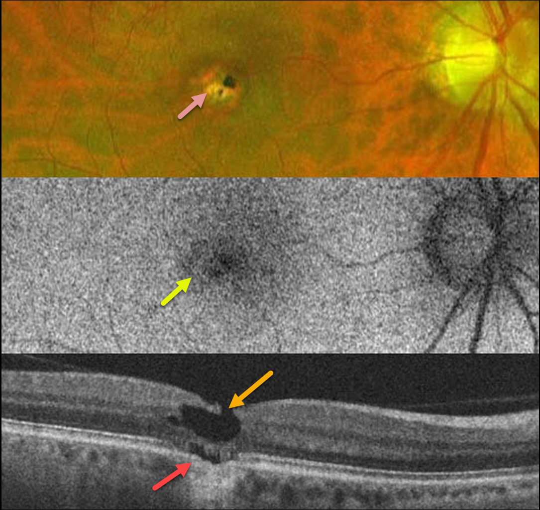
This 36YO male gave a 2-year history of immediate vision loss in his right eye following an accidental 1064nm YAG laser exposure. Vision was 20/80.
Optos color imaging shows a small, round, mostly atrophic foveal scar, which is variably hypoautofluorescent.
OCT scanning shows a degenerative lamellar macular hole (LMH) with focal loss of the underlying outer retinal bands (outer foveal microdefect), RPE loss, and a possible shallow choroidal excavation.
LASER MACULOPATHY
Originally posted on @retina.rocks February 21, 2020
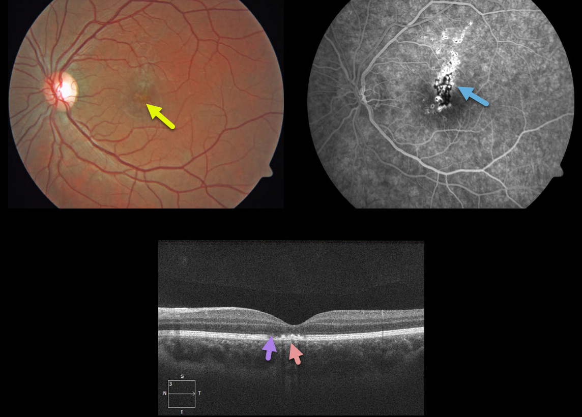
This 18YO patient presented with sudden painless 20/400 vision loss in his left eye. He admitted to staring 3 days earlier directly into his recently purchased green laser pointer.
There are multifocal small subretinal pigment clumps in the left central macula and fluorescein angiogram reveals variable areas of hypo and hyperfluorescence. OCT shows EZ attenuation with hyperreflectivity above the RPE.
Learning Points:
Ocular damage from these lasers depends highly on the laser wavelength, power and duration. The US regulates hand-held laser pointers as a category 3R, which cannot exceed 5 mW. However, a 2013 study looking at 122 laser pointers labeled as 3R found that 90% of green and 44% of red laser pointers actually exceeded the 5mW limit.


