

Lattice Degeneration
<< Back to Cases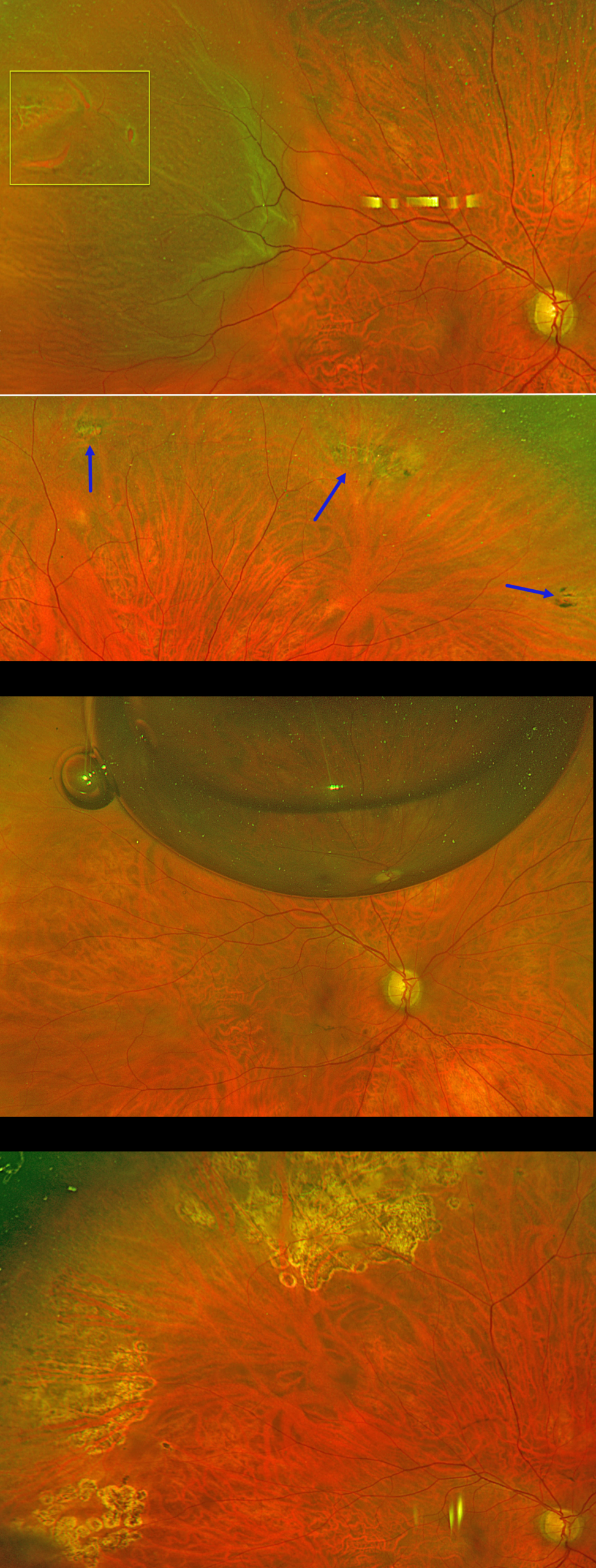
PNEUMATIC RETINOPEXY
Originally posted on @retina.rocks August 13, 2026
This 56YO phakic male presented with floaters and a football-shaped nasal shadow in his right eye for one week. Vision was 20/20 bilaterally.
Optos color RG imaging shows a superotemporal macula-on rhegmatogenous retinal detachment (RRD) from several tiny adjacent breaks within lattice. There were other areas of lattice degeneration superiorly, inferiorly, and inferotemporally within the attached retina. Options for retinal detachment repair were discussed, including pneumatic retinopexy, scleral buckle, and vitrectomy.
The patient chose to undergo pneumatic retinopexy (PnRP) with 0.3cc of pure C3F8. The areas of lattice within the attached retina were lasered just prior to performing the procedure. The retina was completely attached the following day. The causative break was lasered four days later once the detachment flattened. Two months postoperatively, vision was 20/20, and the retina remains attached. Given the diffuse areas of lattice and absence of posterior vitreous detachment in the fellow eye, we performed prophylactic laser retinopexy to the left eye (not shown).
Learning Points:
PnRP was introduced by Hilton and Grizzard in 1986 as an in-office alternative to traditional retinal reattachment surgery (Ophthalmology 1986;93:626-651). It was initially recommended for detachments with one or more breaks within a single clock hour, located within the superior eight clock hours of the fundus.
Forty years after its introduction, the role of PnRP, compared with scleral buckling and vitrectomy, remains controversial. In Canada, where access to operating rooms is more limited than in the US, PnRP is often the initial treatment of choice for appropriate detachments. The Canadian PIVOT trial reported that PnRP offered better final vision, less vertical metamorphopsia, and reduced morbidity compared to vitrectomy (Ophthalmology 2019;126:531-539).
However, in the US, PnRP is not as popular except on a late Friday afternoon (we’re only partially kidding). A recent critical review (Chronopoulos et al, Surv Ophthalmology 2021;66:585-593) argued against PnRP for initial treatment due to the need for “multiple procedures that largely negate its potential cost savings and subjects the patient to prolonged stress and disability…” A recent cost-utility analysis surprisingly found PPV to be the most cost-effective primary procedure (Teja et al, AJO 2023;255:141-154).
Although extensive lattice degeneration is generally considered a contraindication for pneumatic retinopexy, we decided to perform prophylactic treatment. Our patient did well after lasering the areas of lattice prior to performing the pneumatic procedure and was followed closely with scleral depression exams.
LATTICE DEGENERATION WITH SYMPTOMATIC RETINAL TEARS
Originally posted on @retina.rocks May 14, 2026
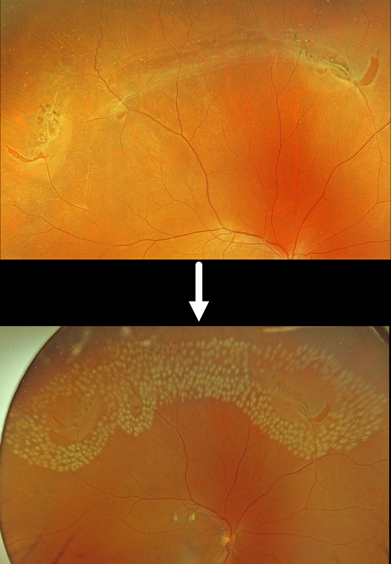
This 55YO male was referred for an acute posterior vitreous detachment with symptomatic retinal tears. Vision was 20/25.
Optos color RGB imaging shows several patches of lattice degeneration extending from the 10 to 1:30 o’clock meridians associated with multiple retinal tears. Some inferior vitreous hemorrhage was noted (not shown). Immediate prophylactic laser photocoagulation was applied to all surrounding the lattice and breaks.
Lattice lesions have a pocket of liquefied vitreous overlying thinned retina with a firm area of surrounding vitreoretinal adhesion. White, fibrosed vessels often crisscross the lesions, giving them their name, “lattice.” This firm vitreoretinal adhesion, along with the often-present atrophic holes, is what can predispose these eyes to retinal breaks and detachment. Although prophylactic treatment is rarely needed, symptomatic breaks, as in our patient, need prompt retinopexy.
LATTICE DEGENERATION WITH SUBCLINICAL DEGENERATIVE RETINOSCHISIS
Originally posted on @retina.rocks March 5, 2026
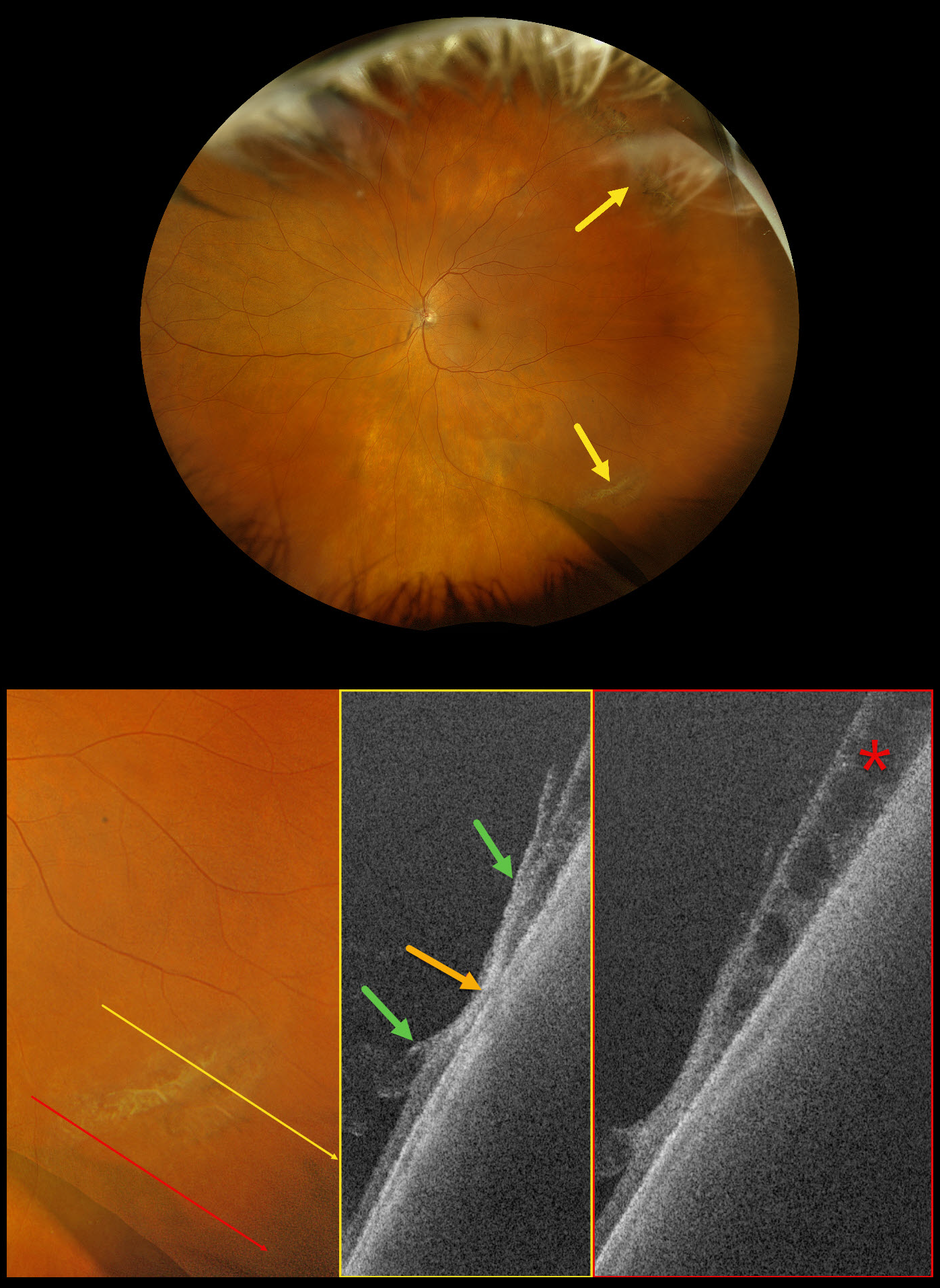
This 73-year-old female was referred for a symptomatic posterior vitreous detachment (PVD) with a retinal break in her left eye. Vision was 20/20.
Optos Silverstone RGB imaging shows supero- and inferotemporal lattice degeneration with some atrophic retinal holes. Swept-source OCT through the lattice shows a thinned, disorganized retina with areas of vitreoretinal adhesion along its margins. A more inferior line scan shows degenerative retinoschisis, which could not be visualized clinically or on the Optos image.
Our patient showed no retinal breaks from her symptomatic PVD, and observation was recommended.
Learning Points:
Ultrawidefield imaging has revolutionized our specialty, making many of us realize how limited our clinical examinations are compared with our imaging devices. In our patient’s case, the RGB image captured the fundus nearly from ora to ora. Swept-source OCT provided a near histologic view of the lattice pathology, including atrophic retina centrally, with a firm area of vitreoretinal adhesion along its margins. And finally, it revealed subclinical degenerative retinoschisis.
ASYMPTOMATIC LATTICE DEGENERATION WITH HOLES
Lattice degeneration SLF-
Originally posted on @retina.rocks February 18, 2025
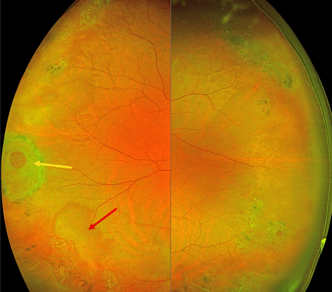
This 42YO female was referred for asymptomatic peripheral retinal findings. Vision was 20/20 OU.
Optos color RG imaging shows extensive bilateral temporal lattice lesions with atrophic holes. A larger hole with surrounding pigment is seen in the right eye, along with a nearby area of white without pressure.
Learning Points:
Lattice lesions have a pocket of vitreous liquefaction overlying the thinned retina with a firm vitreo-retinal adhesion along their borders. White, fibrosed vessels often crisscross the lesions, which leads to the name, lattice. This firm vitreoretinal adhesion, along with the often-present atrophic holes, is what can predispose these eyes to rhegmatogenous retinal detachment (RRD).
Retinal detachment occurs in 1 in 10,000 people per year. Lattice is found in 10% of the general population, especially in high myopes. Since about one-third of patients with retinal detachment have lattice as the cause, the question always arises: Should prophylactic retinopexy be performed? The short answer is that asymptomatic lattice should generally not be treated.
Byer, in a Long-term natural history study of 423 untreated eyes, concluded that prophylactic treatment with or without holes in phakic non-fellow eyes should not be performed (Ophthalmology 1989;96:1396-1402). Folk et al (Ophthalmology 1989;96:72-79, Retina 1990;10:165-169) found no compelling evidence for prophylactic treatment of phakic fellow higher-risk eyes with more extensive lattice or higher myopia. They also found that some eyes developed subsequent non-lattice breaks outside the prophylactic treatment. A recent Cochrane systematic review by Wilkinson concluded that asymptomatic breaks and lattice should not be treated (Cochrane Database of Systematic Reviews 2014;9;CD003170) and the most recent 2019 American Academy of Ophthalmology Preferred Practice Pattern also cautions against prophylactic treatment.
A recent study by Curran et al recommended prophylactic treatment of lattice in fellow retinal detachment eyes, but this was in a retrospective, uncontrolled case series (Retina 2024;44:63-70). Another recent report by Kazan et al (Journal of VitreoRetinal Diseases 2024;8:381-387), in an uncontrolled retrospective series of eyes with “high-risk” lattice, found few post-laser detachments and concluded that treatment “may be effective in decreasing progression to RD…” But 3% of their treated eyes developed RRD over a mean 3- to 4-year follow-up period, which is higher than expected from the natural history of untreated lattice. So, our interpretation of their results is that prophylactic treatment may have actually increased the risk for detachment.
Stickler syndrome is a usually autosomal-dominant genetic disorder characterized by defective vitreous collagen (types II, IX, and XI). It is the most common cause for inherited rhegmatogenous retinal detachment and is the exception to the rule against prophylactic treatment. Prophylactic encircling laser extending from the pre-equator to the ora should be strongly considered since it lowers the risk for retinal detachment and giant retinal tears (Linton et al, Retina 2023;43:88-93).
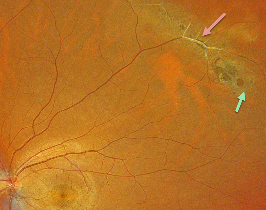
LATTICE DEGENERATION
Originally posted on @retina.rocks December 5, 2024
This healthy 33YO male was referred for asymptomatic lattice degeneration in his left eye. Optos color RGB imaging shows a circumferential patch of midperipheral superotemporal paravenous lattice. The large retinal vessels within the lesion are fibrosed. A small atrophic retinal hole is noted along its inferior edge. Observation was recommended.
Learning Points:
Lattice lesions have a pocket of vitreous liquefaction overlying the thinned retina with a firm vitreo-retinal adhesion along their borders. White, fibrosed vessels often crisscross the lesions, hence the name, lattice. This firm vitreoretinal adhesion, along with the often-present atrophic holes, is what can predispose these eyes to retinal detachment.
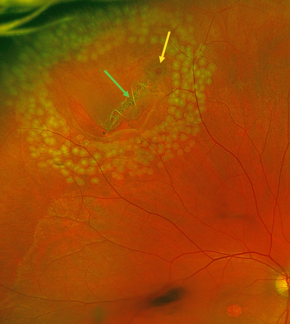
RETINAL BREAK
Justin Grassmeyer, Ambar Faridi, and Brittany Heckerman
Originally posted on @retina.rocks June 5, 2024
This 63YO male with history of idiopathic central serous chorioretinopathy (ICSC) with secondary macular neovascularization OD and lattice degeneration OU presented with new floaters in his right eye. He was scheduled for an anti-VEGF injection for the right eye on a treat-and-extend protocol the same day. Vision was stable at 20/100 OD and 20/20 OS. An acute, related retinal tear was found and immediately demarcated with thermal laser photocoagulation.
Optos color RG imaging immediately following the laser procedure shows the retinal break well surrounded by multiple nearly confluent rows of laser burns. The flap of the tear consists of an entire lattice lesion along with a chronic atrophic hole with surrounding pigment. There is an area of central macular atrophy from the ICSC.
Learning Points:
Lattice lesions have a pocket of liquefied vitreous overlying thinned retina with a firm area of surrounding vitreoretinal adhesion. White, fibrosed vessels often crisscross the lesions, which gives the lesions their name, “lattice.” This firm vitreoretinal adhesion, along with the often-present atrophic holes, is what can predispose these eyes to retinal breaks and detachment. Although prophylactic treatment is rarely needed, symptomatic breaks, as in our patient, need prompt retinopexy.
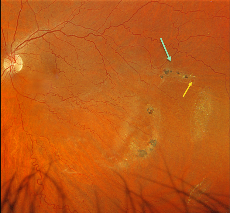
LATTICE DEGENERATION
Originally posted on @retina.rocks February 6, 2024
This 42YO male was referred for asymptomatic findings on retinal examination in his left eye. Vision was 20/20 in his normal OD and 20/20 OS.
Optos color RGB imaging shows patches of variably pigmented lattice degeneration in the inferotemporal midperiphery. At least one atrophic retinal hole is noted in the superior-most lesion.
The retinal veins are also diffusely tortuous except superotemporally, likely indicating a prior spontaneously resolved retinal vein occlusion.
Observation was recommended.
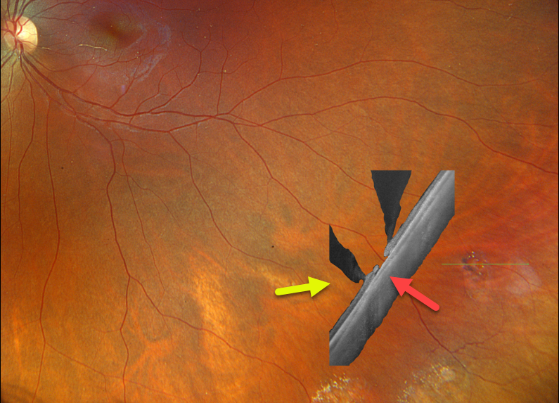
LATTICE DEGENERATION
Originally posted on @retina.rocks January 30, 2024
This healthy 27YO female has a known history of lattice degeneration with atrophic holes. She is completely asymptomatic, and vision is 20/20 OU.
Optos color RGB imaging of her left eye shows patches of inferotemporal lattice degeneration. Optos California Silverstone swept-source OCT through a lesion containing an atrophic retinal hole reveals classic findings, including retinal thinning within the lattice, liquified vitreous overlying the lattice lesion, and adherent vitreous along the edges of the lattice.
Learning Points:
Lattice lesions have a pocket of vitreous liquefaction overlying the thinned retina with a firm vitreo-retinal adhesion along their borders. White, fibrosed vessels often crisscross the lesions, which leads to the name “lattice”. This firm vitreoretinal adhesion, along with the often present atrophic holes, is what can predispose these eyes to retinal detachment.
LATTICE DEGENERATION
Originally posted on @retina.rocks November 9, 2023
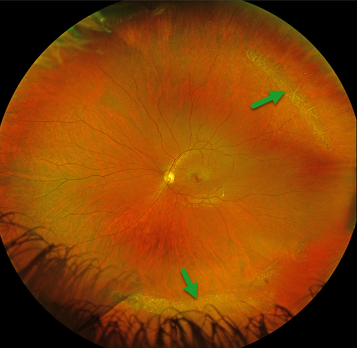
This 35YO female was referred for asymptomatic lattice degeneration. Vision was 20/20 OU.
Optos color RGB imaging of her left eye shows prominent temporal and inferior lattice lesions, some of which are associated with atrophic retinal holes. Similar findings were noted in her right eye (not shown).
Learning Points:
Lattice lesions have a pocket of vitreous liquefaction overlying the thinned retina with a firm vitreo-retinal adhesion along their borders. White, fibrosed vessels often crisscross the lesions, which leads to the name “lattice”. These are particularly well seen in our patient’s superotemporal lesion. This firm vitreoretinal adhesion, along with the often present atrophic holes, is what can predispose these eyes to retinal detachment.
Retinal detachment occurs in 1 in 10,000 people per year. Lattice is found in 10% of the general population, especially in high myopes. Since about one-third of patients with retinal detachment have lattice as the cause, the question always arises: Should prophylactic retinopexy be performed?
The short answer is that asymptomatic lattices generally should not be treated. To put things into perspective, imagine a sports stadium of 30,000 people. Three people will develop a retinal detachment in the coming year. Only one of the 3,000 people in the stadium with a lattice would develop one of those 3 detachments. Assuming prophylactic treatment would be 100% effective and complication-free, 2,999 eyes would be unnecessarily treated to prevent one detachment.
LATTICE DEGENERATION
Originally posted on @retina.rocks June 12, 2023
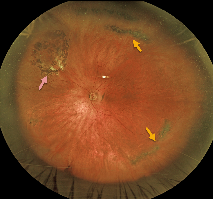
This 68YO male presented with asymptomatic lattice degeneration bilaterally. He remembered getting a laser to one of his eyes many years earlier at an outside practice.
Optos imaging of his left eye shows a near ora to ora view of scattered circumferential pigmented lattice lesions. Laser scarring surrounds an area of lattice with atrophic holes superonasally.
Learning Points:
Lattice lesions have a pocket of vitreous liquefaction overlying the thinned retina with a firm vitreo-retinal adhesion along their borders. White, fibrosed vessels often criss-cross the lesions, which leads to the name, lattice. This firm vitreoretinal adhesion, along with the often-present atrophic holes, can predispose these eyes to retinal detachment.
Retinal detachment occurs in 1 in 10,000 people per year. Lattice is found in 10% of the general population, especially in high myopes.
Since about one-third of patients with retinal detachment have lattice as the cause, the question always arises, especially in patients like this: Should prophylactic retinopexy be performed? The short answer is that asymptomatic lattices generally should not be treated.
To put things into perspective, imagine a sports stadium of 30,000 people. Three people will develop a retinal detachment in the coming year. Only one of the 3,000 people in the stadium with lattice would develop one of those 3 detachments. Assuming prophylactic treatment is 100% effective with no complications, 2,999 eyes would be unnecessarily treated to prevent one detachment.
RHEGMATOGENOUS RETINAL DETACHMENT
Originally posted on @retina.rocks May 29, 2023
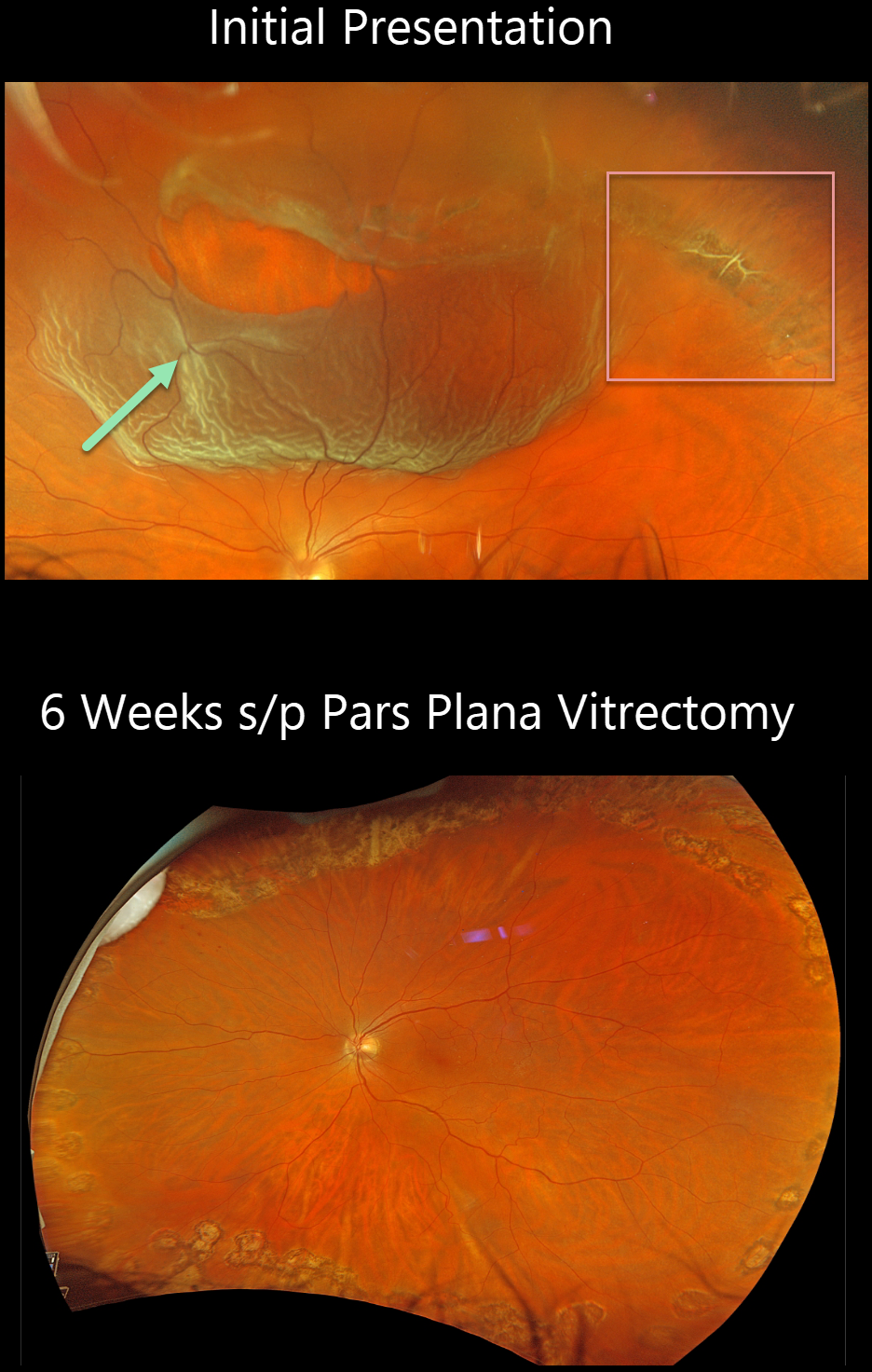
This 55YO male presented with this acute superior macula-on rhegmatogenous retinal detachment (RRD). Vision was 20/30.
Optos color RGB imaging shows a large patch of equatorial lattice degeneration extending from 11 to 2 o’clock. Nasally, the lattice lesion has torn along its lateral and posterior edges, causing the detachment. Several intact bridging retinal vessels are noted within the tear.
The detachment was repaired with vitrectomy the following day and remained attached 6 weeks later, following complete absorption of the intraocular gas bubble.
LATTICE DEGENERATION WITH RETINAL TEAR
Originally posted on @retina.rocks December 13, 2022
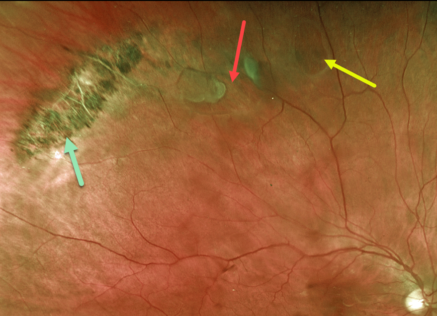
This 64YO male presented with acute flashes and floaters in his right eye. Optos imaging shows a patch of pigmented lattice superotemporally, associated with a retinal tear and atrophic hole. The breaks were surrounded with laser photocoagulation.
Learning Points:
Retinal detachment occurs in 1 in 10,000 people per year. Lattice is found in 10% of the general population, especially in high myopes. Since about one-third of patients with retinal detachment have lattice as the cause, the question always arises: Should prophylactic retinopexy be performed?
The short answer is that asymptomatic lattice generally should not be treated. However, an eye with symptomatic lattice breaks, as in our patient, is at high risk for developing rhegmatogenous detachment and should always be treated.
Excellent references on this controversial topic include: Byer Ophthalmology 1989;96:1396-1402, Folk et al Ophthalmology 1989;96:72-79 and Folk et al Retina 1990;10:165-169.
RHEGMATOGENOUS RETINAL DETACHMENT
Originally posted on @retina.rocks April 15, 2022
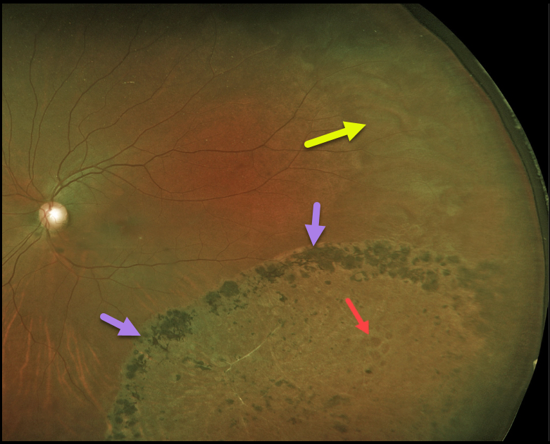
Years earlier, this 44YO female underwent laser demarcation of an asymptomatic inferotemporal rhegmatogenous retinal detachment (RRD). The prior detachment remains well demarcated, but the detachment itself has completely flattened.
Some of the causative lattice-related atrophic holes are seen within the area of prior detachment. White without pressure is also noted in the temporal periphery.
Learning Points:
Demarcation, rather than reattachment, of the retina is a valid option for select cases of RRD. When demarcating these detachments, it is critical that the retinopexy completely surrounds the subretinal fluid and that treatment extends to the ora; otherwise, the detachment can spread through the untreated retina.
Patients need to be followed postoperatively for life since the detachment can occasionally progress through the prior retinopexy.
Interestingly, we have seen that some of these detachments, as occurred in this case, spontaneously flatten following demarcation.
See Vrabec and Baumal, Ophthalmology 2000;107:1063-1067 for an excellent review regarding demarcation laser photocoagulation for selected macula-sparing retinal detachments.
RHEGMATOGENOUS RETINAL DETACHMENT
Originally posted on @retina.rocks April 1, 2022
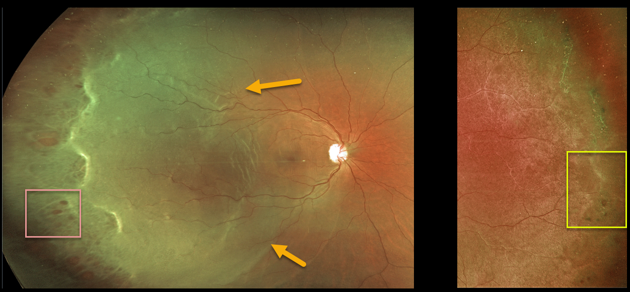
This 32YO female presented with a macula-off rhegmatogenous retinal detachment (RRD) in her right eye. There were innumerable temporal retinal holes.
The detachment was successfully repaired with vitrectomy surgery.
Her asymptomatic left eye showed extensive temporal lattice lesions with atrophic holes, and prophylactic laser was recommended.
RHEGMATOGENOUS RETINAL DETACHMENT
Originally posted on @retina.rocks February 25, 2022
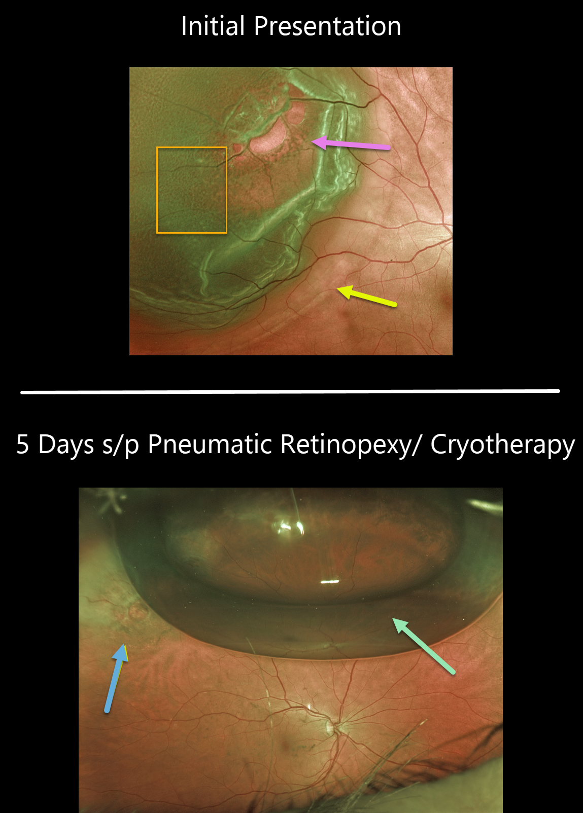
This 63YO female presented with 20/25 vision and a superotemporal retinal detachment. The posterior edge of the detachment was just outside the macular center. There was diffuse fine microcystic retinal edema within the area of detachment, except for outer retinal hydration folds more posteriorly.
The retina was completely attached 5 days later following successful cryotherapy with pneumatic retinopexy. Early cryotherapy scarring is already visible.
Learning Points:
This detachment could have been repaired with any technique, including vitrectomy, scleral buckling, or pneumatic retinopexy. All techniques yield similar final anatomic success, although the debate continues about the relative merit of one over the other.
See the ___References folder in the Retinal detachment (RD) rhegmatogenous (RRD) folder in the Image Library at www.retinarocks.org for a summary of the key journal articles.
LATTICE DEGENERATION
Originally posted on @retina.rocks February 8, 2022
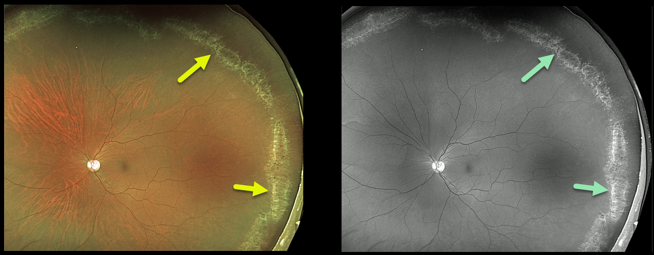
This 36YO male presented with extensive asymptomatic lattice degeneration seen on Optos color photography and Optos green channel.
The circumferential glistening lattice lesions are also called snail tracks. Treatment was not recommended.
Learning Points:
White, fibrosed vessels often crisscross the lesions, which gives rise to the term “lattice.” There is vitreous liquefaction overlying the thinned retina with a firm vitreo-retinal adhesion along the borders of the lesions. This firm vitreoretinal adhesion, along with the often-present atrophic holes, can predispose these eyes to retinal detachment.
Retinal detachment occurs in 1 in 10,000 people per year. Lattice is found in 10% of the general population, especially in high myopes. Since about one-third of patients with retinal detachment have lattice as the cause, the question always arises, especially in patients like this: Should prophylactic retinopexy be performed? The short answer is that asymptomatic lattice generally should not be treated.
LATTICE DEGENERATION
Originally posted on @retina.rocks June 9, 2021
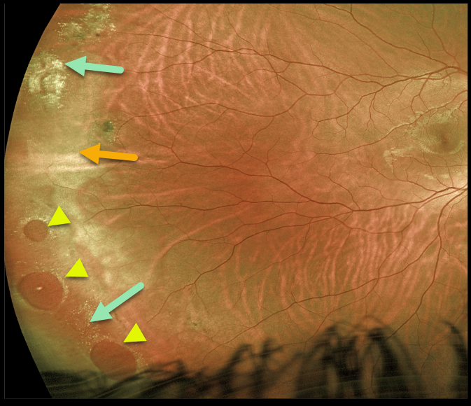
This 24YO male presented with asymptomatic but somewhat confusing clinical findings beautifully captured by this Optos image.
Three large atrophic retinal holes within an area of lattice are found in the inferotemporal periphery, along with a few areas of lattice and scarring superotemporally. There is also associated white without pressure, giving the appearance of a retinal detachment (remember the normally transparent retina turns whitish when it detaches). However, the retina was completely attached, and we are treating him with observation.
Learning Points:
Retinal detachment occurs in 1 in 10,000 people per year. Lattice is found in 10% of the general population, especially in high myopes. Since about one-third of patients with retinal detachment have lattice as the cause, the question always arises, especially in patients like this: Should prophylactic retinopexy be performed?
The short answer is that asymptomatic lattice generally should not be treated. To put things into perspective, consider a pre-COVID, non-social-distancing football stadium with 30,000 people. Three people will develop retinal detachment in the coming year. Only one of the 3,000 people in the stadium with lattice would develop one of those 3 detachments.
Excellent references on this controversial topic include: Byer Ophthalmology 1989;96;1396-1402, Folk et al Ophthalmology 1989;96;72-79, and Folk et al Retina 1990;10:165-169.
LATTICE DEGENERATION
Originally posted on @retina.rocks April 23, 2020
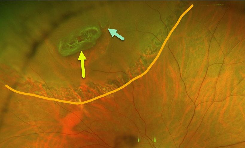
This patient had an asymptomatic peripheral lattice-related retinal detachment. Note that the entire lattice lesion represents the flap of the tear, with an adjacent secondary smaller tear.
The detachment was demarcated with laser photocoagulation and has remained stable since.
Learning Points:
Retinal detachment occurs in 1 in 10,000 people per year. Lattice is found in 10% of the general population, especially in high myopes.
Since about one-third of patients with retinal detachment have lattice as the cause, the question always arises, especially in patients like this: should prophylactic retinopexy be performed? The short answer is that asymptomatic lattice generally should not be treated.
However, this patient had lattice-related retinal detachment and thus underwent laser demarcation.
An especially relevant article for this topic is by Vrabec and Baumal, Ophthalmology 2000;107;1063-1067.
LATTICE DEGENERATION
Originally posted on @retina.rocks April 14, 2020
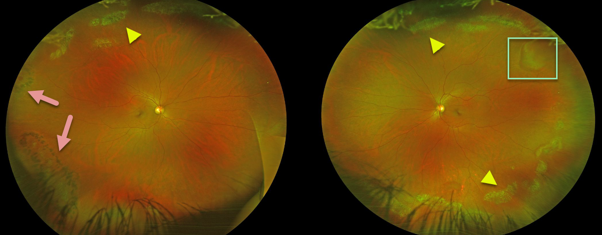
Beautiful Optos images of this patient demonstrate extensive bilateral lattice degeneration.
Prophylactic laser was placed at an outside institution around some of the lattice lesions in the right eye inferotemporally and around an atrophic hole temporally.
There is a pseudo-retinal tear superotemporally in the left eye from a patch of white without pressure.
Learning Points:
White, fibrosed vessels often criss-cross the lesions, which lead to the name, lattice. There is vitreous liquefaction overlying the thinned retina with a firm vitreo-retinal adhesion along the borders of the lesions. This firm vitreoretinal adhesion, along with the often present atrophic holes, is what can predispose these eyes to retinal detachment.
Retinal detachment occurs in 1 in 10,000 people per year. Lattice is found in 10% of the general population, especially in high myopes. Since about one-third of patients with retinal detachment have lattice as the cause, the question always arises, especially in patients like this: should prophylactic retinopexy be performed? The short answer is that asymptomatic lattice generally should not be treated.


