

Leukemic Optic Nerve Infiltration
<< Back to Cases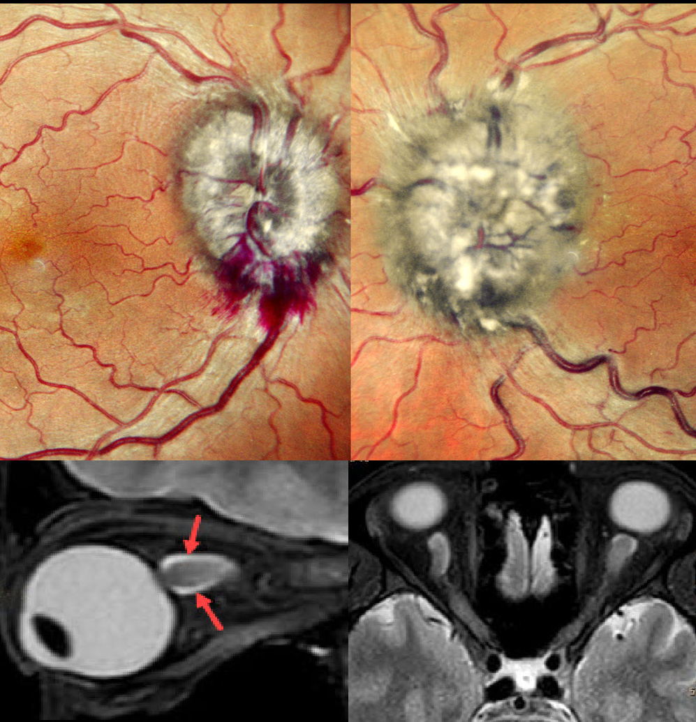
LEUKEMIC OPTIC NERVE INFILTRATION WITH PSEUDOPAPILLEDEMA
Anjana Mirajkar and Manish Nagpal
Originally posted on @retina.rocks July 8, 2024
This 40YO male with a known 10-year history of chronic myeloid leukemia complained of vision loss in his left eye for 3 weeks. Vision was 20/20 OD and hand motion OS.
Fundus examination revealed marked bilateral optic nerve swelling, with the left eye greater than the right eye, and some nerve fiber layer hemorrhages along the right inferior disc margin. MRI scanning was consistent with leukemic infiltration of the optic chiasm and optic nerves. She was referred back to her oncologist.
Learning Points:
Leukemia with central nervous system involvement can rarely present with leukemic optic nerve infiltration. This can masquerade as papilledema (bilateral swollen nerves from elevated intracranial pressure) or nerve edema from inflammatory or infectious causes (Miller et al, Graefe’s 2021;259;1315-1322).


