

Lipemia Retinalis
<< Back to Cases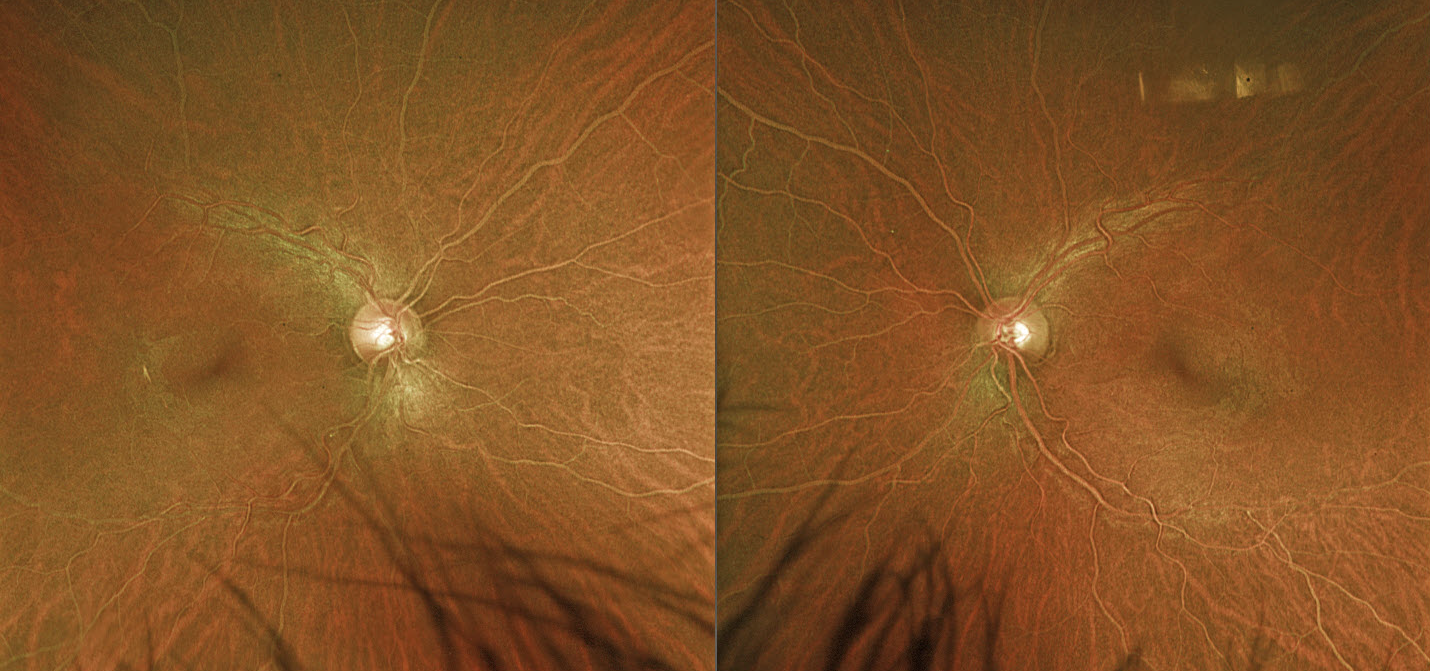
LIPEMIA RETINALIS
Hailey Dunlow
Originally posted on @retina.rocks February 4, 2026
This previously healthy 38YO male was referred for asymptomatic retinal findings. Vision was 20/25 OD and 20/20 OS. Optos color RG imaging shows symmetric, creamy-colored blood vessels bilaterally. Emergent bloodwork showed total cholesterol 1093 mg/dL, triglycerides 9644 mg/dL, and HDL 4 mg/dL. He was immediately referred to his PCP for emergent medical management. He was immediately lost to follow up.
Learning Points:
Lipemia retinalis is a rare retinal manifestation of severe hypertriglyceridemia, characterized by creamy white discoloration of the retinal vasculature. In severe cases, the fundus can turn salmon-colored. Patients are asymptomatic, and vision is normal. However, they are at obvious cardiovascular risk without systemic treatment.
LIPEMIA RETINALIS
Originally posted on @retina.rocks August 21, 2023
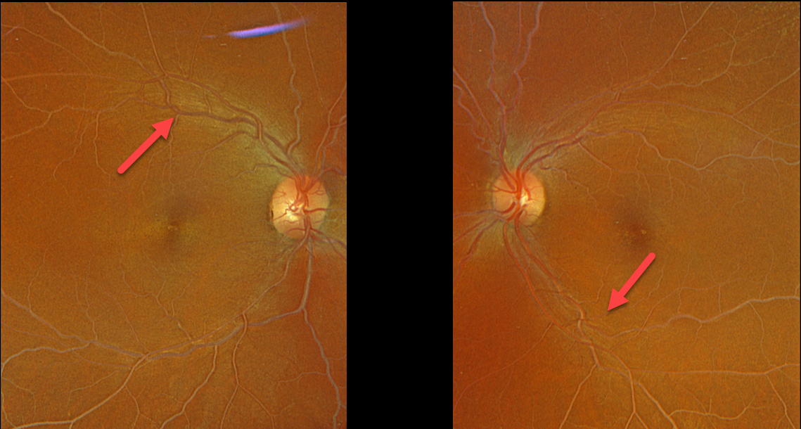
This 47YO female presented with bilateral symmetric creamy colored blood vessels.
Her lipid panel results were: Cholesterol 1039, Triglyceride 821, HDL 7, and LDL too high to measure.
She was immediately referred for medical management of her previously undiagnosed hyperlipidemia.
Learning Points:
Lipemia retinalis is a rare retinal manifestation of severe hypertriglyceridemia, characterized by creamy white discoloration of the retinal vasculature. In severe cases, the fundus can turn salmon-colored.
Patients are asymptomatic and have normal vision. However, they are at obvious cardiovascular risk without systemic treatment.
LIPEMIA RETINALIS
Nayara Pinto and Romano Vasconcelos
Originally posted on @retina.rocks October 11, 2022
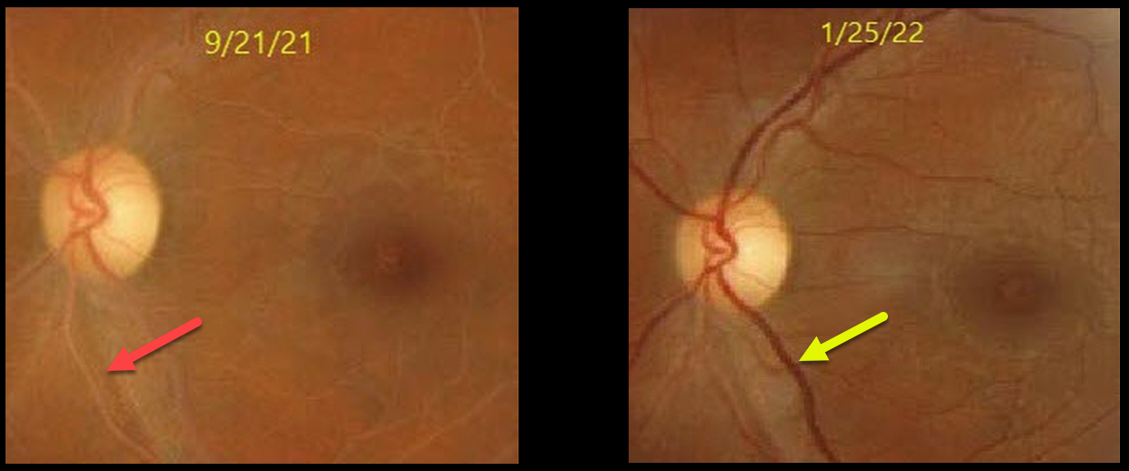
This 28YO female presented with bilateral symmetric creamy colored blood vessels (only left eye shown). Triglycerides were markedly elevated above 1000.
The vascular appearance normalized 4 months following systemic treatment with a relatively normal triglyceride level of 189.
Learning Points:
Lipemia retinalis is a rare retinal manifestation of severe hypertriglyceridemia, characterized by creamy white discoloration of the retinal vasculature. In severe cases, the fundus can turn salmon-colored.
Patients are asymptomatic, and vision is normal. However, they are at obvious cardiovascular risk without systemic treatment.
LIPEMIA RETINALIS
Originally posted on @retina.rocks July 21, 2022
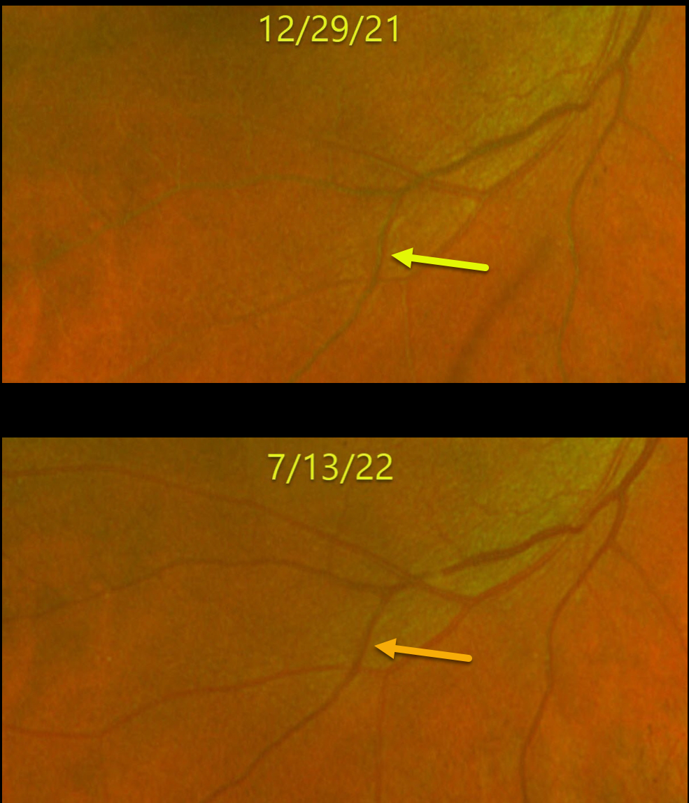
This 51YO male’s retinal vasculature turned creamy white from very high triglycerides. His original triglyceride levels were around 4,200 mg/dL.
After 6 months of systemic treatment, his triglyceride levels decreased to 3,100 mg/dL. His blood returned to a normal red color, and his retinal vessels normalized in appearance.
Learning Points:
Lipemia retinalis is a rare retinal manifestation of severe hypertriglyceridemia, characterized by creamy white discoloration of the retinal vasculature. In severe cases, the fundus can turn salmon-colored.
Patients are asymptomatic, and vision is normal. However, they are at obvious cardiovascular risk without systemic treatment.
LIPEMIA RETINALIS
Grace Wong
Originally posted on @retina.rocks May 10, 2021
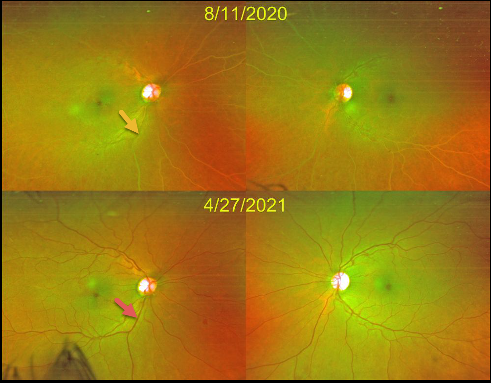
This 49YO female’s retinal vasculature was initially creamy white from very high triglycerides.
After 8 months of systemic treatment, her triglyceride levels normalized, her blood turned back to its normal red color, and her retinal vessels normalized in appearance.
Learning Points:
Lipemia retinalis is a rare retinal manifestation of severe hypertriglyceridemia, characterized by creamy white discoloration of the retinal vasculature. In severe cases, the fundus can turn salmon-colored.
Patients are asymptomatic, and vision is normal. However, they are at obvious cardiovascular risk without systemic treatment.
LIPEMIA RETINALIS
Originally posted on @retina.rocks September 10, 2020
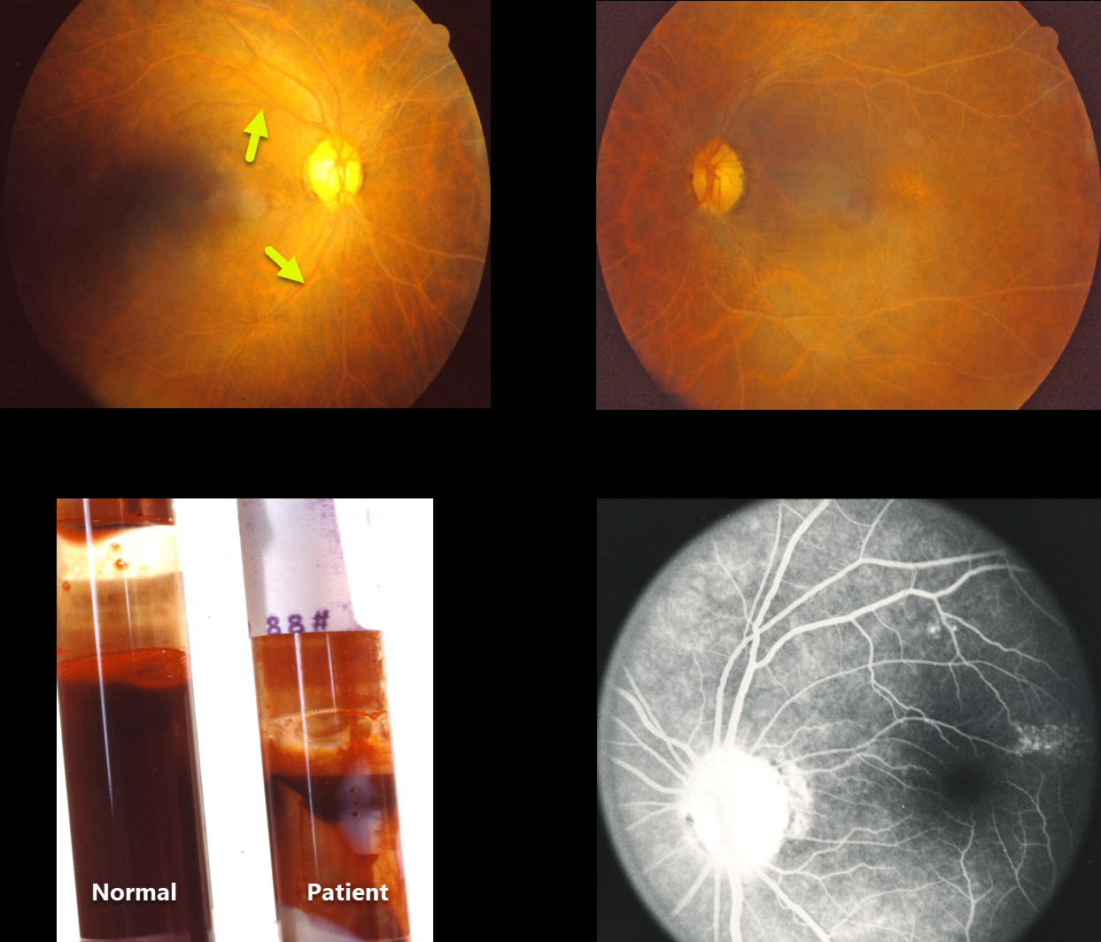
This patient’s blood vessels appear milky and possibly occluded, although fluorescein angiography is normal.
A tube of blood drawn from both the patient and a much younger 25YO future founder of Retina Rocks (in his first year of ophthalmology residency) is displayed for comparison. The patient’s blood shows increased lipid content.
Learning Points:
Lipemia retinalis is a rare manifestation of severe hypertriglyceridemia. The blood vessels, and sometimes the entire posterior pole, may appear milky or even salmon-colored.
Patients are asymptomatic, and vision is normal. However, they are at obvious cardiovascular risk without systemic treatment.


