

Lymphoma
<< Back to Cases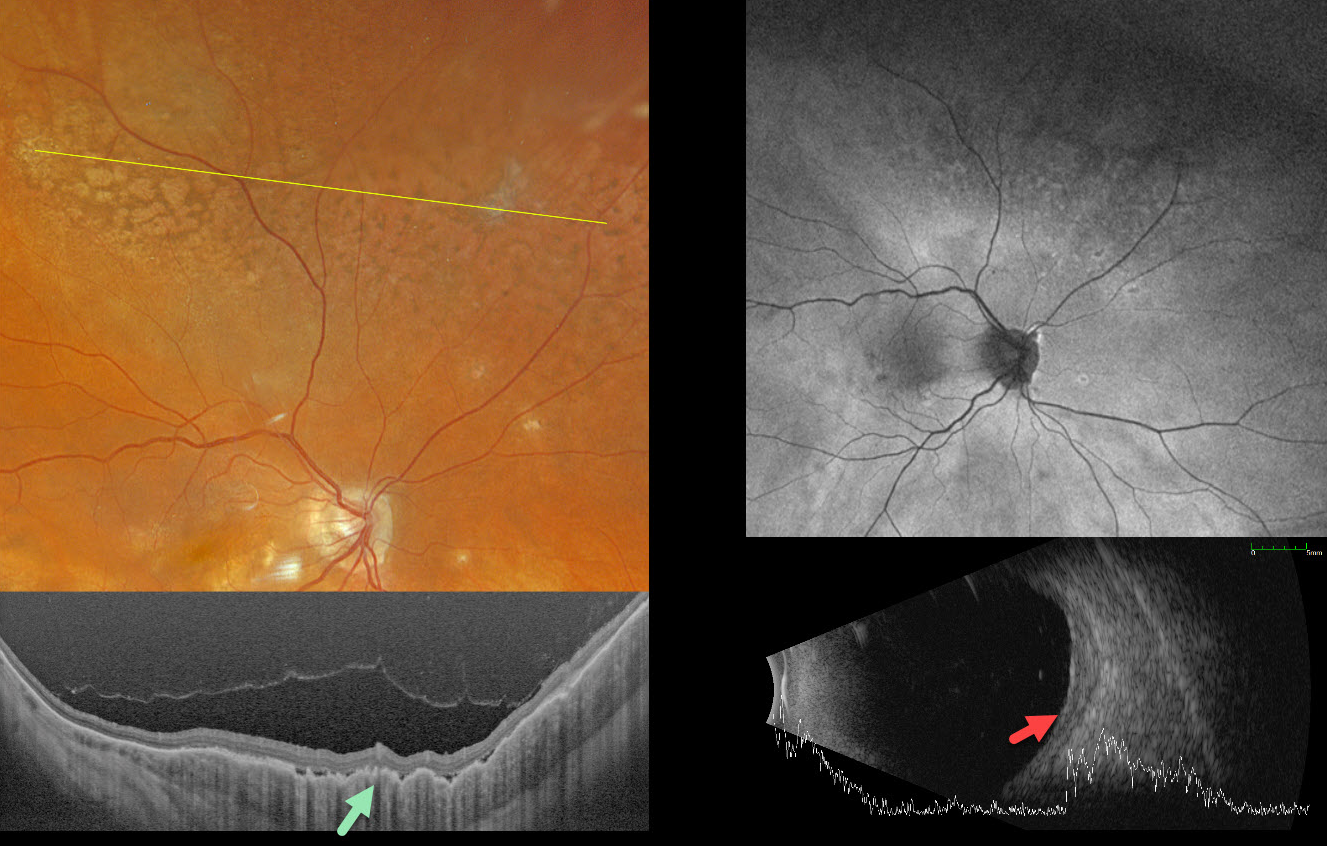
UVEAL LYMPHOMA
Janelle Adeniran
Originally posted on @retina.rocks June 19, 2025
This 69YO female was receiving ongoing bilateral anti-VEGF therapy for neovascular age-related macular degeneration when she presented with new asymptomatic fundus changes in her left eye. There was no significant past medical history.
Optos color RGB imaging shows a leopard spot pattern of superior midperipheral pigmentary changes. Silverstone swept-source OCT shows a markedly thickened choroid with overlying chorioretinal folds. Fundus autofluorescence (FAF) is fairly unremarkable despite these funduscopic findings. The choroid is diffusely thickened on B-scan ultrasonography, with a maximal thickness of 1.90mm.
CT of the chest, abdomen, and pelvis was negative. MRI of the brain and orbits was also negative. We felt this represented primary uveal lymphoma, and this was confirmed with an outside third opinion before beginning serial intravitreal methotrexate injections. After 11 months of treatment, the choroid flattened clinically and on ultrasonography, shrinking to 1.09mm in thickness (not shown).
HODGKIN LYMPHOMA MIMICKING MULTIFOCAL CHOROIDITIS
Originally posted on @retina.rocks December 4, 2024
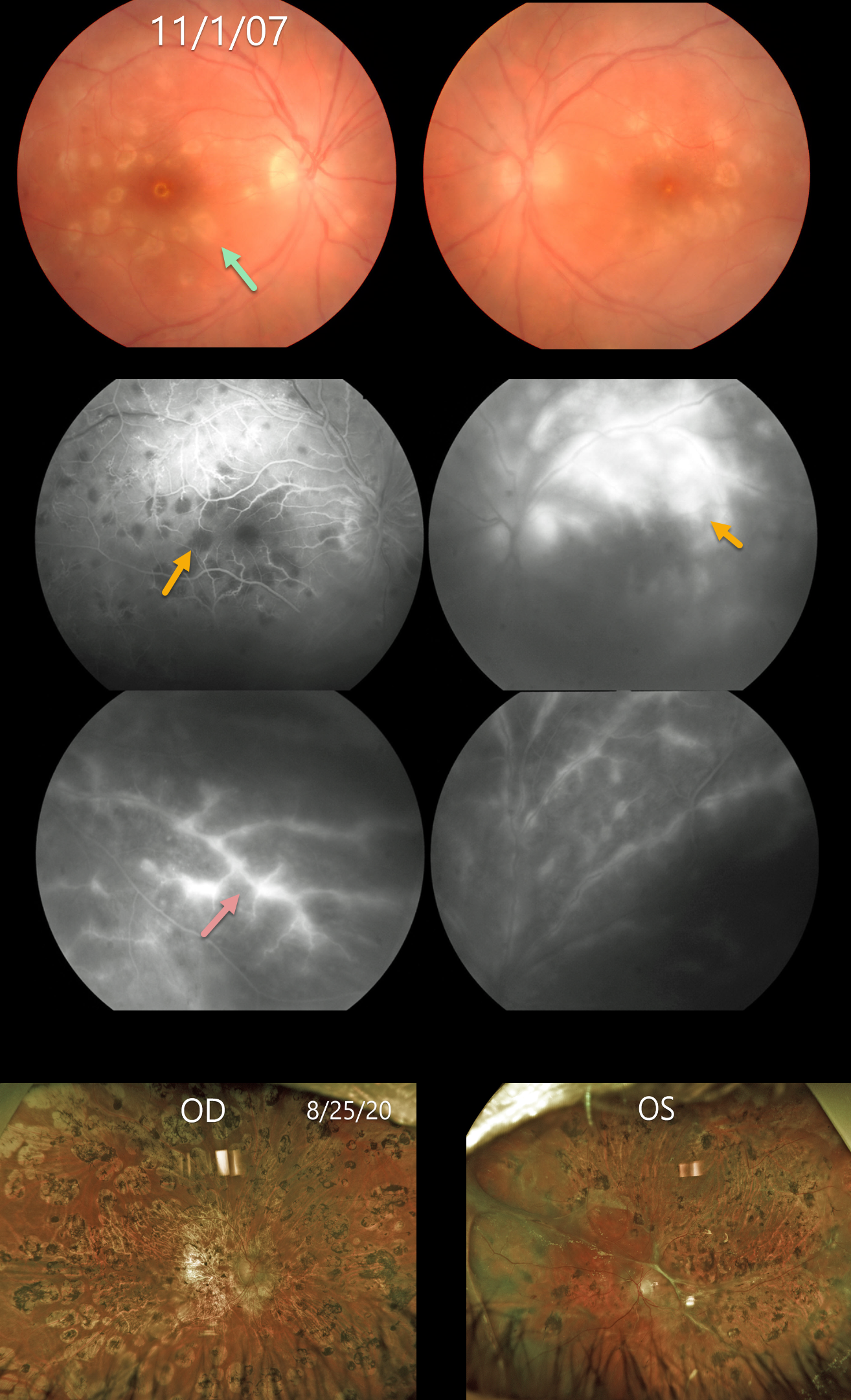
This 18YO female presented on 11/1/07 with acute bilateral panuveitis. Moderate vitreous cells were present along with subretinal multifocal yellow-white choroidal infiltrates. Angiographically, these choroidal lesions blocked early and stained late. A frosted-branch pattern of vascular leakage was noted bilaterally. Following a course of oral prednisone, the acute uveitic findings resolved, leaving behind inactive multifocal scarring. She developed severe recurrent panuveitis a year or two later, leaving her with 20/400 vision bilaterally (images not shown).
Shortly thereafter, she was diagnosed with Hodgkin lymphoma. At almost the exact date of this diagnosis, Baillif et al reported a patient with bilateral angle-closure glaucoma and multifocal choroiditis as the initial presentation for Hodgkin lymphoma (Retinal Cases 2011;5:201-205). The posterior segment findings in this report were virtually identical to those in our patient. The authors felt that the uveitis was a paraneoplastic phenomenon.
She subsequently underwent successful bone marrow transplantation. Her lymphoma and uveitis have both remained in remission since. A rhegmatogenous retinal detachment later developed in her left eye, which was repaired with vitrectomy and silicone oil. Despite successful reattachment, vision subsequently decreased to no light perception. On 8/25/20, vision was 20/400 OD with severe stable bilateral chorioretinal scarring bilaterally.
Learning Points:
Multifocal choroiditis (MFC) is the current preferred term for a group of historically heterogeneous disorders, including multifocal choroiditis, recurrent multifocal choroiditis, multifocal choroiditis and panuveitis, punctate inner choroidopathy, progressive subretinal fibrosis, and pseudo-histoplasmosis (Essex et al, Retina 2013;33:1-4 and Spaide et al, Retina 2013;33:1315-1324). MFC occurs in otherwise healthy adults, often myopic and female. Findings can include variably pigmented multifocal scars throughout the fundus, panuveitis, subretinal fibrosis, and macular neovascularization (MNV). Unlike ocular histoplasmosis, new scars and uveitis can be seen. MNV can be successfully managed with anti-VEGF therapy.
As with any uveitis, the clinician must always include the possibility of a masquerade syndrome as occurred in our patient.
CHOROIDAL METASTASES
Originally posted on @retina.rocks January 23, 2023
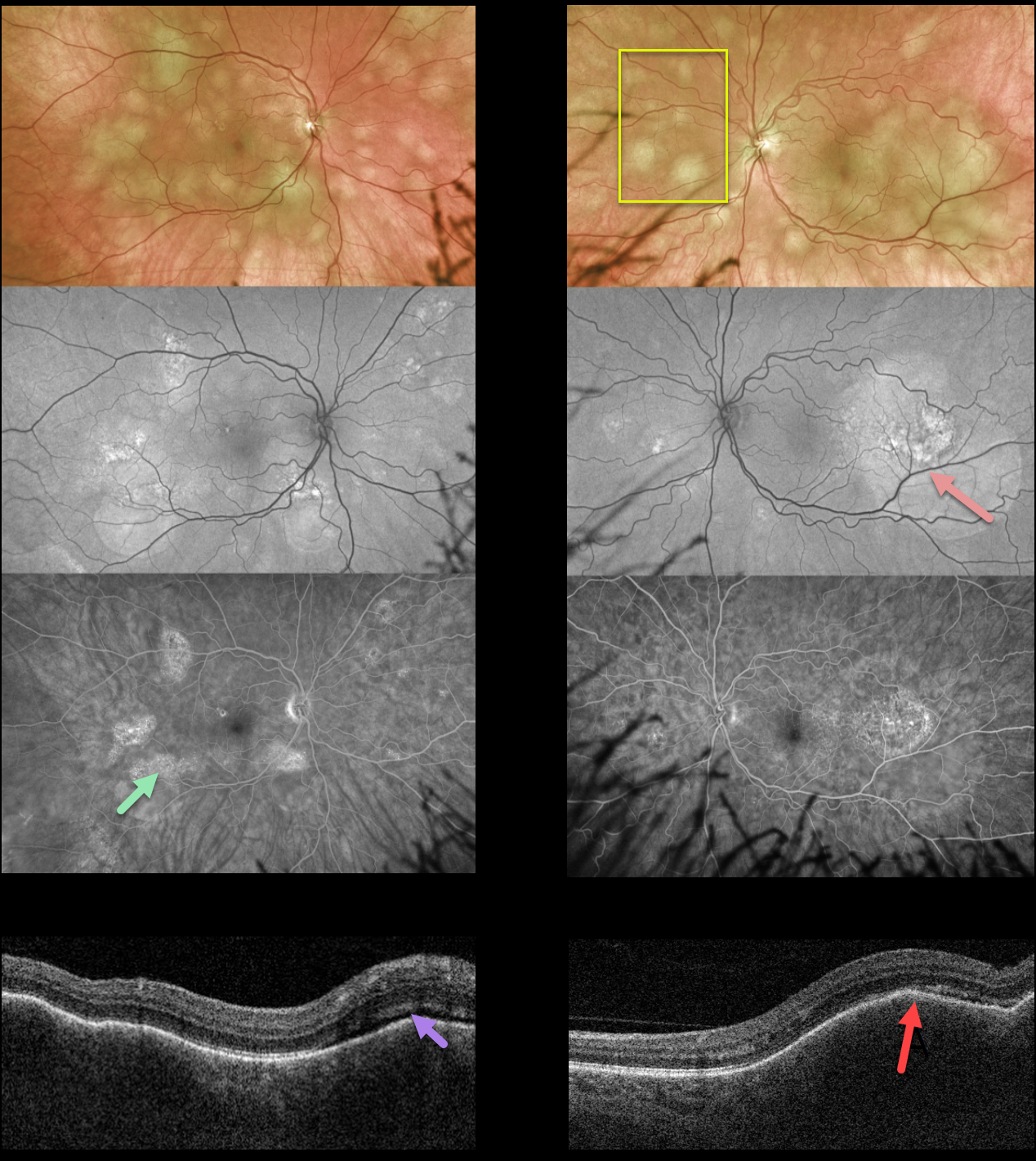
This 58YO female presented with 2 weeks of blurred vision in her left eye. There was no past medical history. Vision was 20/40 OD and 20/200 OS.
Optos color imaging shows multifocal, creamy-yellow subretinal lesions bilaterally. The lesions are more confluent, extending beneath the left fovea.
The smaller of these lesions is fairly silent on fundus autofluorescence, although the larger ones show hyper-FAF. These lesions angiographically show variable late staining with window defects.
Swept-source OCT shows bilateral thickened and undulating choroid with trace overlying subretinal fluid.
She was referred to the emergency room and was subsequently diagnosed with widely metastatic lung adenocarcinoma.
Learning Points:
Although uveal melanoma is the most common primary intraocular malignancy, metastatic choroidal tumors are the most common intraocular malignancies. Lung and breast cancer are the most common sources.
The extensive multifocal lesions in each of our patients’ eyes are unusual, since most patients have 1-2 choroidal lesions (Shields et al Retina 2020;40:204-213).
LYMPHOMA
Originally posted on @retina.rocks January 5, 2022
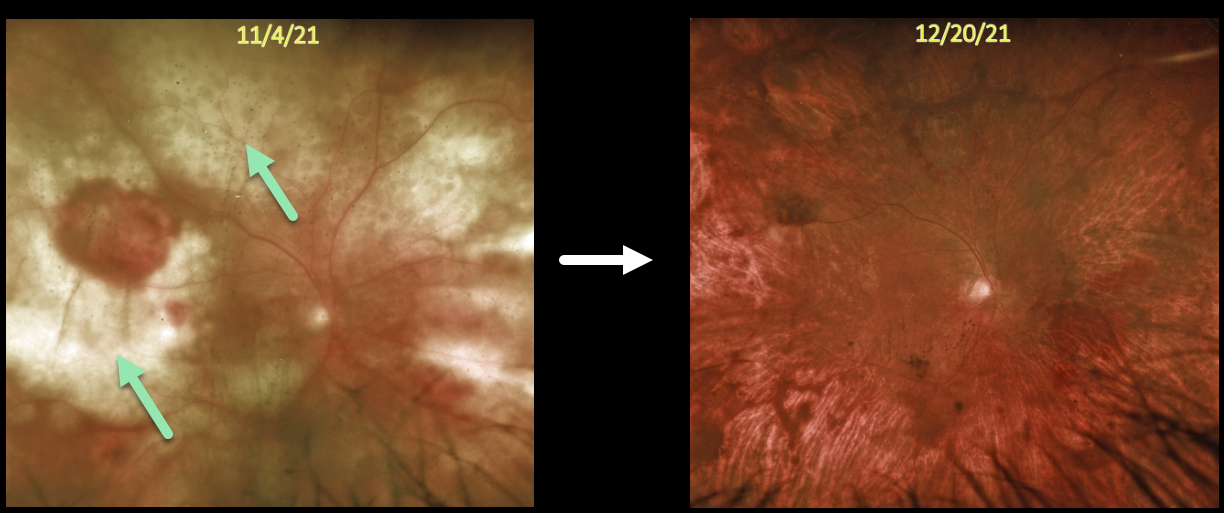
This 73YO man presented with subacute vision loss and these retinal findings. There were variable yellow-white, mostly confluent, lumpy subretinal infiltrates throughout the right posterior pole. The left fundus was normal.
He had a remote history of bladder cancer, which was in remission. He had recently completed chemotherapy for non-Hodgkin lymphoma, including a left retroperitoneal biopsy confirming a diffuse large B-cell lymphoma. We felt the findings in his right eye were consistent with metastatic lymphoma to the choroid.
Systemic chemotherapy supplemented with weekly intravitreal methotrexate was started. About 6 weeks later, vision remained at counting fingers, but there was a dramatic, complete clinical resolution of the tumor.


