

Macular Dystrophies + Acquired Vitelliform Lesions
<< Back to Cases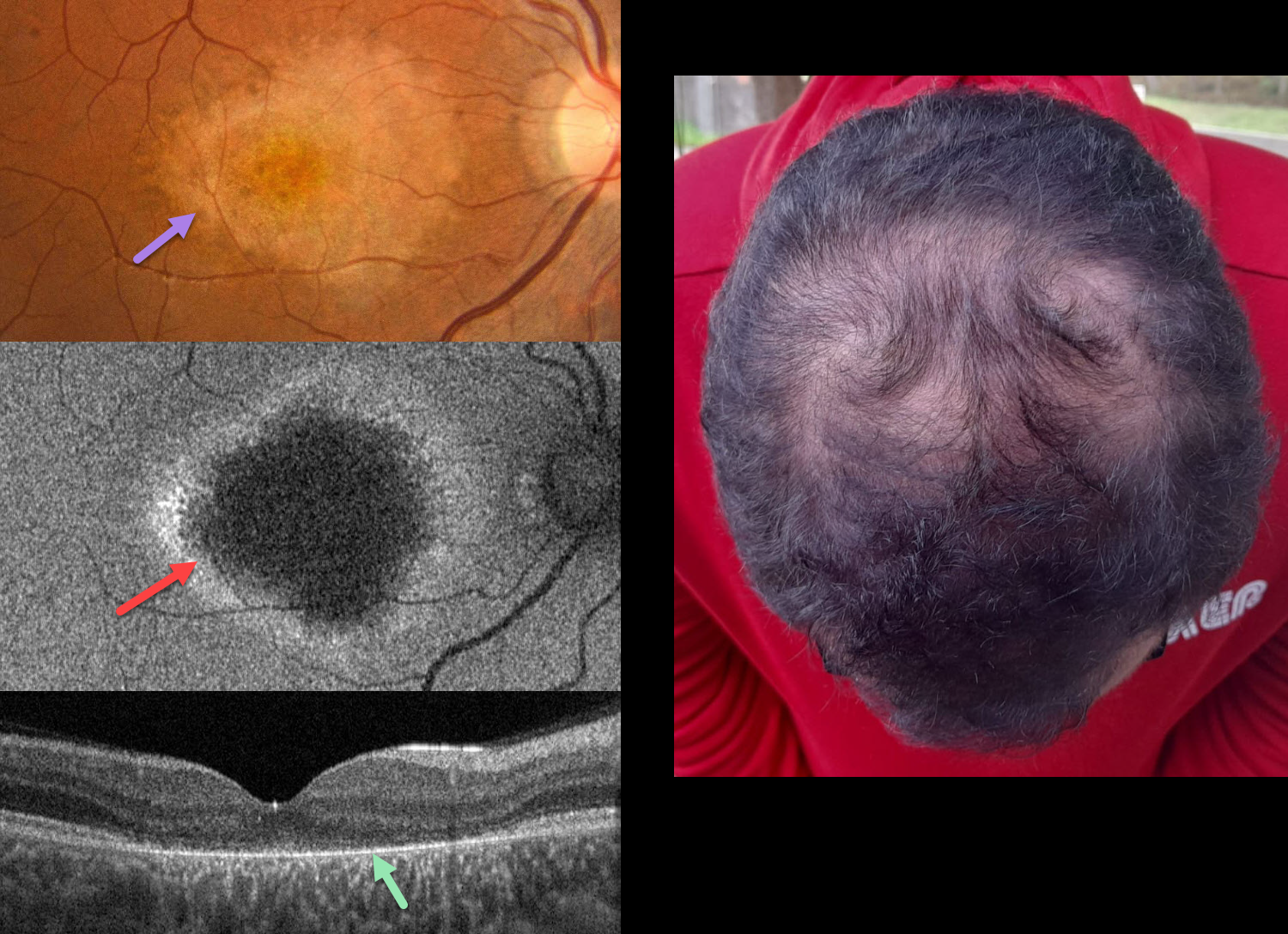
HYPOTRICHOSIS WITH JUVENILE MACULAR DEGENERATION
João Pedro Marques
Originally posted on @retina.rocks July 15, 2026
This 19YO male was referred for progressive vision loss and hemeralopia since early childhood. Ocular family history was negative, although his parents were consanguineous. Vision was 20/40 OU.
Color photography of his right eye shows an oval to tear-drop shaped area of central and nasal macular atrophy with clumps of more distal subretinal pigmentation. Fundus autofluorescence (FAF) shows central hypo-FAF with a ring of peripheral hyper-FAF. OCT scanning shows near-complete loss of the outer retinal bands external to the outer nuclear layer. Similar findings were noted in his left eye (not shown). The hair is thinned along the top of his head. Genetic testing revealed a likely pathogenic homozygous variant in the CDH3 gene (16q22.1).
Learning Points:
Hypotrichosis with juvenile macular dystrophy is a rare autosomal recessive disorder caused by biallelic mutations in the CDH3 gene, which encodes for P-cadherin, a calcium-dependent cell adhesion protein expressed in hair follicles and RPE. It is characterized by congenital sparse, short scalp hair (with normal facial and body hair). Progressive macular chorioretinal atrophy begins in childhood, often causing severe central visual impairment by the second to third decade of life.
This case was previously published in Ophthalmology Retina 2021;5:593.
BUTTERFLY-SHAPED PATTERN MACULAR DYSTROPHY
João Pedro Marques
Originally posted on @retina.rocks January 27, 2026
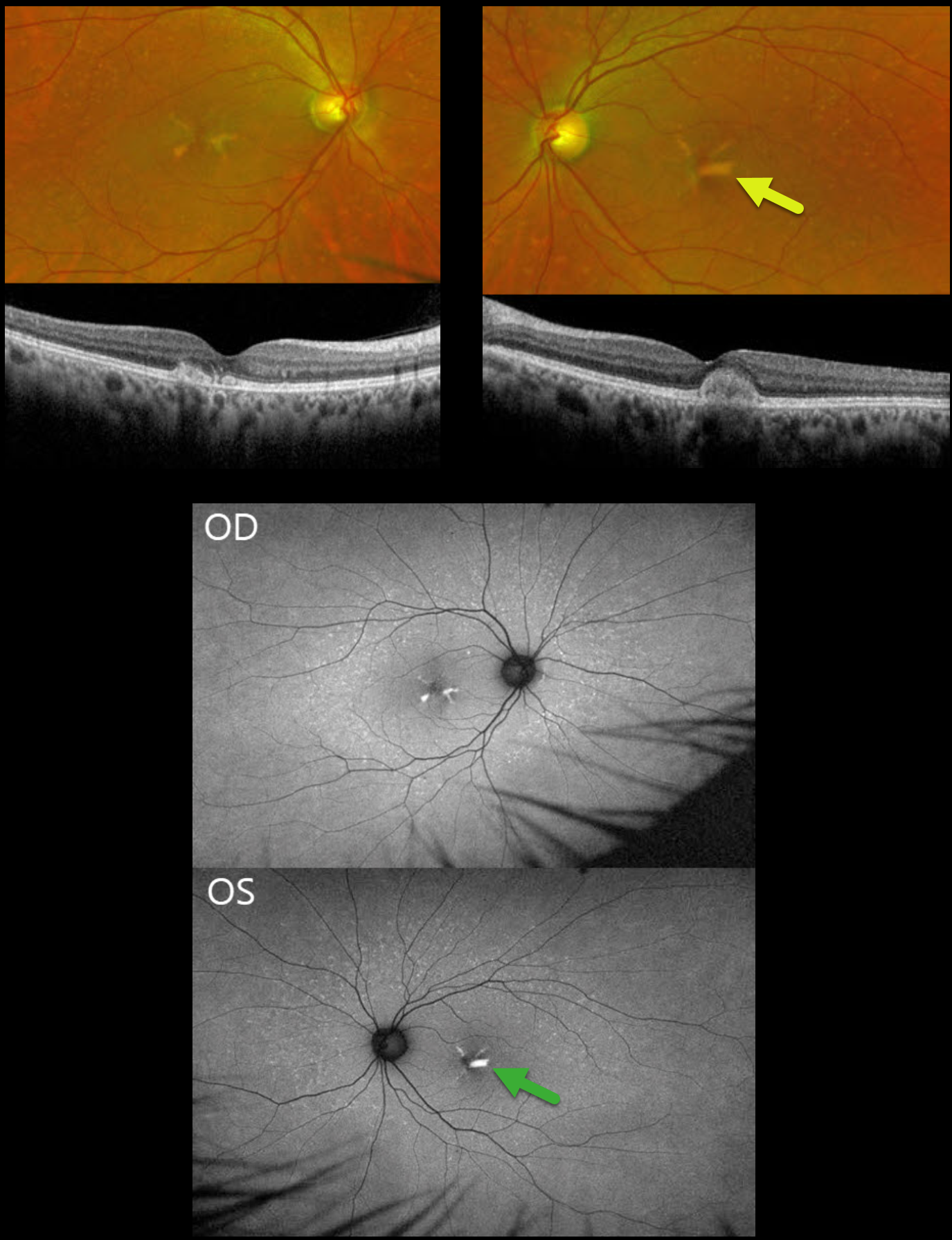
This asymptomatic 53YO male was referred because of a positive family history of two brothers with genetically confirmed PRPH2-associated pattern dystrophy. His father was legally blind by age 65 from an unknown cause. Vision was 20/20 OU.
Optos color RG imaging shows multiple drusen-like deposits in each posterior pole, as well as larger yellow subfoveal deposits that mimic the wings of a butterfly. On OCT, the subfoveal lesions show central hyperreflective deposits in the outer retina. These obscure the outer band details OD. The lesion is more focal in his OS, indenting the overlying retina. On fundus autofluorescence (FAF), these deposits are hyper-FAF.
Learning Points:
Pattern macular dystrophies are a genetically heterogeneous group of disorders characterized by variably shaped yellow subretinal deposits, the most common of which, in our experience, is adult-onset foveomacular vitelliform dystrophy. Causative mutations, including BEST1, PRPH2, IMPG1, and IMPG2. PRPH2 mutations account for 25% of pattern dystrophies, which are inherited as an autosomal dominant trait (Alashwal et al, AJO 2024;271:7-30).
ANGIOID STREAKS WITH MNV AND PATTERN DYSTROPHY PHENOTYPE
Shraddha Raj Shrivastava and Manish Nagpal
Originally posted on @retina.rocks January 6, 2026
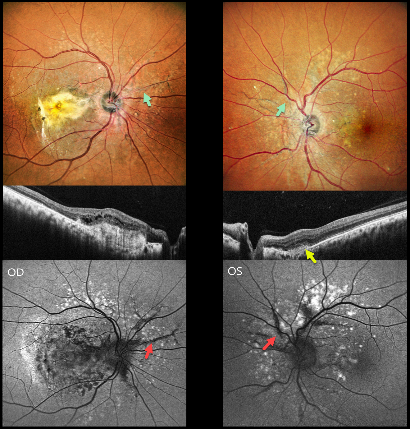
This healthy 42YO male presented with 10 months of vision loss in his right eye and 1 month of mild blurred vision in his left eye. Vision was counting fingers OD and 20/20 OS.
Pseudocolor SLO imaging shows angioid streaks extending around and radiating outwards from each optic nerve. There are drusen-like changes around each posterior pole, along with clumps of subretinal pigment temporally.
OCT scanning shows hyperreflective subretinal fibrosis with mild overlying cystic retinal edema OD. A much smaller area of subretinal hyperreflectivity (indicating a type 2 macular neovascularization, MNV) with overlying subretinal hyperreflective material is noted OS. The angioid streaks on fundus autofluorescence (FAF) are hypo-FAF due to RPE atrophy. The drusen-like changes are hyper-FAF.
Learning Points:
Retinal findings in pseudoxanthoma elasticum (PXE) most commonly include angioid streaks and a subretinal peaux d’orange appearance, most pronounced in each temporal macula. However, pattern dystrophy-like changes, as in our patient, can sometimes be the predominant phenotype (Murro et al, Graefe’s 2020;258:1881-1892). As with typical pattern dystrophies, the findings are dynamic and best captured with multimodal imaging.
PXE is caused by a mutation in the ABCA6 protein and is virtually always transmitted as an autosomal recessive disorder, although autosomal dominant transmission may rarely occur (Plomp et al, Am J Med Genet A 2004;126:403-412).
Our patient had no clinical signs of PXE and denied any family history of this disease. He was referred to his primary care doctor for further evaluation. Intravitreal anti-VEGF therapy was started OS.
PATTERN MACULAR DYSTROPHY
Nilesh Kumar
Originally posted on @retina.rocks November 17, 2025
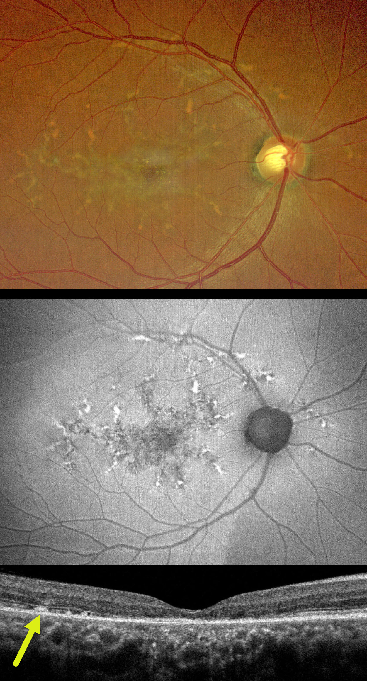
This healthy 50YO male presented with 10 months of bilateral vision loss. Family history was negative. Vision was 20/80 OU.
Color photography of his right eye shows submacular pigment and orange-yellow flecks radiating outwards from the fovea towards the peripheral macula and around the nerve. These lesions on fundus autofluorescence (FAF) are variably hyper- and hypo-FAF. OCT shows variable outer retinal loss. The subretinal flecks appear as hyperreflective deposits above the RPE. Identical findings are present in his left eye (not shown).
Learning Points:
Autosomal dominant pattern macular dystrophies present in mid-life with macular subretinal orange-yellow or pigmented deposits that accumulate in various patterns. Usually caused by mutations in the PRPH2 gene, the condition is classified by the pattern created by these deposits. However, they likely represent different phenotypes of the same disorder. The most common clinical presentation is adult-onset foveomacular vitelliform dystrophy, which presents as symmetrical, small, subfoveal, yellow-pigmented lesions. Our patient’s appearance is characteristic of butterfly-shaped pigment dystrophy. The visual prognosis is generally good, although some patients may develop vision loss due to secondary atrophy or macular neovascularization.
PSEUDOXANTHUM ELASTICUM WITH ANGIOID STREAKS AND PATTERN DYSTROPHY PHENOTYPE
Mattie Adams
Originally posted on @retina.rocks June 4, 2025
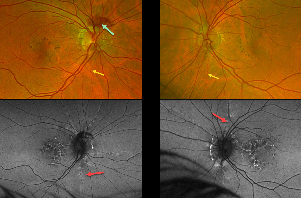
This 53YO female was referred for asymptomatic macular findings. Her mother has a history of pseudoxanthoma elasticum (PXE). Vision was 20/25 OD and 20/40 OS.
Optos color RG imaging shows coarse subretinal pigment clumps bilaterally. Subretinal blood is noted along the superior aspect of the right nerve from a peripapillary choroidal neovascularization. A few barely visible angioid streaks radiate from each nerve.
Fundus autofluorescence (FAF) shows variable hyper- and hypo-FAF flecks. The angioid streaks appear as hyper-FAF lesions radiating outwards from each nerve.
Learning Points:
Retinal findings in PXE most commonly include angioid streaks and a subretinal peaux d’orange appearance, most pronounced in each temporal macula. However, pattern dystrophy-like changes, as in our patient, can sometimes be the predominant phenotype (Murro et al, Graefe’s 2020;258:1881-1892). As with typical pattern dystrophies, the findings are dynamic and best captured with multimodal imaging.
PXE is caused by mutations in the ABCA6 gene, and our patient tested positive for homozygous pathogenic ABCA6 mutations. PXE is virtually always transmitted as an autosomal recessive disorder, although autosomal dominant transmission may rarely occur (Plomp et al, Am J Med Genet A 2004;126A:403-412).
ADULT-ONSET FOVEOMACULAR VITELLIFORM DYSTROPHY
Tejaswita Verma and Manish Nagpal
Originally posted on @retina.rocks February 4, 2025
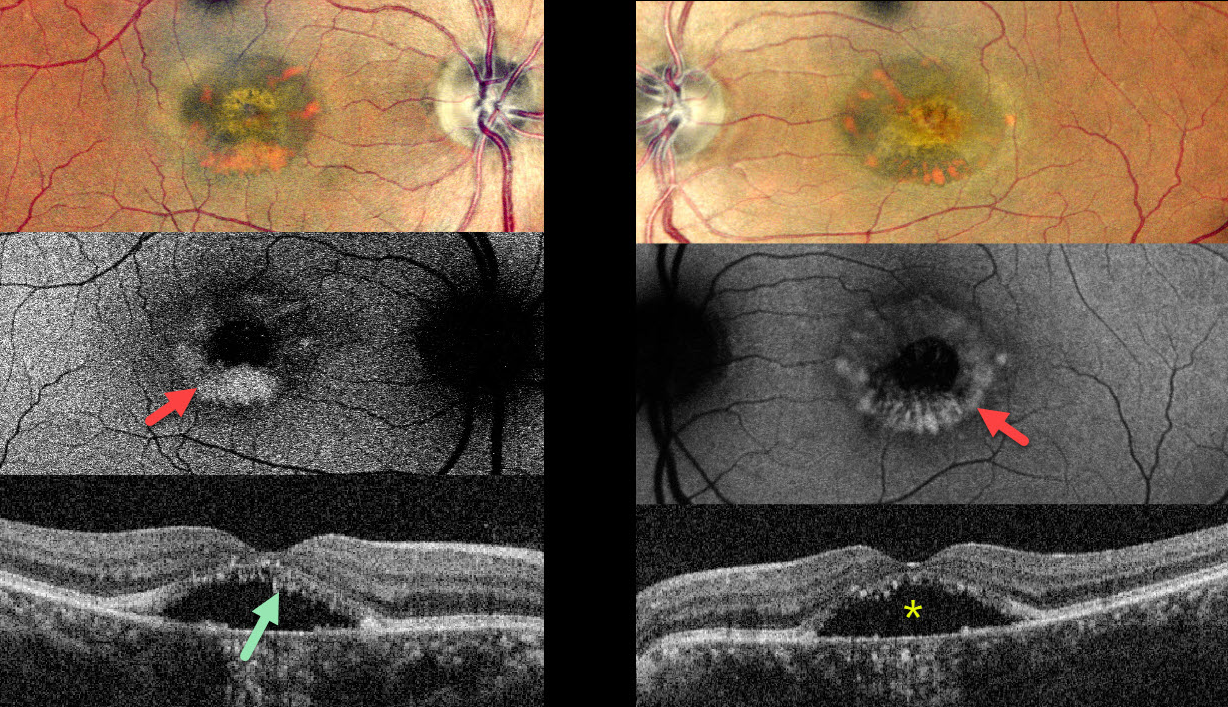
This 62YO female complained of stable bilateral blurred vision for seven years, having been diagnosed with central serous chorioretinopathy elsewhere. Vision was 20/30 OD and 20/40 OS.
Pseudocolor SLO imaging shows symmetrical, partially scrambled vitelliform lesions. The lesions are hyperautofluorescent along their outer edges and hypoautofluorescent centrally. OCT shows shaggy hyperreflective material lining the outer retina overlying foveal subretinal fluid.
Learning Points:
Adult-onset foveomacular vitelliform dystrophy is characterized by bilateral, symmetrical, grayish-yellow, one-third to one-half disc diameter subfoveal lesions with a central pigmented spot. In our experience, it is the most common pattern of dystrophy and closely resembles Best disease, although the lesions are generally smaller and diagnosed in adulthood. Causative mutations include BEST1, PRPH2, IMPG1 and IMPG2.
For a great review article, see Chowers et al, Progress in Retinal and Eye Research 2015;47:64-85.
ANGIOID STREAKS
Anand Temkar and Manish Nagpal
Originally posted on @retina.rocks November 21, 2024
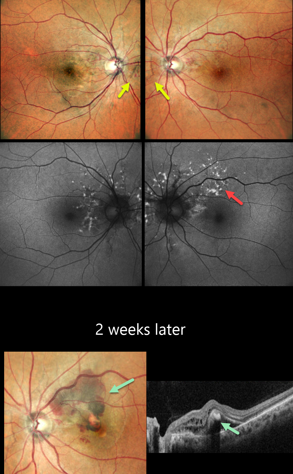
This 45YO male presented for a second opinion for bilateral wavy vision for 2 months. He was diagnosed elsewhere with macular neovascularization (MNV) and received two prior anti-VEGF injections (last 10 days earlier). Vision was 20/20 OU.
Pseudocolor SLO imaging shows subtle bilateral peripapillary angioid streaks associated with a few retinal hemorrhages. Fundus autofluorescence (FAF) shows numerous mostly peripapillary hyper-FAF flecks. Initial observation was recommended.
He returned two weeks later with new symptoms OS, and vision decreased to 20/120. New submacular fluid and blood are noted clinically and on OCT, and monthly anti-VEGF therapy was restarted. Unfortunately, he was lost to follow-up after 3 injections.
Learning Points:
Angioid streaks are orange-yellow, irregular, crack-like dehiscences in Bruch’s membrane associated with atrophic degeneration of the overlying RPE. These patients, especially those with pseudoxanthoma elasticum (PXE), are at significant risk for MNV as occurred in each of our patient’s eyes. MNV responds best to anti-VEGF therapy, and treatment is usually ongoing as with the typical neovascular AMD protocols. However, long-term vision is often poor due to RPE and choroidal atrophy (Mori et al, Graefe’s 2020;258:1863-1869).
Our patient had no family history of PXE or suggestive skin lesions, so skin biopsy or genetic testing was not performed. However, the funduscopic changes and macular dystrophy-like FAF findings are fairly pathognomonic for PXE.
PSEUDOXANTHUM ELASTICUM WITH ANGIOID STREAKS AND PATTERN DYSTROPHY PHENOTYPE
Originally posted on @retina.rocks July 11, 2024
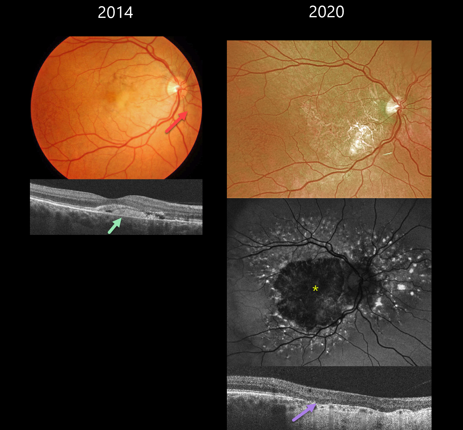
This 39YO male with a known history of pseudoxanthoma elasticum presented on 2/19/14. Findings were bilateral at this, and subsequent visits, so only his right eye images will be shown and discussed.
Color photography shows nasal yellowish subfoveal pigmentary changes with a peaux d’orange appearance temporally. Some peripapillary angioid streaks are noted. OCT shows hyperreflective vitelliform material and fluid below the retina, with some of this material also infiltrating the outer retina.
He has been followed without treatment, and when examined on 7/17/20, vision was 20/200 OD and 20/80 OS. Optos color RG imaging shows a variably atrophic macula. The atrophy on fundus autofluorescence is hypo-FAF. There is a fairly dramatic pattern of hyper-FAF spots radiating out from this atrophy. OCT shows outer retinal atrophy and no fluid. When last examined on 7/6/23, vision was 20/200 OU.
Learning Points:
Retinal findings in PXE most commonly include angioid streaks and a subretinal orange skin (peaux d’orange) appearance, most pronounced in each temporal macula. However, pattern dystrophy-like changes, as in our patient, can sometimes be the predominant phenotype (Murro et al, Graefe’s 2020;258:1881-1892). As with typical pattern dystrophies, the findings are dynamic and best captured with multimodal imaging.
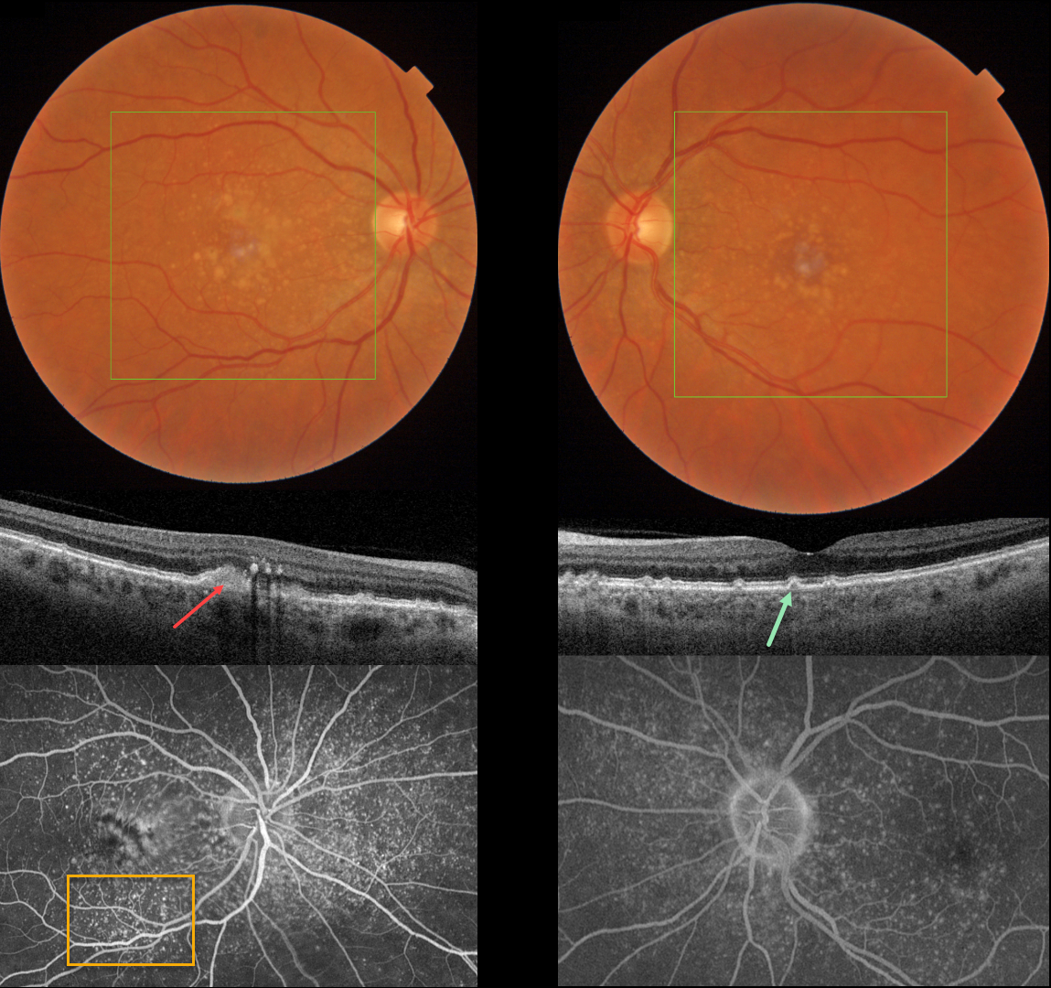
CUTICULAR DRUSEN WITH VITELLIFORM LESION
Originally posted on @retina.rocks May 28, 2024
This 57YO female was referred for asymptomatic macular changes. Vision was 20/20 bilaterally.
Triton color imaging shows bilateral confluent central soft drusen and smaller, more peripheral macular drusen. Swept-source OCT shows bilateral, small arrowhead-shaped cuticular drusen. A hyperreflective vitelliform lesion is noted OD, along with some hyperreflective deposits in the outer plexiform layer. Optos fluorescein angiography shows extensive bilateral hyperfluorescent cuticular drusen along with a staining vitelliform lesion OD.
Learning Points:
Originally described by Gass as basal laminar drusen (AJO 1985;99:445-459), cuticular drusen were initially thought to represent nodular thickening of the RPE basement membrane. They appear clinically as numerous, round, yellow-white ‘hard drusen’, and on OCT as small, arrowhead-shaped drusen located above Bruch’s membrane and below the RPE (Spaide and Curcio, Retina 2010;30:1441-1454).
Our patient’s angiographic appearance echoes Dr. Gass’ original description of “a beautiful stars-in-the-sky or Milky-Way pattern” of hyperfluorescence. Cuticular drusen are classically associated with avascular vitelliform lesions (Finger et al, Retina 2010;30:1455-1464), as in this case, but can also develop macular neovascularization.
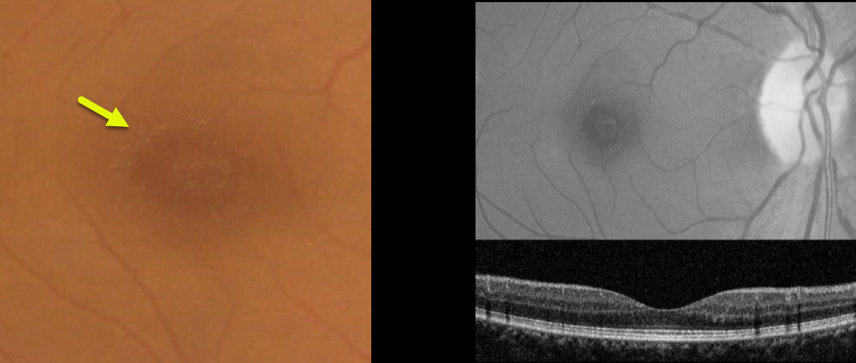
BENIGN YELLOW DOT MACULOPATHY
Asma Samsudeen and Ashish Sharma
Originally posted on @retina.rocks March 21, 2023
This 49YO female presented with asymptomatic macular changes. Vision was 20/30 OD and 20/40 OS. There was no past medical history and no family history of eye disease.
Color imaging shows tiny yellow perifoveal dots, which are somewhat better seen with red-free imaging. OCT scanning is unremarkable.
Learning Points:
Benign yellow dot maculopathy was reported as a new nonprogressive macular phenotype by Borman et al (Ophthalmology 2017;124:1004-1013).
Patients present with normal vision and asymptomatic tiny yellow foveal dots. These dots are hyperautofluorescent, but OCT scanning is generally normal. There is no known causative gene.
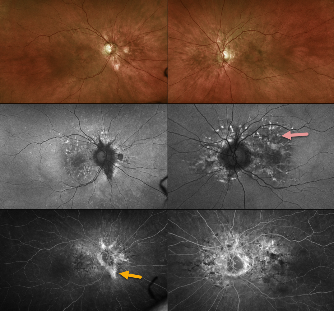
PSEUDOXANTHUM ELASTICUM WITH ANGIOID STREAKS AND PATTERN DYSTROPHY PHENOTYPE
Originally posted on @retina.rocks March 20, 2023
This 55YO female has been followed for years with macular pattern dystrophy-type changes due to pseudoxanthoma elasticum (PXD). Vision is 20/25 OU.
Color imaging shows variable mild pigmentary changes. Optos fundus autofluorescence imaging shows variable hyperautofluorescent flecks with variable angiographic staining. Some staining angioid streaks are noted in her right eye.
Learning Points:
Retinal findings in PXE most commonly include angioid streaks and a subretinal orange skin (peaux d’orange) appearance, most pronounced in each temporal macula.
However, pattern dystrophy-like changes, as in our patient, can sometimes be the predominant phenotype in some PXE patients (Murro et al, Graefe’s 2020;258:1881-1892). As with typical pattern dystrophies, the findings are dynamic and best captured with multimodal imaging.
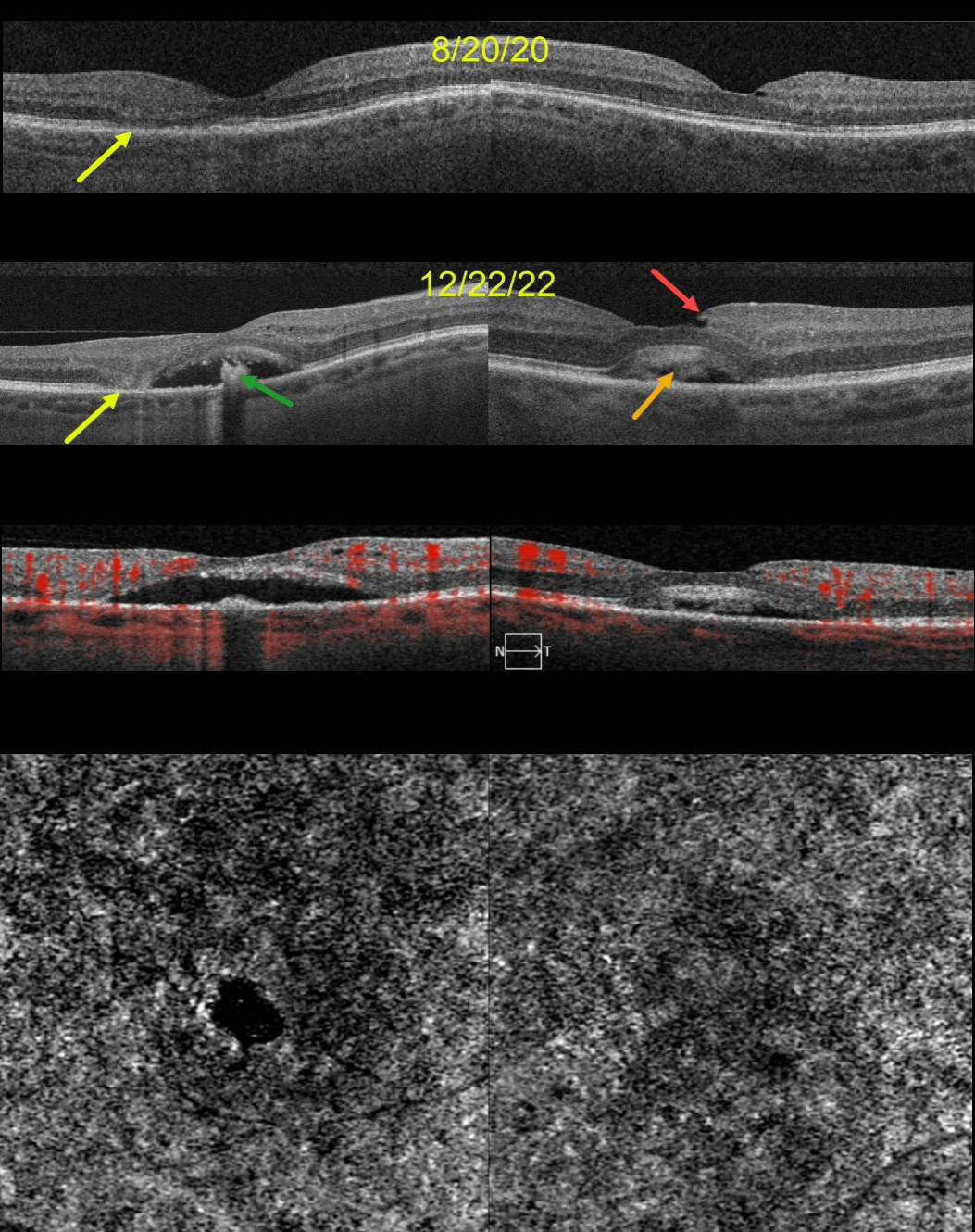
ACQUIRED VITELLIFORM LESION
Originally posted on @retina.rocks January 30, 2023
This 68YO female was seen on 8/20/20. Vision was 20/70 OD and 20/40 OS. Although there were no visual complaints, OCT scanning showed bilateral defects in the EZ bands, along with some temporal outer retinal atrophy OD.
She returned two years later, complaining of gradual vision loss OU, although vision was stable at 20/70 OD and 20/30 OS. OCT scanning shows bilateral acquired vitelliform lesions (AVL). The lesion OD is mostly scrambled with a hyporeflective space between the RPE and outer retina, along with a central hyperreflective clump on top of the RPE. There is now complete outer retinal atrophy temporally.
The AVL in her left macula shows variable hyperreflective material. An unrelated small partial lamellar macular hole is noted temporally. OCT angiography (OCTA) shows no flow signals from either AVL.
Learning Points:
Vision loss from AVLs most commonly results from lesions collapsing due to secondary atrophy or macular neovascularization, which can develop in about 8% of eyes (Balaratnasingam et al., AJO 2016;172:28-38).
The absence of OCTA flow signals in either eye confirmed that these lesions were avascular, and we continue to follow her.
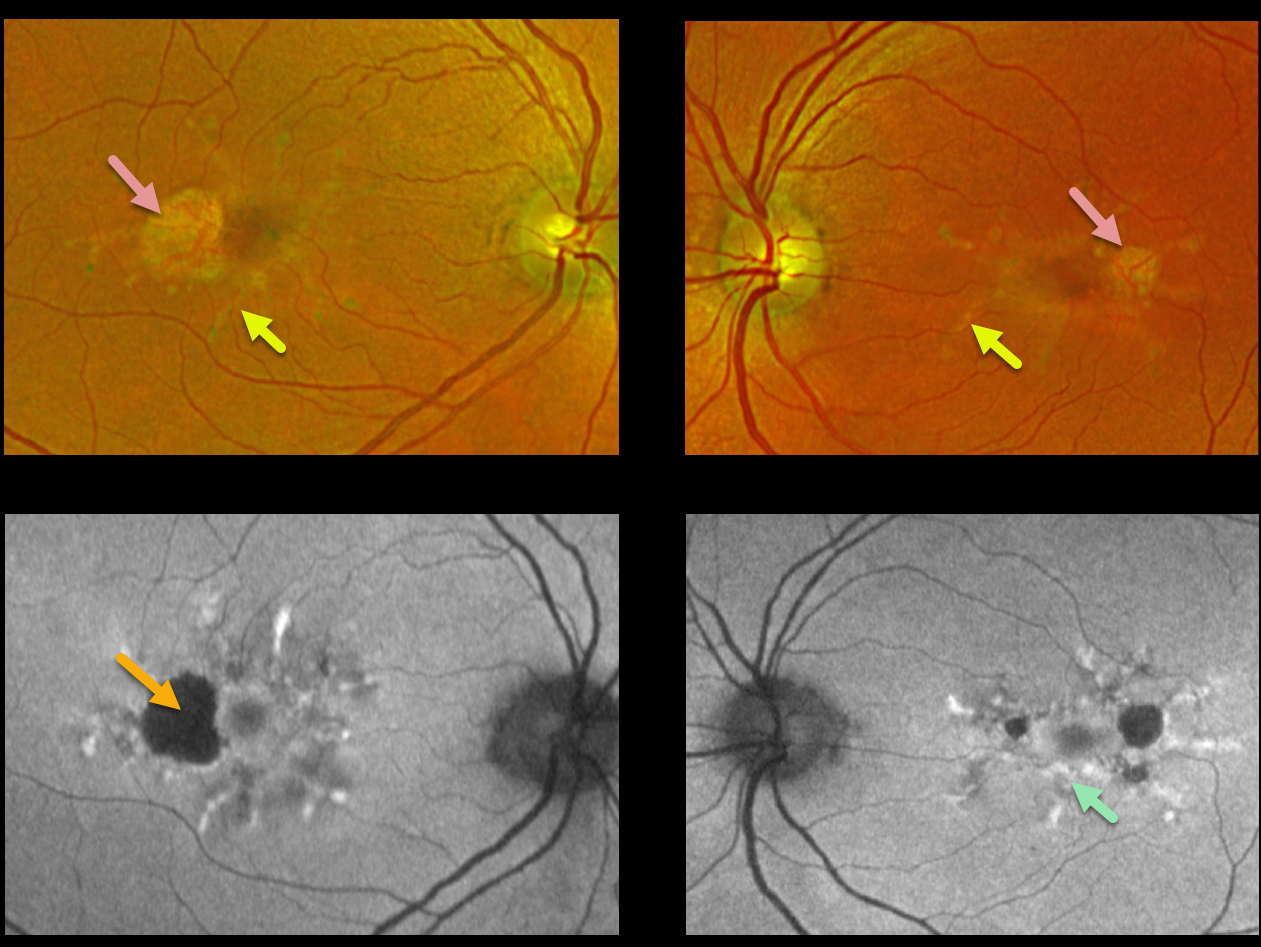
MACULAR DYSTROPHY
Nicole Wood
Originally posted on @retina.rocks February 22, 2022
This 62YO female presented with 20/20 vision OU and no visual symptoms. Optos imaging shows bilateral radiating subretinal yellowish macular lesions with temporal atrophy.
Fundus autofluorescence (FAF) shows variable hyper-FAF from these lesions, along with hypo-FAF within the areas of atrophy.
Learning Points:
Pattern macular dystrophies are associated with numerous mutations, including ABCA4, PRPH2, BEST1, PROM1, CERKL, CNGB3, and CRB1, among others (see Del Pozo-Valero et al, Invest Ophthalmol Vis Sci 2022;63(2):11).
These patients tend to maintain fairly good vision, although outer retinal and RPE atrophy can cause central vision loss, as can the rare development of macular neovascularization. Patients should therefore regularly monitor their vision with an Amsler grid.
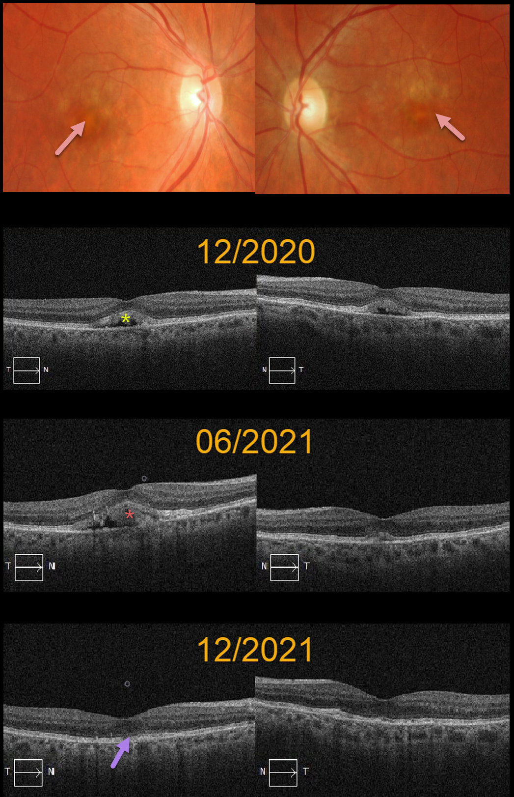
ADULT-ONSET FOVEOMACULAR VITELLIFORM DYSTROPHY
Originally posted on @retina.rocks January 19, 2022
This 73YO female presented in 12/20 with bilateral central vitelliform lesions and more peripheral yellowish macular lesions, consistent with a probable macular dystrophy.
OCT shows bilateral subretinal hyperreflective lesions with underlying hyporeflectivity. She was asymptomatic with 20/25 vision OU. There was no family history of macular dystrophy. Genetic testing was negative, including for BEST1.
Six months later, the vitelliform lesion increased OD and decreased OS. Another six months later, the vitelliform lesions collapsed OU, and vision remained 20/25.
Learning Points:
Vitelliform lesions are commonly found in association with other disorders, including retinal dystrophies such as Best disease, macular dystrophies, cuticular drusen, AMD, and vitreomacular traction.
The yellow subretinal material is felt to be debris from shed photoreceptor outer segments. These lesions markedly autofluoresce on fundus autofluorescence imaging, which unfortunately, is unavailable for this patient.
A great reference for learning more about AVLs is Freund et al, Retina 2011;31:13-25.
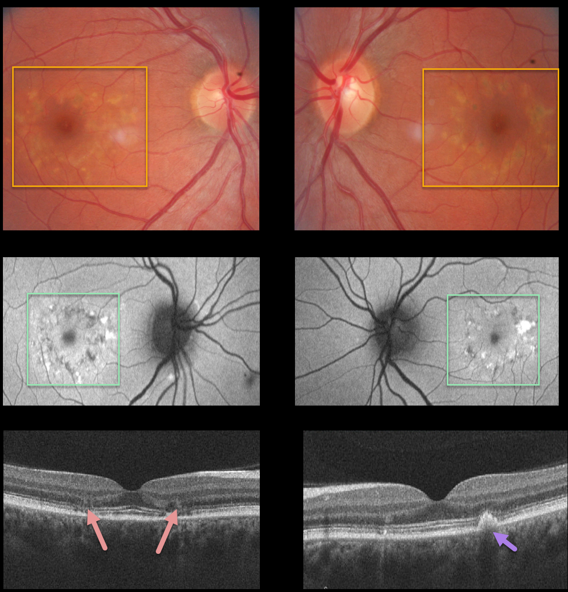
MACULAR DYSTROPHY
Originally posted on @retina.rocks October 20, 2021
This 34YO female, a local optometrist’s sister, was referred for these asymptomatic bilateral macular changes consistent with a macular dystrophy.
Fundus images show symmetrical yellowish subretinal pigmented drusen-like changes. Fundus autofluorescence reveals that these lesions are variably hyper- and hypoautofluorescent.
OCT scans show some variable ellipsoid zone loss and outer retinal atrophy, as well as subretinal hyperreflective material. Her brother had similar findings. Unfortunately, we have no further clinical information.
Learning Points:
Pattern macular dystrophy is a group of disorders characterized by pigment deposition of the macula and caused by mutations in the RDS/peripherin gene. The following are considered types of pattern dystrophies: adult-onset foveomacular vitelliform dystrophy, butterfly-shaped pigment dystrophy, reticular dystrophy, multifocal pattern dystrophy simulating Stargardt disease, and fundus pulverulentus.
Pattern macular dystrophy has also been found in systemic disorders such as pseudoxanthoma elasticum, myotonic dystrophy, and McArdle Disease.
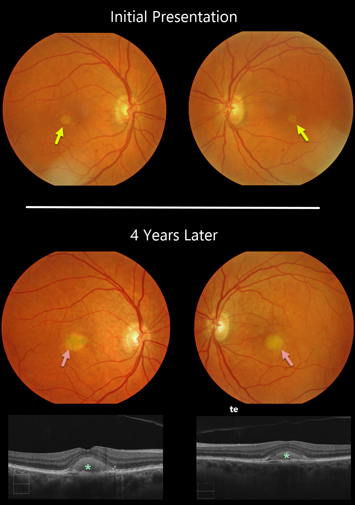
ADULT-ONSET FOVEOMACULAR VITELLIFORM DYSTROPHY
Originally posted on @retina.rocks September 13, 2021
Our patient shows classic findings for adult-onset foveomacular vitelliform dystrophy (Adult Best’s), including small, round, symmetrical subfoveal yellow spots. The vitelliform lesions enlarged over four years, which is seen on OCT as subretinal hyperreflective material between the RPE and outer retina. This material often indents the overlying retina.
Learning Points:
Adult Best’s is associated with a mutation in the BEST1 gene, which encodes the bestrophin-1 protein. Bestrophin-1, a calcium-activated chloride channel, is primarily found in the basolateral plasma membrane of the RPE.
BEST1 mutations cause a variety of phenotypes, including autosomal recessive bestrophinopathy, best vitelliform macular dystrophy, and autosomal dominant vitreoretinochoroidopathy.
These patients tend to maintain fairly good vision. Outer retinal and RPE atrophy can cause central vision loss, as can the rare development of macular neovascularization. Patients should therefore regularly monitor their vision with an Amsler grid.
MACULAR DYSTROPHY
Originally posted on @retina.rocks July 29, 2021
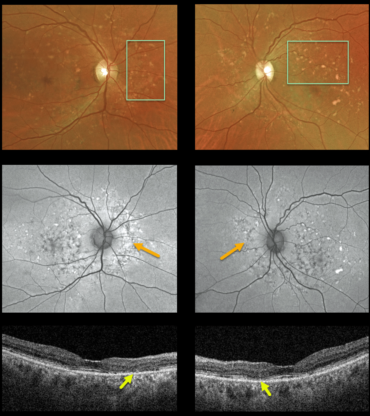
This 74YO female came in to see us for a second opinion for age-related macular degeneration. She was visually asymptomatic and denied prior Elmiron (pentosan polysulfate sodium, PPS) use. Vision was 20/30 OU.
Bilateral drusenoid/fleck-like yellow-orange subretinal lesions are scattered throughout the macula and peripapillary regions. These are variably hyper- or hypofluorescent. OCT scanning shows variable outer retinal atrophy without drusen.
Learning Points:
We believe these findings likely represent a type of macular dystrophy. However, she declined genetic testing.
PPS, a medication used to treat interstitial cystitis, can also mimic the findings in age-related macular degeneration and macular dystrophies. Peripapillary hypoautofluorescence, more densely packed macular autofluorescent changes, and earlier central macular involvement suggest PPS toxicity over other causes (see Barnes et al, Ophthalmology Retina 2020;4:1196-1201). Thus, it is important to perform thorough history-taking and document all medications the patient is taking.
BEST DISEASE
Originally posted on @retina.rocks June 3, 2021
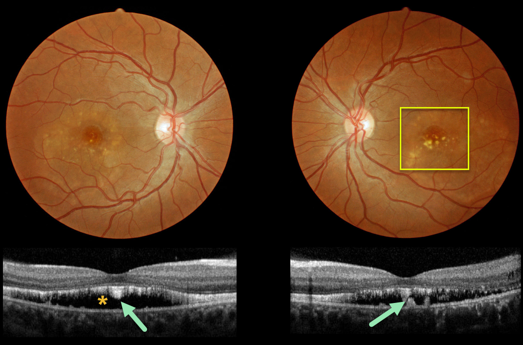
This patient has progressed to the more atrophic ‘scrambled egg’ appearance of Best’s disease.
OCT scanning shows the common hyporeflective subretinal space left behind after the yellow subretinal material absorbs, which often flattens over time.
Shaggy hyperreflective material, likely representing shed photoreceptors and residual vitelliform material, is noted along the posterior retinal surface and on top of the RPE.
Learning Points:
Best vitelliform macular dystrophy, or Best’s disease, is associated with a mutation in the BEST1 gene. This inherited disorder usually presents in childhood or early adulthood with a bilateral foveal “egg-yolk” appearance.
ADULT-ONSET FOVEOMACULAR VITELLIFORM DYSTROPHY
Originally posted on @retina.rocks April 5, 2021
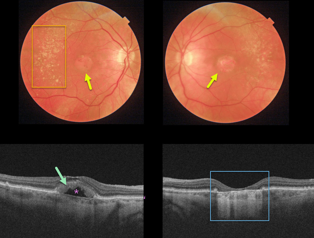
This 80YO female was 20/400 in her right eye and counting fingers in her left eye due to probable adult-onset foveomacular vitelliform dystrophy (Adult Best’s). Funduscopically, there are bilateral round atrophic foveal lesions with peripheral drusen.
OCT of the right eye shows hyperreflective outer retinal material overlying a pocket of subretinal fluid. The underlying RPE is atrophic with bare Bruch’s membrane. OCT of the left eye shows complete outer retinal and RPE atrophy.
Learning Points:
Adult Best’s is associated with a mutation in the BEST1 gene, which encodes the bestrophin-1 protein.
Bestrophin-1, a calcium-activated chloride channel, is primarily found in the basolateral plasma membrane of the RPE. BEST1 mutations cause a variety of phenotypes, including autosomal recessive bestrophinopathy, best vitelliform macular dystrophy, and autosomal dominant vitreoretinochoroidopathy.
ANGIOID STREAKS
Originally posted on @retina.rocks April 1, 2021
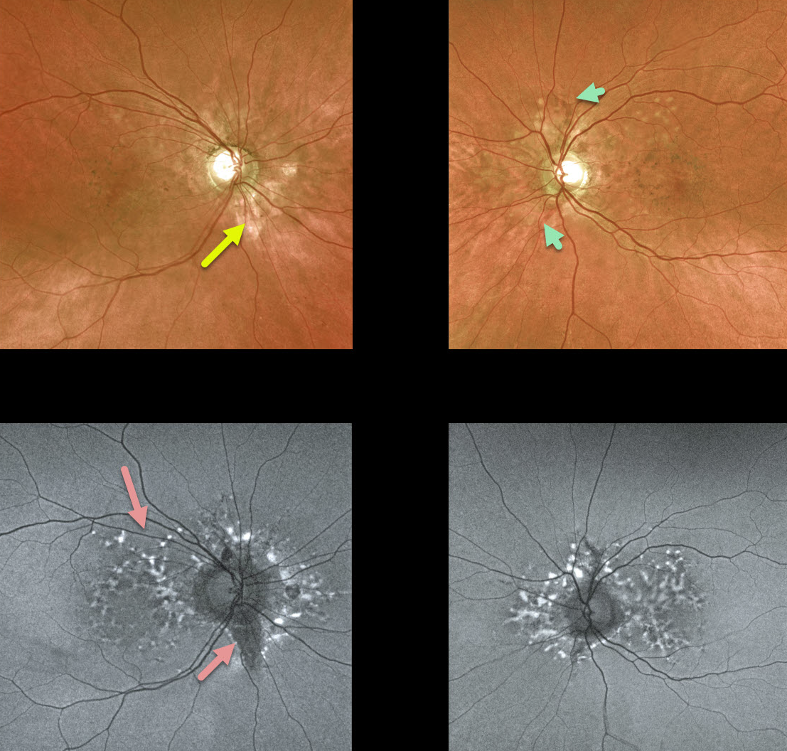
We have been following this 54YO female with pseudoxanthoma elasticum (PXE) for many years. Vision is 20/20 bilaterally, and fortunately, she remains asymptomatic.
She has bilateral macular peripapillary pigmentary changes. Angioid streaks, always tough to find unless the clinician actively looks for them, are not prominent, although a few are visible in her left eye.
Fundus autofluorescence (FAF) shows more dramatic changes, including scattered hyper- and hypo-FAF peripapillary and macular spots.
Her sister became legally blind many years ago from bilateral macular neovascularization (MNV).
Learning Points:
Patients with angioid streaks and pseudoxanthoma elasticum (PXE) can present with retinal findings similar to those of pattern dystrophies.
These patients are at significant risk for macular neovascularization and need to be followed and treated as needed with anti-VEGF agents, as in patients with AMD.
ADULT-ONSET FOVEOMACULAR VITELLIFORM DYSTROPHY
Originally posted on @retina.rocks November 18, 2020
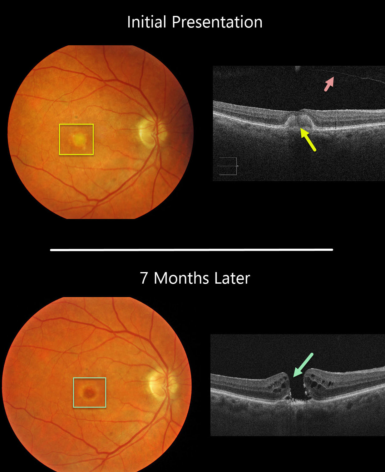
This 77YO female was initially examined with 20/60 vision OD, 20/30 vision OS, and bilateral vitelliform lesions (only right eye pictured) consistent with Adult Best (adult-onset foveomacular vitelliform dystrophy). A complete posterior vitreous detachment (PVD) was noted in her right eye.
Seven months later, vision decreased to counting fingers OD due to a new full-thickness macular hole. This patient was asymptomatic due to good vision in her left eye and has opted against surgery for now.
Learning Points:
Macular holes are most commonly caused by a partial PVD with active vitreomacular traction. However, they can occasionally develop without traction following a prior PVD or vitrectomy surgery. Macular holes have also been reported as a rare complication of adult Best’s disease.
Adult-onset foveomacular vitelliform dystrophy is associated with a mutation in the BEST1 gene, which encodes the bestrophin-1 protein.
Bestrophin-1, a calcium-activated chloride channel, is primarily found in the basolateral plasma membrane of the RPE. BEST1 mutations cause a variety of phenotypes, including autosomal recessive bestrophinopathy, best vitelliform macular dystrophy, and autosomal dominant vitreoretinochoroidopathy.
PIGMENT EPITHELIAL DETACHMENT (PED)
Originally posted on @retina.rocks September 30, 2020
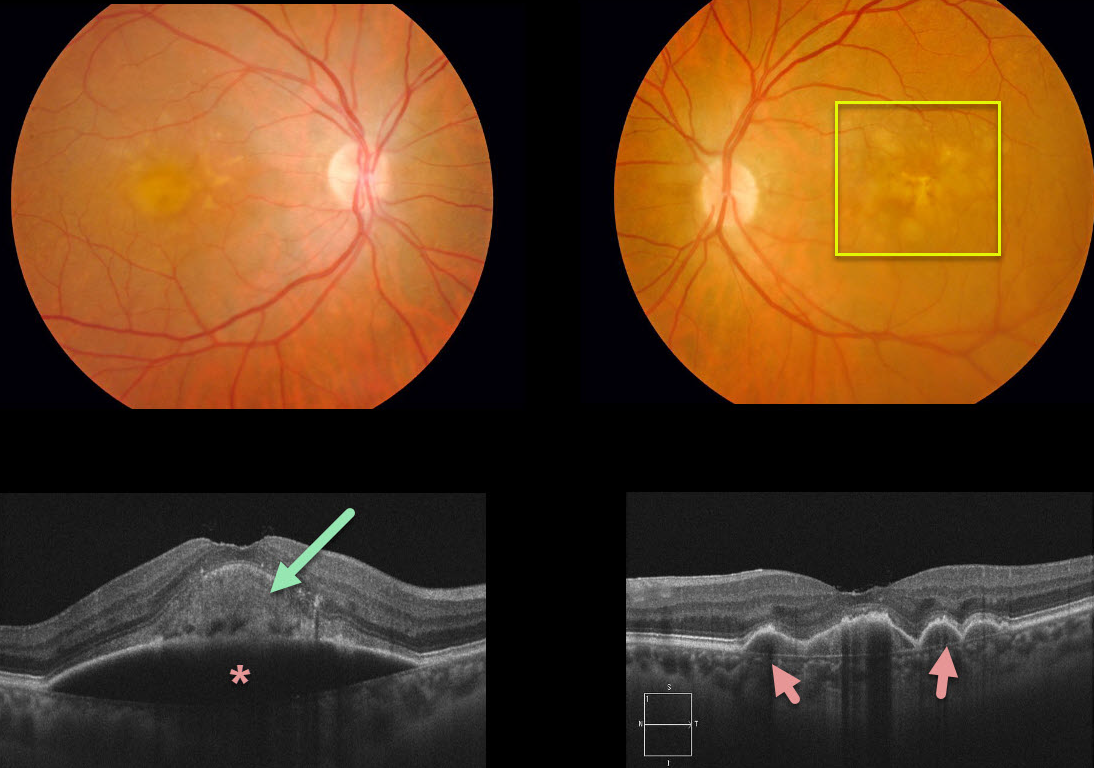
This patient had been followed for age-related macular degeneration (AMD) with bilateral confluent soft drusen. She then developed a large acquired vitelliform lesion overlying a retinal pigment epithelial detachment (PED) in her right eye. There are several PEDs also shown in the left eye.
Learning Points:
Vitelliform lesions are commonly found with other disorders, including retinal dystrophies (Best’s disease and macular dystrophies), cuticular drusen, AMD, and vitreomacular traction.
The yellow subretinal material is felt to be debris from shed photoreceptor outer segments. These lesions markedly autofluoresce on fundus autofluorescence.
A great reference to learn more about AVLs is Freund et al Retina 2011;31:13-25.
MACULAR DYSTROPHY
Originally posted on @retina.rocks August 19, 2020
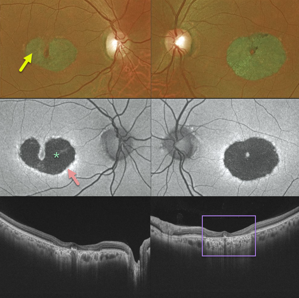
Our patient has a bilateral bull’s-eye maculopathy with central foveal sparing. Vision was 20/30 OD and 20/60 OS.
Fundus autofluorescence shows central hypo-FAF from loss of the central RPE. The thin surrounding hyper-FAF ring likely represents diseased, at-risk RPE cells. The OCT scans show pericentral outer retinal and RPE loss.
Learning Points:
Our patient no doubt has some type of inherited macular disorder. Some possibilities include central areolar chorioretinal dystrophy or a cone-rod dystrophy.
Central areolar chorioretinal dystrophy is a genetic condition associated with mutations in the RDS/peripherin gene, which encodes for a cell surface glycoprotein found in the rod and cone outer segments. The RPE and choriocapillaris atrophy over time, eventually forming a round, atrophic lesion in the macula. Vision can vary from 20/20 to 20/200.
ADULT-ONSET FOVEOMACULAR VITELLIFORM DYSTROPHY WITH SECONDARY MACULAR HOLE
Originally posted on @retina.rocks May 28, 2020
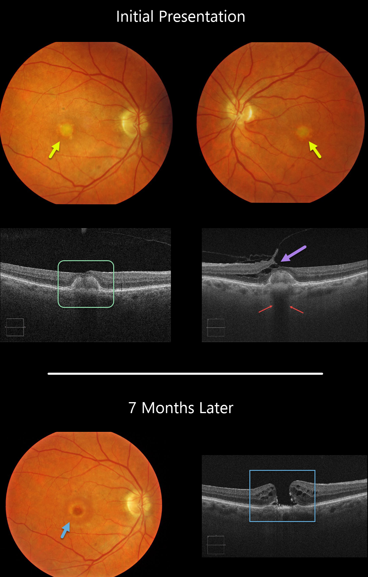
Our patient shows classic findings for adult-onset foveomacular vitelliform dystrophy (Adult Best’s) including small, round, symmetrical subfoveal yellow spots, which is seen on OCT as subretinal hyperreflective material between the RPE and outer retina.
Additionally, the vitelliform lesions provide a great example of shadowing of the underlying choroid. Shadowing occurs when a hyperreflective structure prevents the OCT laser from penetrating to illuminate deeper structures.
A bonus finding in the left eye is focal nasal vitreofoveal traction lifting the retina, splitting the outer plexiform layer and forming a partial tractional lamellar macular hole.
Learning Points:
There are numerous causes for the subfoveal accumulation of yellow material (acquired vitelliform lesions. Adult-onset foveomacular vitelliform dystrophy (Adult Best’s) is associated with a mutation in the BEST1 gene, which encodes for the bestrophin-1 protein.
Bestrophin-1, a calcium-activated chloride channel, is primarily found in the basolateral plasma membrane of the RPE. BEST1 mutations cause a variety of varied phenotypes, also including autosomal recessive bestrophinopathy, best vitelliform macular dystrophy, and autosomal dominant vitreoretinochoroidopathy.
These patients tend to maintain fairly good vision. Outer retinal and RPE atrophy can cause central vision loss, as can the rare development of macular neovascularization. Patients should therefore regularly monitor their vision with an Amsler grid.


