

Macular Hole
<< Back to Cases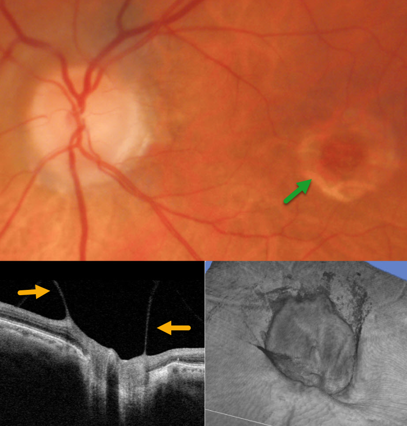
VITREOPAPILLARY TRACTION WITH CHRONIC MACULAR HOLE
Originally posted on @retina.rocks April 15, 2026
This 81YO male presented for his yearly examination without new complaints. Vision in his left eye was stable at 20/200 due to a chronic macular hole.
Triton color photography shows a full-thickness macular hole with a surrounding large rim of pigment loss. On slit lamp biomicroscopy, a partial PVD extended to the nerve with an elevated, partially attached Weiss ring. Vitreopapillary traction was confirmed on B-scan and 3D OCT. Continued observation was recommended.
Learning Points:
The normal vitreous undergoes a decades-long process of orchestrated degeneration, including liquefaction and gradual separation from the retinal surface. The posterior vitreous detachment (PVD) begins in the perifoveal retina early in life, followed by a foveal PVD, then a peripheral PVD, and then finally a complete PVD when the vitreous separates from the optic nerve (Johnson AJO 2010;149:371-382). The integrity of the internal limiting membrane (ILM) is integral to normal vitreoretinal adhesion, where a thinner ILM has a firmer adhesion than a thicker ILM. The ILM represents the basement membrane of the Müller cell. Hence, an area with fewer Müller cells reaching the inner retina will naturally have a firmer vitreoretinal adhesion. Since the optic nerve is completely devoid of Müller cells, this is the area of the posterior pole that has the firmest vitreoretinal adhesion, and this is the reason why the vitreous tends to remain so firmly attached to it.
Persistent vitreopapillary traction is a rarely reported phenomenon (Gabriel et al, Neuro-ophthalmology 2020;44:213-218), probably due to its lack of symptoms and often subclinical findings. In our patient’s case, the traction caused retinal thickening and hemorrhage, somewhat mimicking a peripapillary macular neovascularization. In our experience, these eyes usually remain asymptomatic and can be observed, although vitrectomy is very successful in relieving the traction and improving vision.
TRACTIONAL RETINAL DETACHMENT WITH PSEUDO-GIANT MACULAR HOLE
The European VitreoRetinal Society (EVRS) and Shishir Verghese
Originally posted on @retina.rocks January 16, 2026
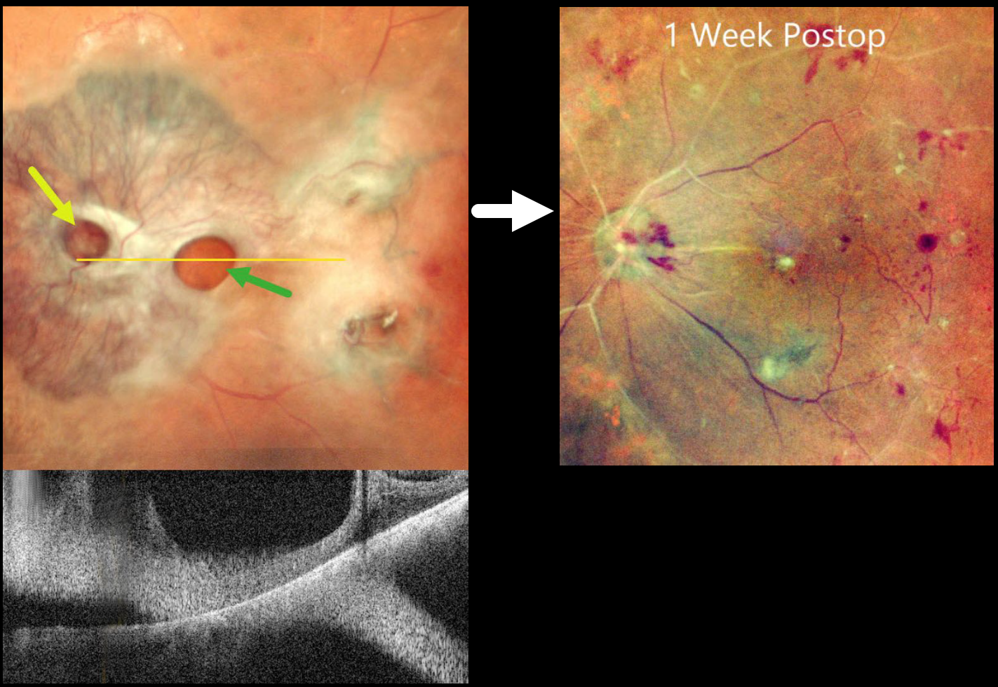
This 65YO diabetic female presented with counting fingers bilaterally from proliferative diabetic retinopathy with macular tractional retinal detachments.
Preoperative pseudocolor SLO imaging of her left eye shows a complex fibrovascular membrane covering the posterior pole. Pseudo giant-macular holes are noted overlying the disc and macula. OCT scanning confirms the lack of a true macular hole. One week following vitrectomy, membrane peeling, and air injection, vision improved to 20/200, and the retina was completely attached with no residual traction.
Learning Points:
A giant macular hole, also reported in Alport syndrome (Shah and Weinberg Ophthalmic Genetics 2010;31(2):93-97), is an exceedingly rare traumatic event (Hernandez-Da Mota Case Rep Ophthalmol 2011;2:283-286). The unusual anatomy of our patient’s fibrovascular diabetic membrane simulated a giant macular hole.
PRIOR CHRONIC ICSC WITH NEW TRACTIONAL MACULAR HOLE-RRD WITH POSTOPERATIVE MENISCUS MICROPYON
Originally posted on @retina.rocks October 29, 2025
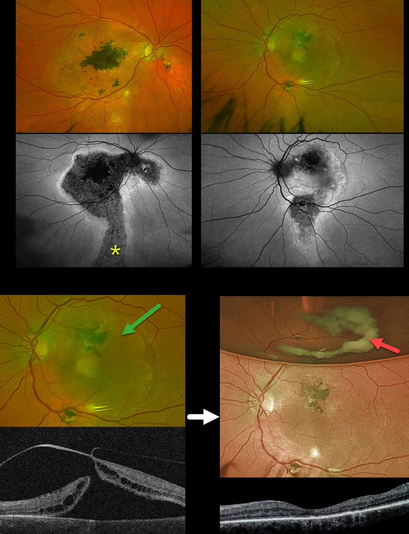
This 58YO male was referred for a retinal detachment in his left eye. He has a long history of idiopathic central serous chorioretinopathy (ICSC). He was previously treated elsewhere for neovascular AMD OD. For many years, he has been receiving intravitreal Avastin OS every 1-2 months. Vision was counting fingers OD and 20/400 OS.
Optos color RG imaging shows bilateral macular scarring with confluent hyperpigmented foveal changes and intraretinal pigment migration, especially in his right eye. Inferior hypopigmented gutters are most obvious on fundus autofluorescence (FAF) as hypo-FAF with surrounding hyper-FAF margins. A rhegmatogenous retinal detachment (RRD) occupies the entire left macula. Triton swept-source OCT shows a full-thickness macula hole, active vitreoretinal traction on the temporal edge of the hole, and a macular detachment.
The tractional macular hole with secondary RRD in our patient’s left eye is completely unrelated to his poorly treated ICSC. One month following vitrectomy, vision improved to 20/100. The retina was completely attached, and the macular hole was closed. A meniscus micropyon is adherent to the inferior meniscus of the gas bubble.
Learning Points:
Photodynamic therapy (PDT) is currently the standard of care for ICSC with symptomatic acute or chronic subretinal fluid. Unfortunately, Visudyne is rarely available outside the US, which has led to the adoption of unproven therapies, including mineralocorticoid receptor antagonists (eplerenone), beta blockers, and intravitreal anti-VEGF agents. Recent meta-analyses and systematic reviews confirm that these alternative therapies are ineffective (Felipe et al Int J Retina Vitreous 2022;8;34 and Kim et al Ophthalmology 2025;132:343-353). Despite the clear lack of efficacy in the literature, specialists both within and outside the US continue to use these drugs (Vankatesh et al Int J Retina Vitreous 2023;9;61). Our patient’s macular findings are a testament to what can happen when proven therapies are ignored.
Russell et al recently reported a unique ophthalmoscopic sign, a meniscus micropyon, that follows PPV with gas tamponade for all causes (Retina 2024;44:1766-1776). Features they noted that distinguish this sterile micropyon from post-PPV fibrin include a delayed appearance, hyperautofluorescence, and absence of anterior chamber or vitreous fibrinous strands/sheets. Vitreous biopsy in two of their cases revealed predominantly white blood cells. The micropyon appearance was associated with epiretinal proliferation (80%). Recurrent RRD from proliferative vitreoretinopathy (PVR) developed 33% of eyes, 12% re-detached without frank PVR, and 18% developed worsening epiretinal membranes.
Our patient’s meniscus micropyon disappeared as the bubble resolved. Three months postoperatively, vision was 20/60, and the retina remained completely reattached without PVR.
COMBINED TRACTIONAL-RHEGMATOGENOUS RETINAL DETACHMENT WITH MACULAR HOLE
Sucheta Kulkarani, Anita Gaikwad and Anjana Mirajkar
Originally posted on @retina.rocks July 24, 2025
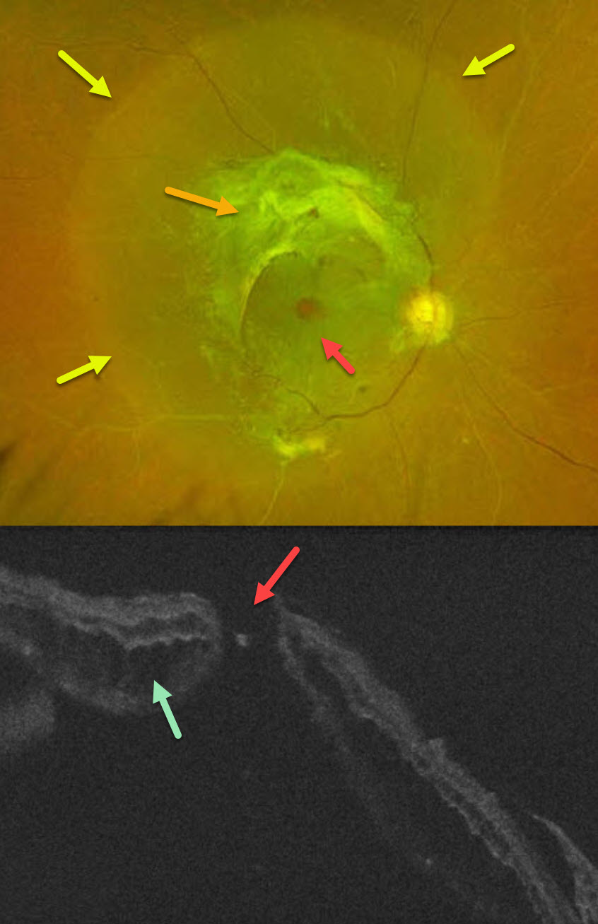
This 49YO male with type 2 diabetes noticed decreased vision in his right eye for 4 months. A single anti-VEGF injection was given elsewhere in his right eye around the time of his vision loss. Vision was 20/400 OD and 20/30 OS.
Optos color RG imaging OD shows a combined tractional-rhegmatogenous retinal detachment from dense fibrovascular proliferation and a full-thickness macular hole. OCT shows diffuse cystic edema within the detached retina, predominantly in the outer nuclear layer, and a macular hole.
Learning Points:
Tractional macular holes as a cause of combined tractional-rhegmatogenous diabetic retinal detachments are a rare event in our experience. Unfortunately, our patient was lost to follow-up immediately. We expect him to lose central vision in his left eye soon if he does not seek care.
ACUTE CHOROIDAL RUPTURES WITH SUBRETINAL BLOOD + TRAUMATIC MACULAR HOLE
Originally posted on @retina.rocks July 16, 2025
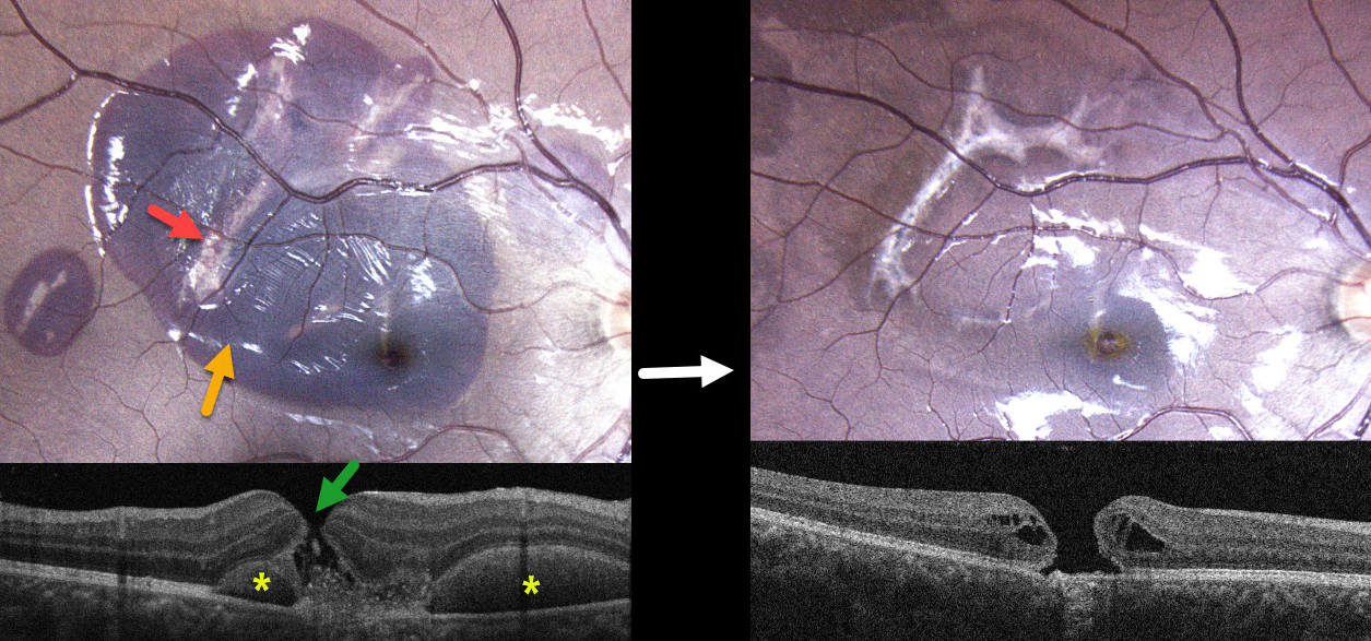
This healthy 17YO male was in a motor vehicle accident 2 weeks earlier. He was not wearing a seat belt and suffered a right upper lid laceration and closed head injury. He noticed immediate vision loss in his right eye. Vision was 20/200 OD and 20/20 in his normal OS.
Color photography shows a large macular and a smaller distal temporal submacular hemorrhage. Multiple choroidal ruptures are visible beneath this blood. OCT scanning shows a central full-thickness macular hole with speckled hyperreflective subretinal material. The submacular blood is hyperreflective with posterior shadowing on either side of the macular hole.
Learning Points:
An acute submacular hemorrhage following blunt trauma always indicates an underlying choroidal rupture as its source. The final vision is generally good unless the rupture extends through the macular center. These patients need long-term follow-up due to the increased risk of macular neovascularization.
Since fresh traumatic holes can spontaneously flatten, especially in pediatric cases (Liu et al, AJO 2020;210:174-183), an initial period of observation is recommended before considering vitrectomy. When examined about 6 months following his injury, all blood had resolved, and the temporal foveal choroidal rupture appears to spare the macular center. We will likely recommend vitrectomy if the hole fails to close with several more months of observation.
TRAUMATIC MACULAR SCARRING AND MACULAR HOLE
Liubov Sitaruk and Ophthalmocoach
Originally posted on @retina.rocks June 10, 2025
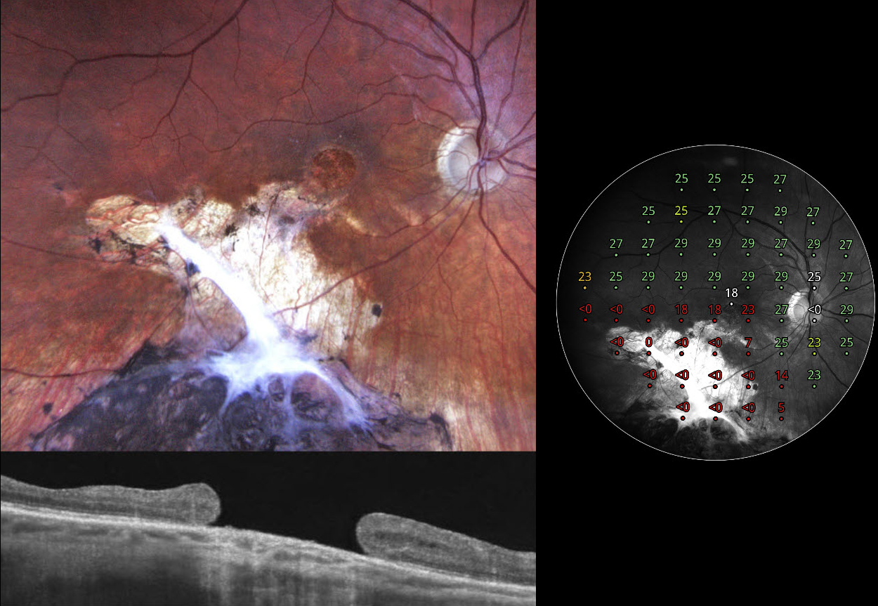
This 36YO Ukrainian male sustained a firearm-related facial injury during military combat in 2014 while in Eastern Ukraine. At the time, he was treated in a frontline hospital, where the left eye underwent primary enucleation due to a non-reconstructible globe rupture. His right eye sustained a penetrating injury with scleral rupture, which was primarily repaired. Over the following months, he underwent pars plana vitrectomy with endolaser and silicone oil due to complications, likely including retinal detachment and/or vitreous hemorrhage. Given the complexity of the trauma, the monocular status, and probable limited access to regular follow-up during wartime, silicone oil was likely retained for an extended period, potentially several years, until its removal at an undetermined later date. On our 2024 examination, he had no new visual complaints, and vision was 20/100.
Fundus photography shows atrophic inferior macular chorioretinal scarring, which becomes hyperpigmented as it extends into the inferior midperiphery. A large macular hole is noted funduscopically and on OCT. Macular perimetry shows marked depression within the area of scarring with preservation of some central sensitivity. Observation was recommended.
GIANT MACULAR HOLE WITH TOTAL RETINAL DETACHMENT
Anand Temkar and Manish Nagpal
Originally posted on @retina.rocks February 10, 2025
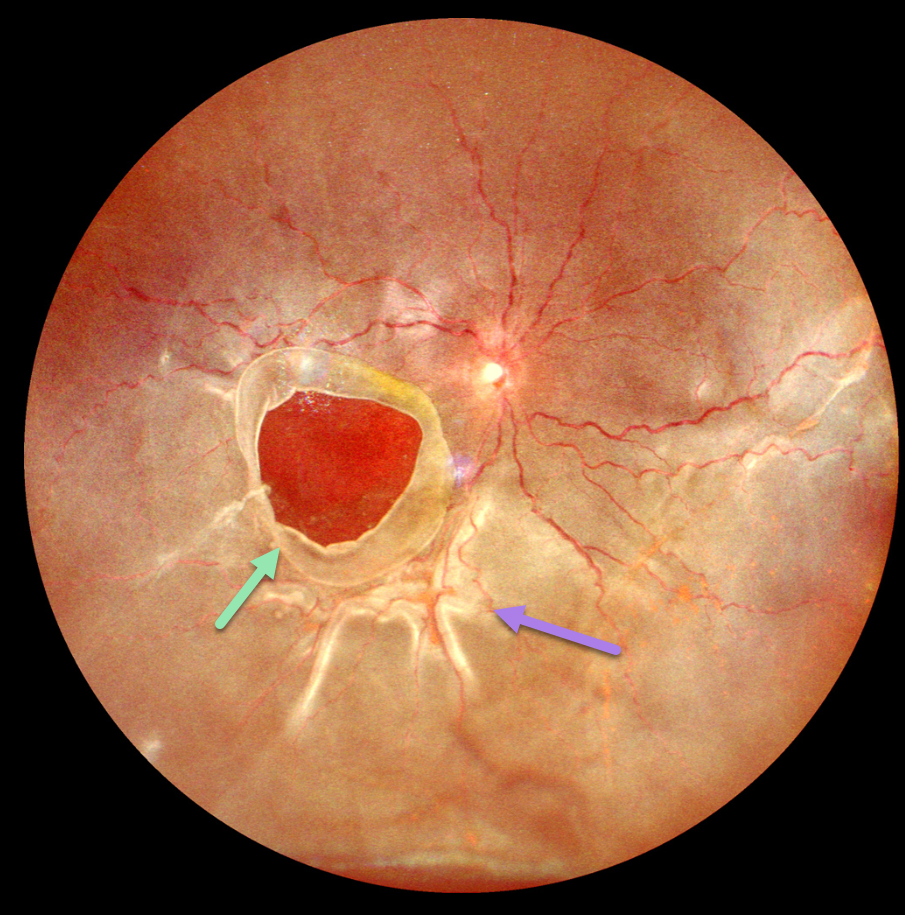
This 10YO boy presented with 1 week of vision loss in his left eye. The parents gave a vague history of trauma with a tennis ball 2 years earlier. Vision was counting fingers OD and 20/20 in his normal OS.
Ultrawidefield imaging shows a giant macular hole occupying nearly the entire macula. The hole’s edges are rolled, and proliferative vitreoretinopathy with star folds is present within the total rhegmatogenous retinal detachment (RRD).
Emergent vitrectomy surgery with silicone oil was scheduled.
Learning Points:
Blunt ocular trauma can result in numerous sequelae, including commotio retina, retinal breaks and detachment, choroidal rupture, and macular hole. A giant macular hole, also reported in Alport syndrome (Shah and Weinberg Ophthalmic Genetics 2010;31(2):93-97), is an exceedingly rare traumatic event (Hernandez-Da Mota Case Rep Ophthalmol 2011;2:283-286).
Traumatic macular holes are thought to result from a vitreous coup contrecoup injury or a jet stream of anterior chamber aqueous humor shooting through Cloquet canal into the premacular bursa.
RETINAL ARTERIAL MACROANEURYSM (RAM) WITH MACULAR HOLE
Will Gibson
Originally posted on @retina.rocks January 13, 2025
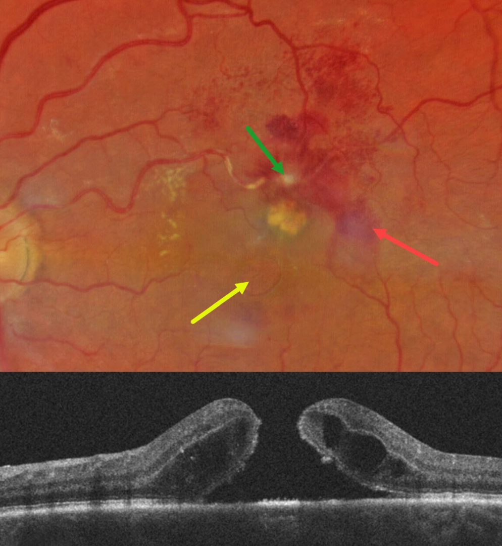
This 67YO white female presented for her six-month diabetic follow-up with a few weeks of vision loss in her left eye. Vision was 20/80 OS and 20/20 in her normal OD.
Color photography shows a fibrosed retinal arterial macroaneurysm (RAM), in the superior macula. Faint subretinal blood is noted temporally, with mostly inner retinal blood superotemporally. The artery is sheathed just proximal to the RAM. A large macular hole with surrounding subretinal fluid is present, accompanied by a lipid clump between the hole and the RAM. Faint lipid is also noted in the nasal macula. She was referred to a retinal specialist for further management.
Learning Points:
Macular hole is a rare finding with retinal arterial macroaneurysms (Sagara et al, Br J Ophthalmol 2009;93:1337-1340). Subretinal and sub-internal limiting membrane blood appears to increase the risk of macular hole.
TRAUMATIC MACULAR HOLE
Originally posted on @retina.rocks December 18, 2024
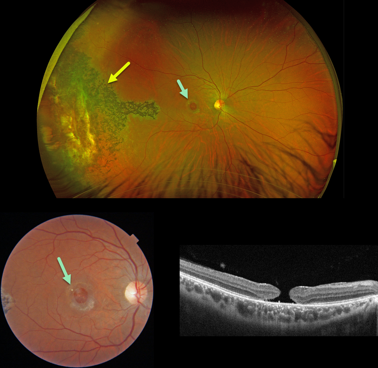
This 31YO male suffered a paint gun injury to his right eye when he was 12YO. Vision has since been stable and poor in this eye. Vision was 20/200.
Optos color RG imaging shows temporal peripheral scarring with intraretinal pigment migration. A large macular hole with surrounding pigmentary changes is noted on Triton color imaging and swept-source OCT.
Learning Points:
Since fresh traumatic holes can spontaneously flatten, especially in pediatric cases (Liu et al, AJO 2020;210:174-183), an initial period of observation is recommended before considering vitrectomy. Given the size and chronicity of our patient’s hole, surgery was not considered.
Photoreceptor loss from numerous causes, including blunt trauma, retinal detachment, macular telangiectasia, and retinitis pigmentosa (RP), can lead to intraretinal RPE migration, a shared phenotype. Unilateral RP is exceedingly rare and requires normal electrophysiologic testing with long-term observation to ensure that delayed findings are present in the fellow eye.
PROLIFERATIVE VITREORETINOPATHY (PVR) WITH MACULAR HOLE
Anjana Mirajkar and Manish Nagpal
Originally posted on @retina.rocks August 14, 2024
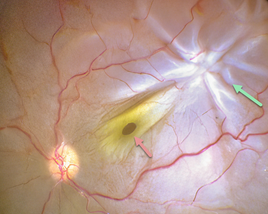
This intraoperative photograph shows a rhegmatogenous retinal detachment (RRD) complicated by proliferative vitreoretinopathy (PVR) with a star fold superotemporally. A full-thickness macular hole surrounded by its luteal pigment is dragged towards the PVR. The macular hole and detachment were repaired with vitrectomy.
Learning Points:
PVR, which consists of cellular membranes growing on either retinal surface, develops in about 5-10% of retinal detachments. RRD with a concurrent non-causal macular hole is found in about 2% detachments (Iros et al, Retina 2023;43:581-584). Although most eyes achieve anatomic success, final vision is usually poor. In our patient, unfortunately, there is no further clinical information, so it is unclear whether the macular hole caused the detachment or was secondary to traction from the star fold.
MACULAR PUCKER WITH ECTOPIC MACULAR HOLE
Originally posted on @retina.rocks July 25, 2024
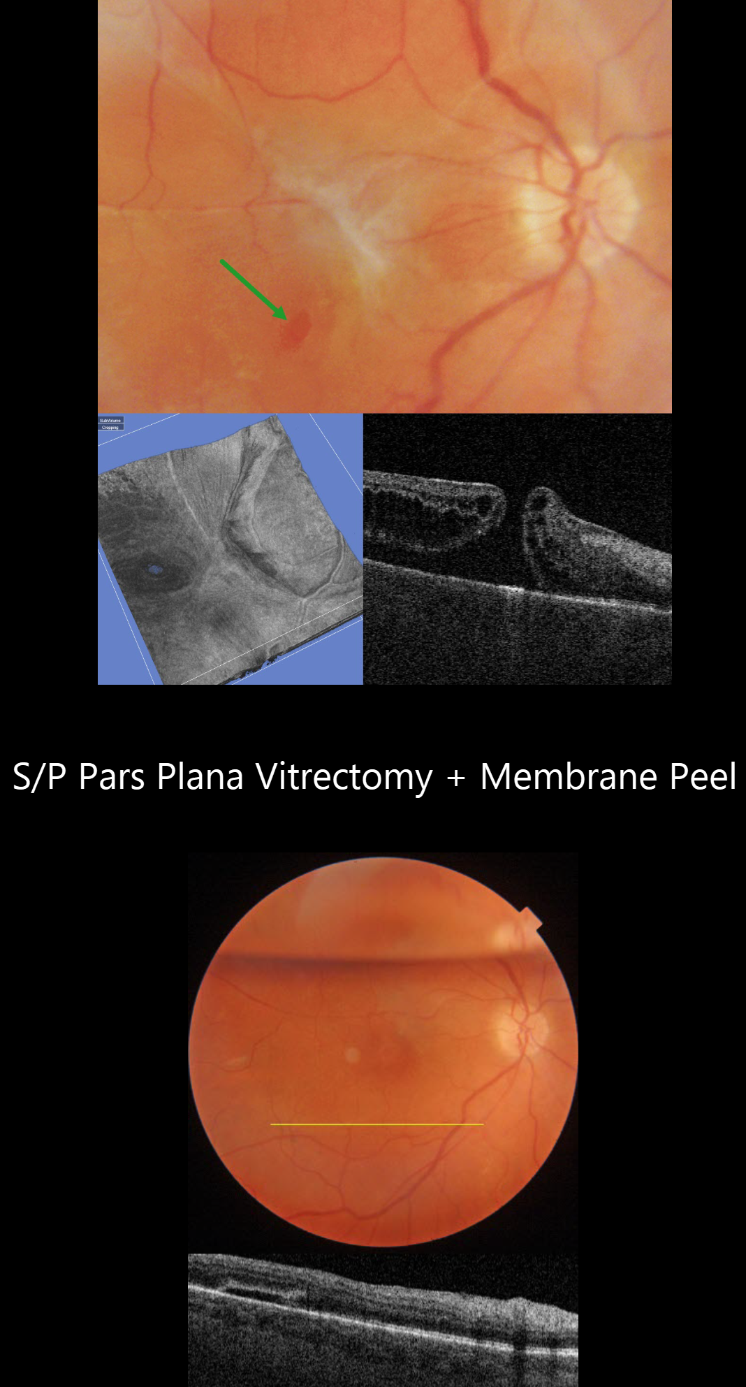
This 52YO female underwent vitrectomy for a hand motion, macula-off rhegmatogenous retinal detachment on 1/25/21. Despite successful reattachment, vision was counting fingers postoperatively, in part due to a severe macular pucker identified on 8/4/21. An ectopic secondary macular hole was present in the inferotemporal macula.
Vitrectomy surgery with membrane peeling was performed on 10/14/21. One week postoperatively, the ectopic hole was closed, and the macula was completely free of epimacular tissue. When last examined on 3/24/23, vision was 20/70.
MACULAR HOLE WITH PSEUDO BULLS-EYE MACULOPATHY
Originally posted on @retina.rocks June 24, 2024
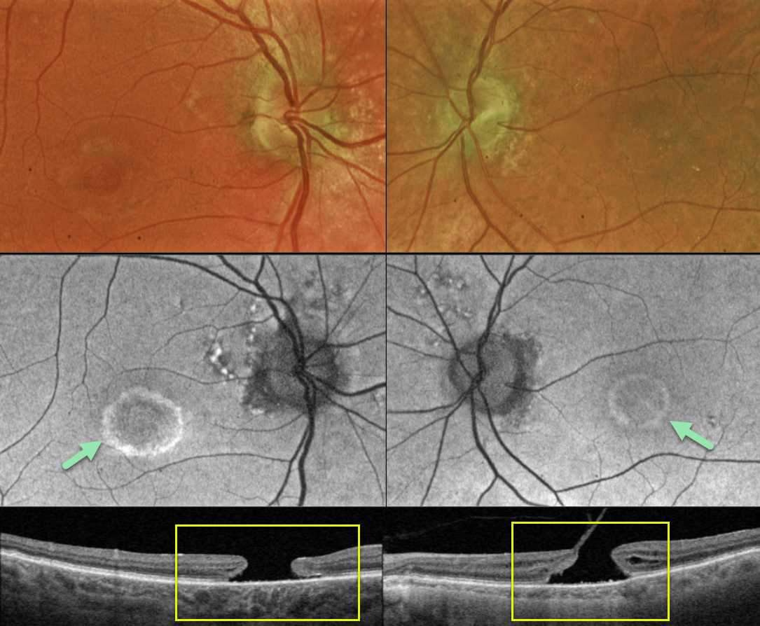
This 83YO male presented with a history of chronic bilateral vision loss. Vision was 20/150 OD and 20/200 OS.
Optos color RG imaging shows a bull’s eye pattern of bilateral pigmentary changes along with a large macular hole OU. Fundus autofluorescence (FAF) shows the bull’s-eye lesions as hyper-FAF. Triton swept-source OCT confirms bilateral macular holes.
Our patient has bilateral chronic macular holes with secondary RPE depigmentation, resulting in a bull’s-eye appearance. Due to the chronicity of the holes, observation was recommended.
Learning Points:
Bull’s eye maculopathy is characterized by a rim of subretinal pigment loss and outer retinal atrophy, most commonly found in inherited retinal diseases (usually ABCA4 disorders) and hydroxychloroquine/chloroquine toxicity.
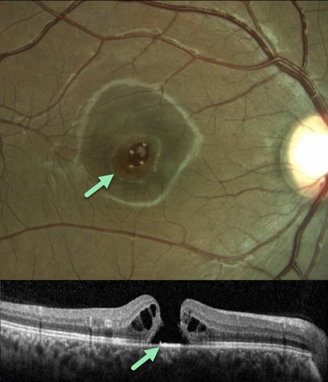
LASER-INDUCED MACULAR HOLE
Tareq Alsulami and Naser Alsaedi
Originally posted on @retina.rocks June 17, 2024
This 14YO boy presented with vision loss in his right eye following self-directed exposure to a blue wavelength laser pointer. Vision was 20/200 in his right eye and 20/20 in his normal left eye.
Color imaging and OCT scanning show an irregular full-thickness macula hole. The hole closed following vitrectomy surgery, but vision failed to improve.
Learning Points:
Laser pointers can cause significant macular injury, including solar maculopathy-like outer foveal microdefects, hemorrhage, pseudo macular dystrophy, vitelliform lesions, and macular scarring (Bhavsar et al, Surv Ophthalmology 2021;66:231-260). There are also numerous reports of a secondary full-thickness macular hole, as developed in our patient.
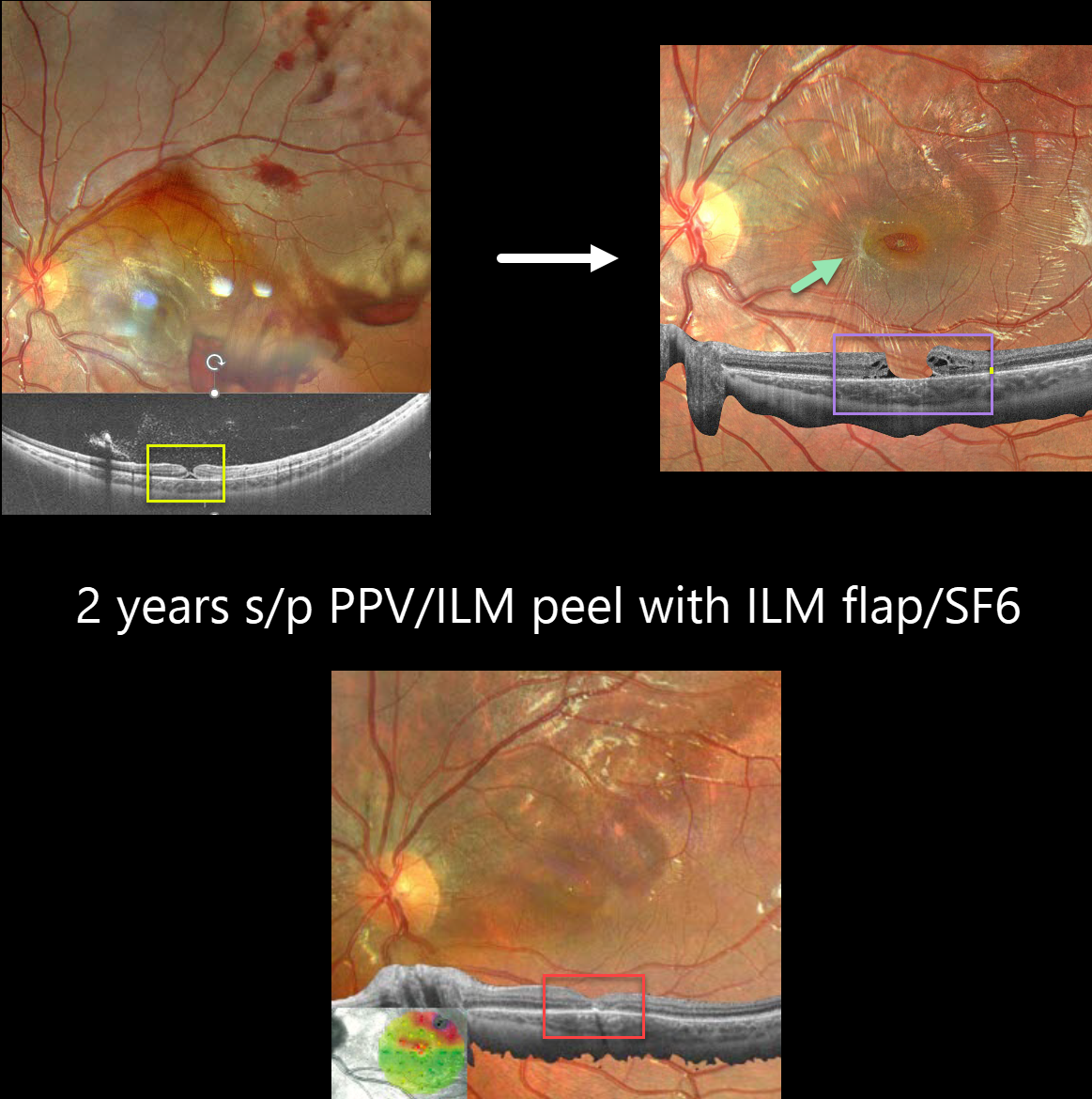
TRAUMATIC MACULAR HOLE
Barbara Parolini, Veronika Matello, Giulia Freschi, and Roberta Penzani
Originally posted on @retina.rocks April 9, 2024
This 10YO boy was referred for vision loss immediately after getting hit in his left eye by a soccer ball 7 days earlier. Vision was 20/70.
Fundus photography shows scattered intra- and preretinal hemorrhages with a large confluent area of commotio retina superiorly and temporally.
Widefield OCT shows a partial-thickness macular hole with the external retina intact but elevated. Three months later, vision was 20/100, and an irregular, full-thickness macular hole with surrounding macular pucker was seen.
25-gauge vitrectomy with ILM peeling, ILM flap, SF6 gas tamponade, and 3 days face-down positioning was performed. Two months later, vision improved to 20/40 with complete closure of the hole. Two years later, vision was 20/25.
Learning Points:
This patient illustrates several sequelae of blunt trauma, including retinal/preretinal blood, commotio retinae, and macular hole formation. Since fresh traumatic holes can spontaneously flatten, especially in pediatric cases (Liu et al, AJO 2020;210:174-183), an initial period of observation is recommended before considering vitrectomy.
In our patient’s case, the hole did not resolve, and surgery provided good anatomic and long-lasting visual outcomes.
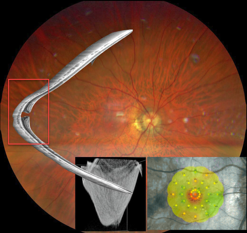
STAPHYLOMA + MACULAR HOLE
Barbara Parolini and Veronika Matello
Originally posted on @retina.rocks February 13, 2024
This Eyecare Clinic (in Brescia, Italy) case was submitted by Barbara Parolini, Veronika Matello, Giulia Freschi, and Roberta Penzani.
This 48YO highly myopic male presented for a sudden drop in vision. Vision was 20/100 OD and 20/20 in OS.
Color photography shows a blunted macular reflex. Widefield OCT shows a conical-shaped staphyloma of the entire posterior pole with a full-thickness macular hole. Microperimetry shows a decreased but not absent foveal sensitivity.
According to the Myopic Traction Maculopathy Staging System (Parolini et al, Eur J Ophthalmol 2021;31:1299-1312), this eye is in Stage 1c. The left eye showed a similar staphyloma but without a macular hole (not shown). The patient declined surgery.
Learning Points:
A macular staphyloma consists of a concave posterior bowing of the eye wall in patients with high myopia. These myopic patients also have a markedly thinned choroid throughout the posterior pole.
Due to stretching of the posterior retinal structures, these eyes are at increased risk of numerous findings, including macular schisis/traction and macular neovascularization.
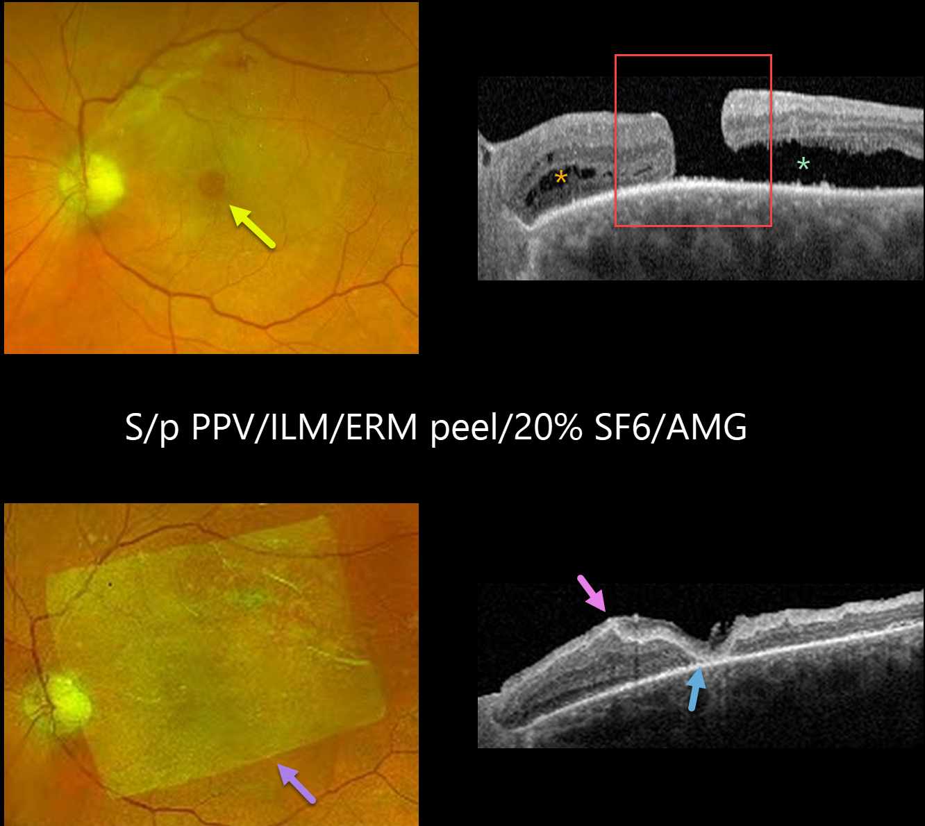
MACULAR HOLE
Yuenpang Cheung, Stephanie Choi and Tongalp Tezel
Originally posted on @retina.rocks November 28, 2023
This 77YO male had a history of failed vitrectomy with internal limiting membrane (ILM) peeling and SF6 gas for a long-standing full-thickness macular hole, and presented for a second opinion. Vision was 20/125.
Optos color RG imaging shows a persistent macular hole with some underlying subretinal fluid. There is a residual epiretinal membrane (ERM) along the superior arcade.
OCT shows the full-thickness macular hole with outer retinal cysts nasally and subretinal fluid temporally. Some ellipsoid zone (EZ) and outer retinal changes are visible.
The patient was eager to try additional surgery and underwent a vitrectomy with ERM/ILM peel, endolaser, 20% SF6, and amniotic membrane graft (AMG) placement. At 9 weeks post-op, the patient’s vision had improved to 20/80.
Fundus imaging shows a sheet of AMG over the macular hole and several superior laser scars. OCT scanning shows the macula hole to be closed under the AMG with EZ and outer retinal changes, but resolved temporal subretinal fluid.
Learning Points:
Approximately 90% of macular holes close with a single surgery. However, larger holes over 400 microns and more chronic holes can be challenging to close. Several techniques have been described to treat these refractory holes, including a broader ILM peel, instruments such as a Flex Loop to loosen up the edges of the hole, amniotic membrane grafts, or various variations of inverted ILM flaps to drape the hole, and even autologous full-thickness retinal tissue transplantation.
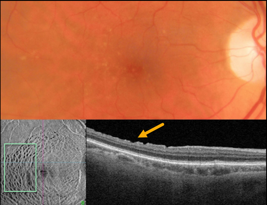
DISSOCIATED RETINAL NERVE FIBER LAYER
Originally posted on @retina.rocks October 17, 2023
This 75YO male underwent successful macular hole surgery 5 years earlier. Vision was 20/40.
Color photography shows scattered mixed drusen. B-scan OCT shows small irregular defects in the inner temporal macula. En face OCT shows extensive dimpling that parallels the nerve fiber layer.
Learning Points:
Pars plana vitrectomy with membrane peeling is frequently performed for the management of many vitreomacular interface disorders, including vitreomacular traction, macular hole, and epiretinal membrane.
While ILM peeling decreases epiretinal membrane recurrence and enhances macular hole closure, it also causes mechanical trauma to the inner retina, as evidenced by dissociated optic nerve fiber layer.
Many mechanisms have been proposed, including Müller cell damage from direct contact with surgical instruments, damage to the deep inner retinal layers, damage from visualization dye, or tractional thinning of the temporal retina.
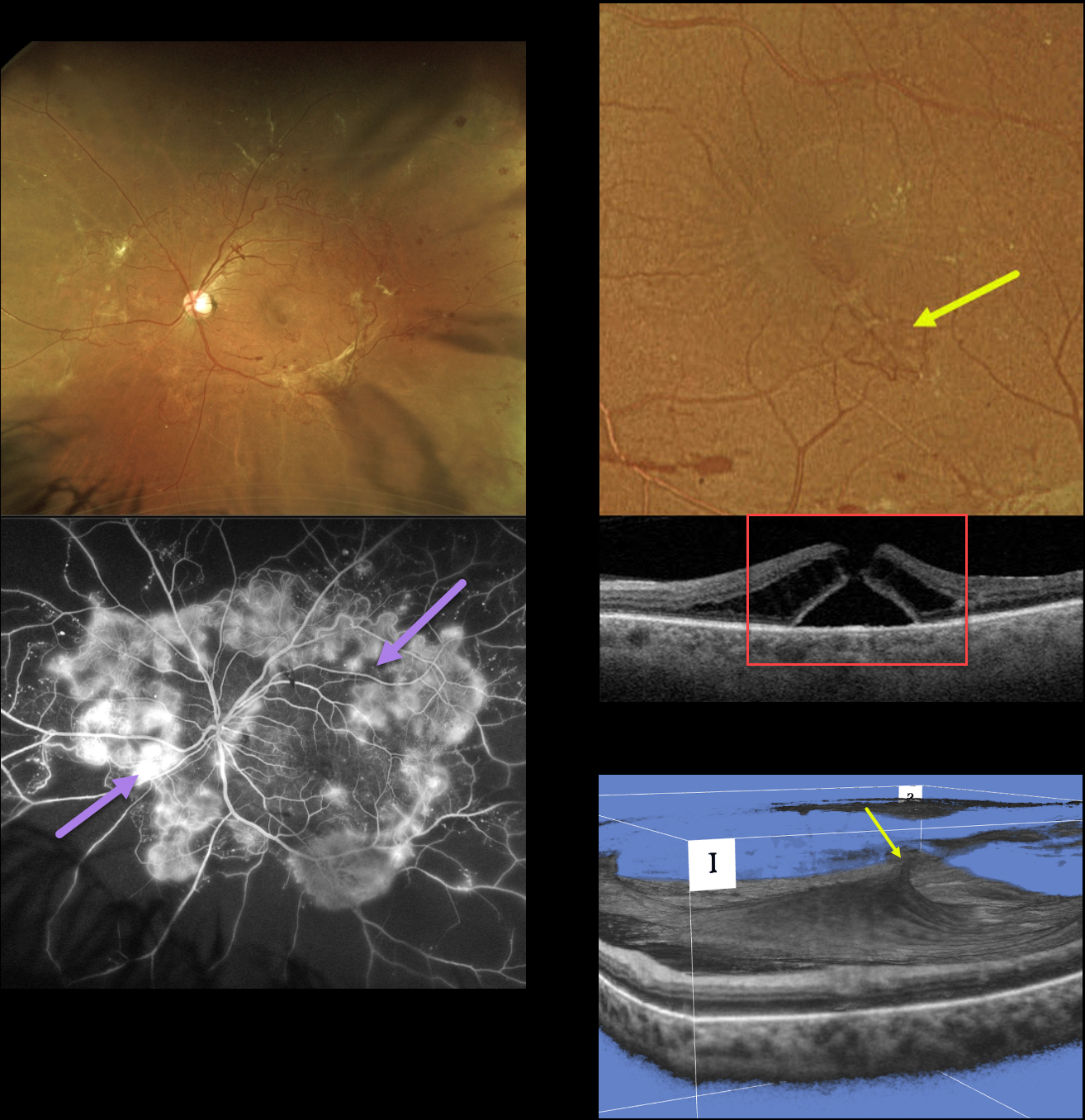
MACULAR HOLE DUE TO DIABETIC TRACTION
Originally posted on @retina.rocks September 21, 2023
This 49YO female with type 2 diabetes presented with a several-year history of blurred vision in her left eye. Vision was 20/40 OD and 20/60 OS.
Optos RG imaging shows variably fibrosed retinal neovascularization encircling the posterior pole. Fluorescein angiography shows active leakage from these vessels.
An area of small neovascularization is present in the inferior temporal macula, which caused a tractional full-thickness macular hole. A 3D OCT view best shows this diabetic traction.
Vitrectomy surgery was scheduled for her left eye. Primary vitrectomy will be performed for her right eye as well once the left eye has stabilized.
Learning Points:
In 1988, Dr. Gass described his concept for the pathogenesis of idiopathic macular holes (Arch Ophthalmol 1988;106:629-639). His classification system, from stage 1 for an impending macular hole through stage 4 for a fully developed macular hole with complete posterior vitreous detachment, was based on his concept of localized, mostly tangential traction caused by the shrinking foveal vitreous cortex. This classic paper heralded modern macular hole surgery for a previously untreatable condition.
The advent of OCT technology, however, has shown that macular hole development is more often due to focal vitreomacular traction, as our case beautifully illustrates.
The current classification system, described by the International Vitreomacular Traction Study Group, emphasizes the characteristics that determine treatment choice and visual prognosis (Ophthalmology 2013;120:2611-2619). This includes size (small <= 250 microns, medium >250 microns and <= 400 microns, large >400 microns), presence or absence of VMT, and cause (primary from VMT or secondary).
Our patient emphasizes why this new classification system was implemented, since it provides information that guides our treatment. Our patient’s hole is secondary to diabetic traction, which obviously requires a very different vitrectomy procedure compared to a primary hole.
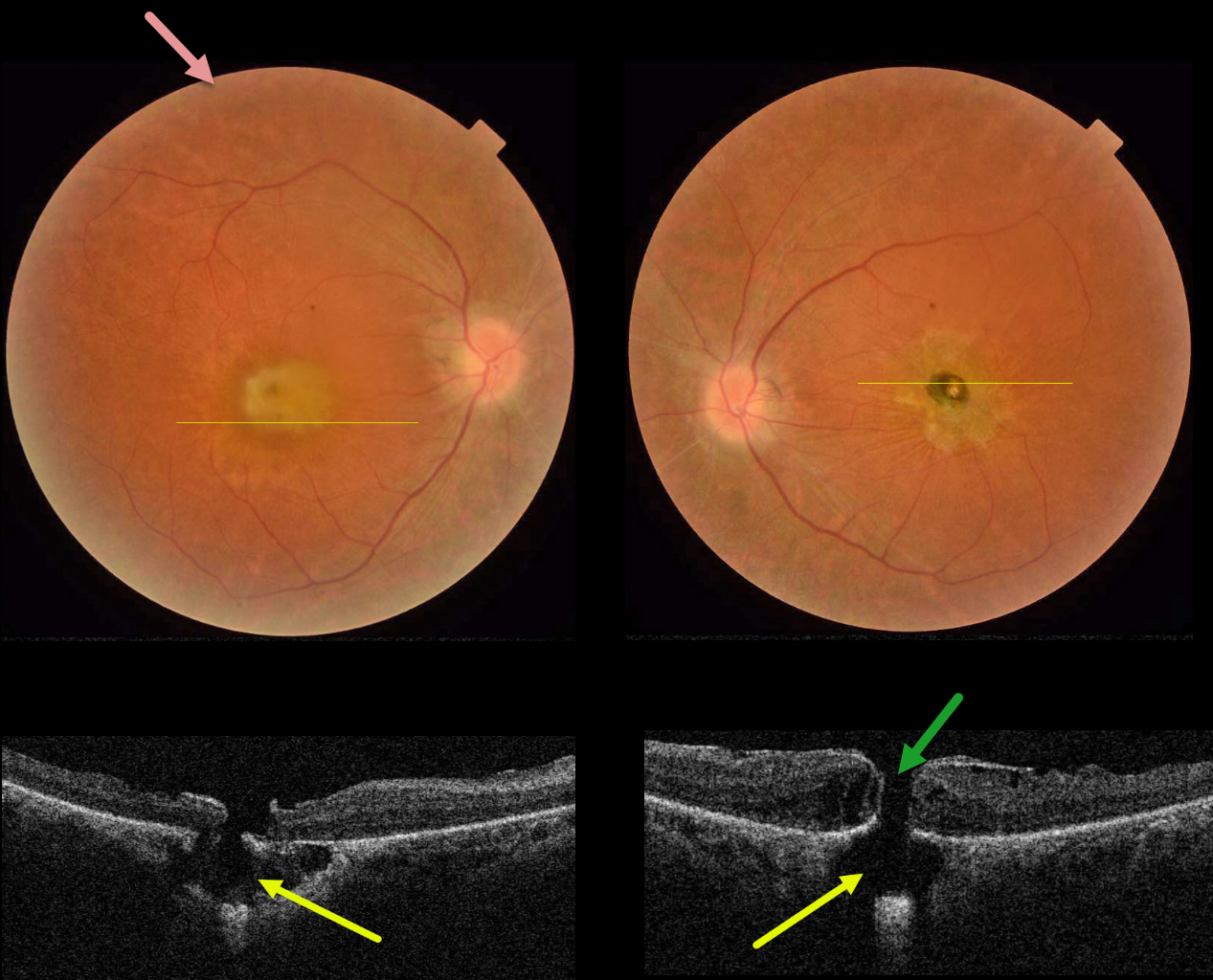
RETINITIS PIGMENTOSA WITH CHOROIDAL CAVITATIONS
Omar Mulki and Faisal Fayyad
Originally posted on @retina.rocks August 16, 2023
This 32YO female presented with a known history of severe vision loss from retinitis pigmentosa (RP). Her parents are first-degree cousins, and there was no family history of RP. Vision was hand motion OD and counting fingers OS.
Fundus photography shows bilateral irregular yellow central macular pigmentary changes, with a central hyperpigmented spot OS.
OCT scanning through each macula shows a full-thickness macular hole with an underlying defect in the RPE. Each macular hole is suspended over a choroidal cavitation.
Surgical repair was offered with appropriate counseling regarding the poor visual prognosis, and the patient elected to be observed.
Learning Points:
Initially described by Freund et al (Arch Ophthalmol 2003;121:197-204), intrachoroidal cavitations are most commonly found in high myopes as a peripapillary yellow-orange subretinal lesion.
Ornek and Ornek reported a patient with pathologic myopia and a unilateral macular hole communicating with a choroidal cavitation (Retinal Cases 2020;14:328-330).
Although macular holes can rarely be associated with RP (Jin et al, Retina 2008;28:610-614), to the best of our knowledge, ours is the first occurrence of RP with bilateral macular holes and underlying choroidal cavitations.
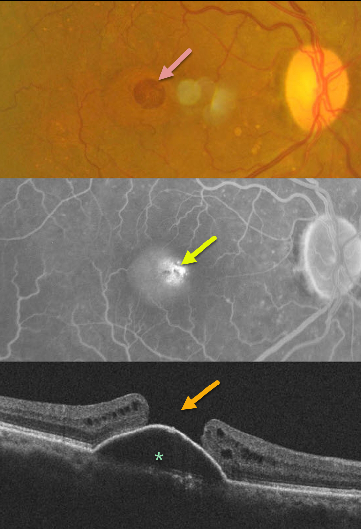
MACULAR HOLE
Shilpi Narnaware and Prashant Bawankule
Originally posted on @retina.rocks August 8, 2023
This 60YO female presented with 1 month of vision loss in her right eye. Vision was 20/100 OD and 20/40 OS.
Fundus photography of her right eye shows a large macular hole with some nasal pachydrusen. There was an old branch retinal vein occlusion without macular drusen in her left eye (image not shown).
Fluorescein angiography shows central pooling beneath a retinal pigment epithelial detachment (PED) with increased transmission through the macular hole. OCT scanning shows a PED with an overlying large macular hole.
This case presents a unique and interesting challenge in that macular hole surgery likely would not close the hole, given the underlying PED. She received 3 monthly Lucentis injections in her right eye, with no change in the PED.
Our thought would be to try a different anti-VEGF agent, such as Eylea, or consider adding photodynamic therapy for a possible pachychoroid-related avascular PED.
A similar case showed spontaneous closure of a macular hole following anti-VEGF therapy (see Chakraborty and Sheth, Indian J Ophthalmol Case Rep 2023;3:439-440).
If our patient’s macular hole remained open once the PED flattened, then macular hole surgery would be performed.
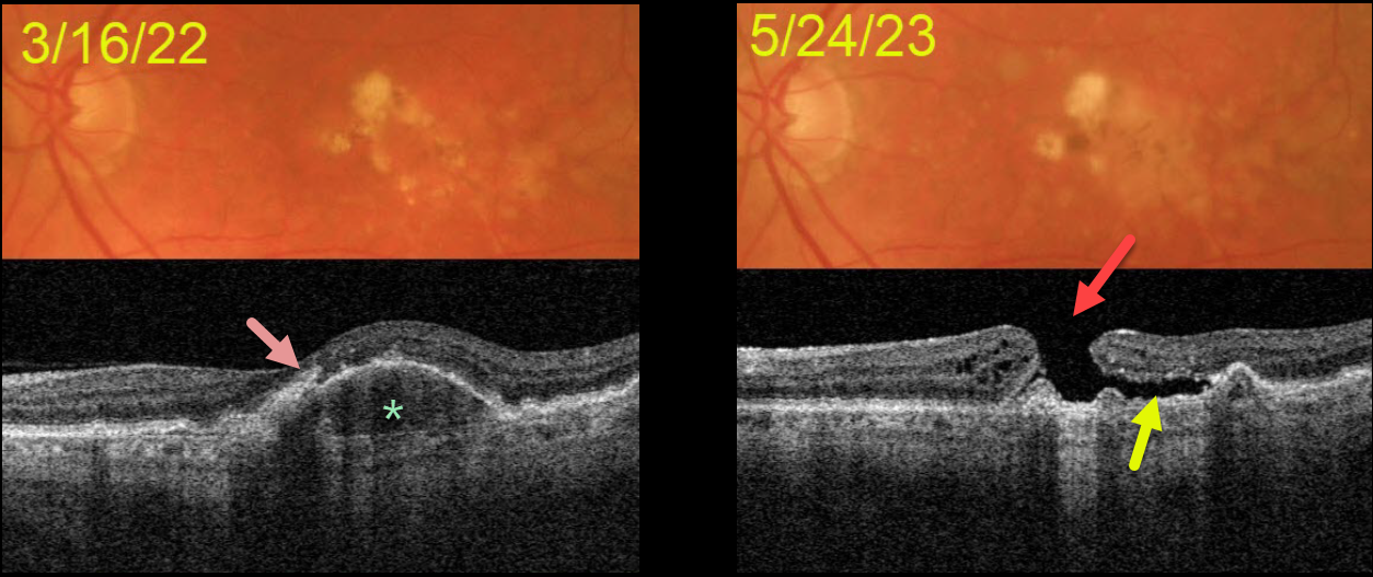
MACULAR HOLE AFTER COLLAPSE OF PED
Originally posted on @retina.rocks July 25, 2023
This 72YOM was examined with asymptomatic intermediate AMD bilaterally. Vision was 20/40 OD and 20/50 OS.
Color imaging of his left eye initially shows mostly large, soft drusen and pigment clumps. Swept source OCT shows a RPE detachment (PED) with overlying hyperreflective foci.
He returned over a year later without new symptoms, despite vision dropping to counting fingers. OCT shows a large macular hole overlying the collapsed PED. Shallow subretinal fluid lies below the temporal aspect of the hole.
The development of a macular hole following spontaneous collapse of a PED is quite unusual. Fortunately, vision in his right eye remains at 20/40 with intermediate dry AMD.
Since our patient was completely asymptomatic, he did not want macular hole surgery, and we will monitor him twice yearly.
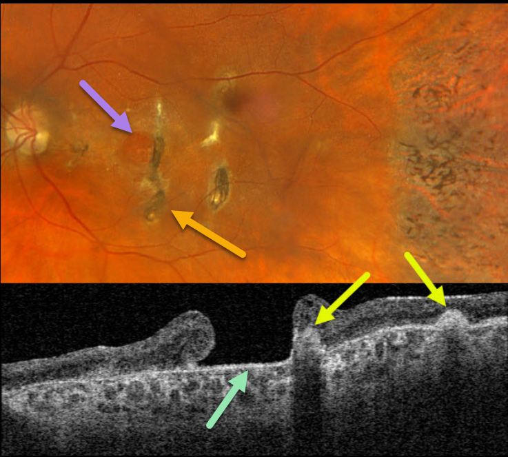
TRAUMATIC MACULAR HOLE + CHOROIDAL RUPTURE
Originally posted on @retina.rocks July 11, 2023
This 57YO female has a long history of vision loss in her left eye following blunt trauma years earlier. Vision was hand motion OS.
Optos color RGB imaging shows a large chronic macular hole with surrounding pigmentary changes. There are several hyperpigmented choroidal ruptures just temporal to the hole. Chorioretinal scarring with intraretinal pigment migration is noted in the temporal periphery.
Swept-source OCT shows a large macular hole. The choroidal ruptures appear as elevated hyperreflective subretinal mounds with posterior shadowing.
Learning Points:
This patient illustrates several sequelae of blunt trauma, including macular hole formation, choroidal rupture, and intraretinal pigment migration with a pseudo-sector retinitis pigmentosa appearance due to commotio-induced photoreceptor loss.
Since fresh traumatic holes can spontaneously flatten, especially in pediatric cases (Liu et al, AJO 2020;210:174-183), an initial period of observation is recommended before considering vitrectomy.
Given the chronicity of our patient’s hole, surgery was not considered.
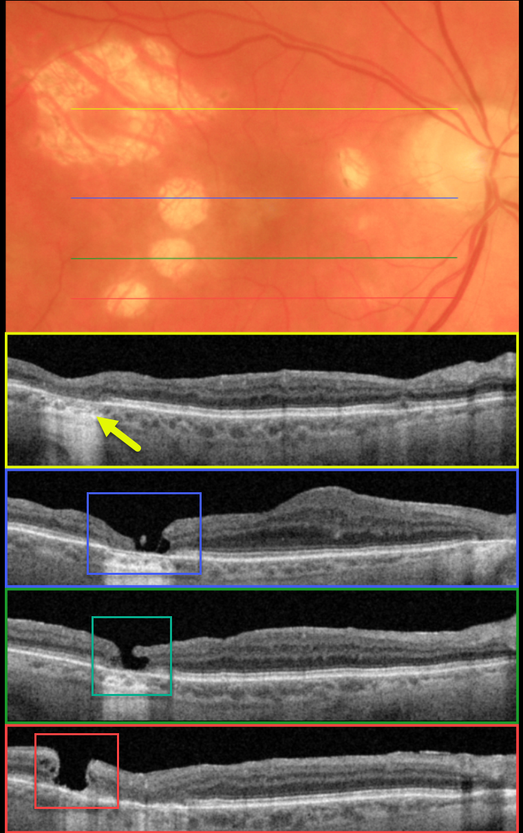
IATROGENIC MACULAR HOLE
Originally posted on @retina.rocks May 3, 2023
This 79YO female presented for a routine examination. ‘Successful’ macular hole surgery was performed elsewhere at least several years earlier.
Optos color imaging shows multiple atrophic scars in the temporal macula with a smaller scar nasally. Swept-source OCT scanning through the most superior, larger scar shows outer retinal atrophy.
OCT scans through each of the 3 smaller temporal scars show independent macular holes. Scanning of the central macula shows a thickened retina just nasal to fixation.
According to our patient, the single vitrectomy was uneventful, but her imaging suggests otherwise. It seems the surgeon made at least 5 deep retinal touches with the intraocular forceps while attempting to peel the internal limiting membrane. The larger superotemporal scar was probably a touch that was surrounded by endolaser.
The 3 ectopic macular holes may have developed intra- or post-operatively. Fortunately, her central macular hole closed, her vision was 20/40, and she remains thrilled with her surgical results.
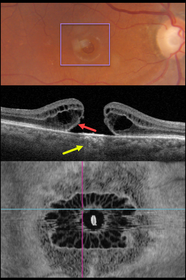
MACULAR HOLE
Originally posted on @retina.rocks January 12, 2023
This 81YO female presented with 20/200 vision in her right eye from a large full-thickness macular hole.
Swept-source OCT shows the classic appearance of a hole from a foveal dehiscence with cystic thickening. Outer retinal bands are noted along the posterior aspect of the hole’s elevated edges, indicating the lack of true tissue loss. This retinal defect results in increased light transmission into the choroid. En face imaging shows a unique and beautiful perspective of the hole.
Vitrectomy surgery was recommended.
Learning Points:
As described by Govetto et al (AJO 2020;212:43-56), idiopathic macular holes have a sunflower appearance showing features of both exudative and tractional cystoid spaces. The macular hole is hyporeflective centrally with multiple hyporeflective cystoid spaces radiating from the center.
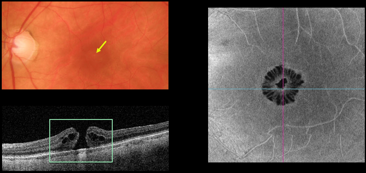
MACULAR HOLE
Originally posted on @retina.rocks November 11, 2022
This 65YO female presented with 20/200 vision in her left eye from a symptomatic full-thickness macular hole. The hole is barely visible funduscopically.
Swept-source OCT shows the classic appearance of a hole from a foveal dehiscence with cystic thickening. En face imaging shows a unique and beautiful perspective of the hole.
Learning Points:
As described by Govetto et al (AJO 2020;212:43-56), idiopathic macular holes have a sunflower appearance showing features of both exudative and tractional cystoid spaces.
The macular hole is hyporeflective centrally with multiple hyporeflective cystoid spaces radiating from the center.
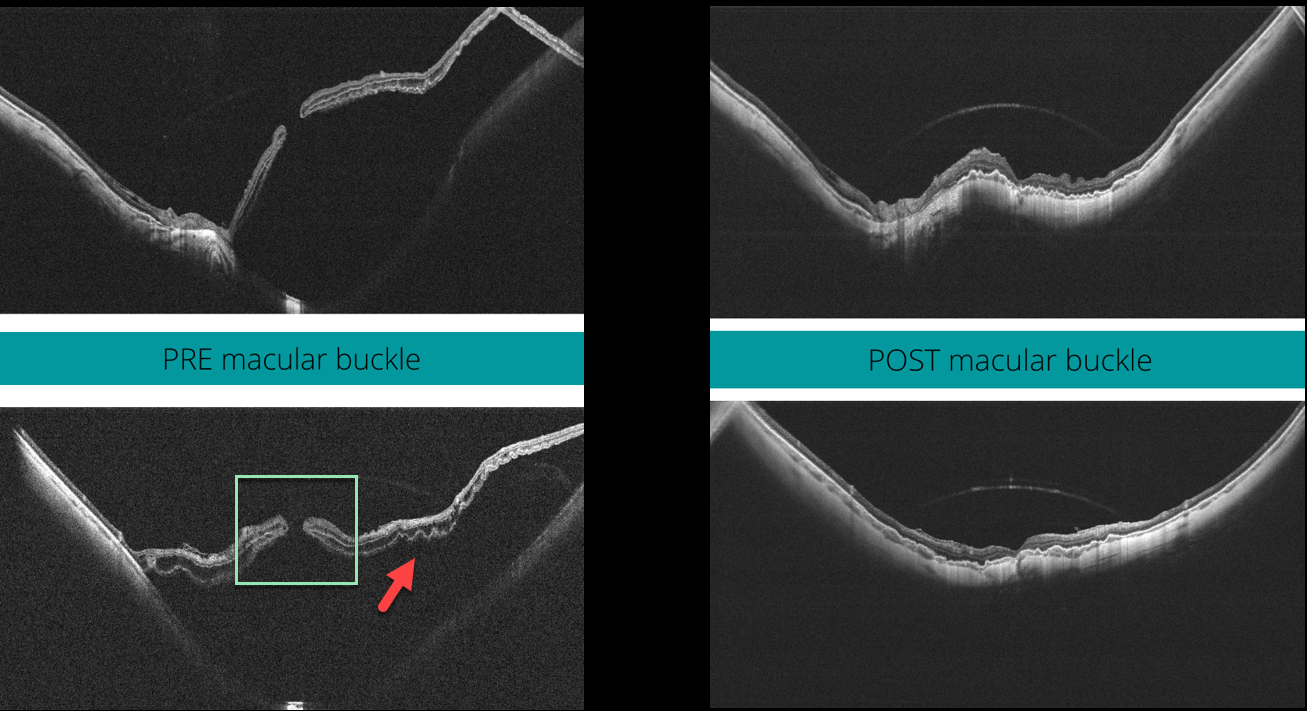
MACULAR HOLE
Veronika Matello and Barbara Parolini
Originally posted on @retina.rocks October 25, 2022
This 58YO highly myopic female presented with 20/400 vision in her left eye. The axial length was 32.45 mm, and her refractive error prior to LASIK surgery was 13D.
Widefield OCT scanning (Canon Xephilio SQ) shows a large macular retinal detachment with (or associated to) a full-thickness macular hole, defined as stage 4c according to the Myopic Traction Maculopathy Staging System (Parolini et al, Eur J Ophthalmol 2021;31:1299-1312).
Pars plana vitrectomy with an ILM flap and macular buckling was performed. One month postoperatively, the retina was flat, and the macular hole closed. Vision was 20/80.
Learning Points:
In our experience, these detachments are totally tractional, with the macular hole developing before or after the retina detaches. The macular buckle treats both the macular schisis and detachment, whereas the vitrectomy with ILM peeling closes the macular hole.
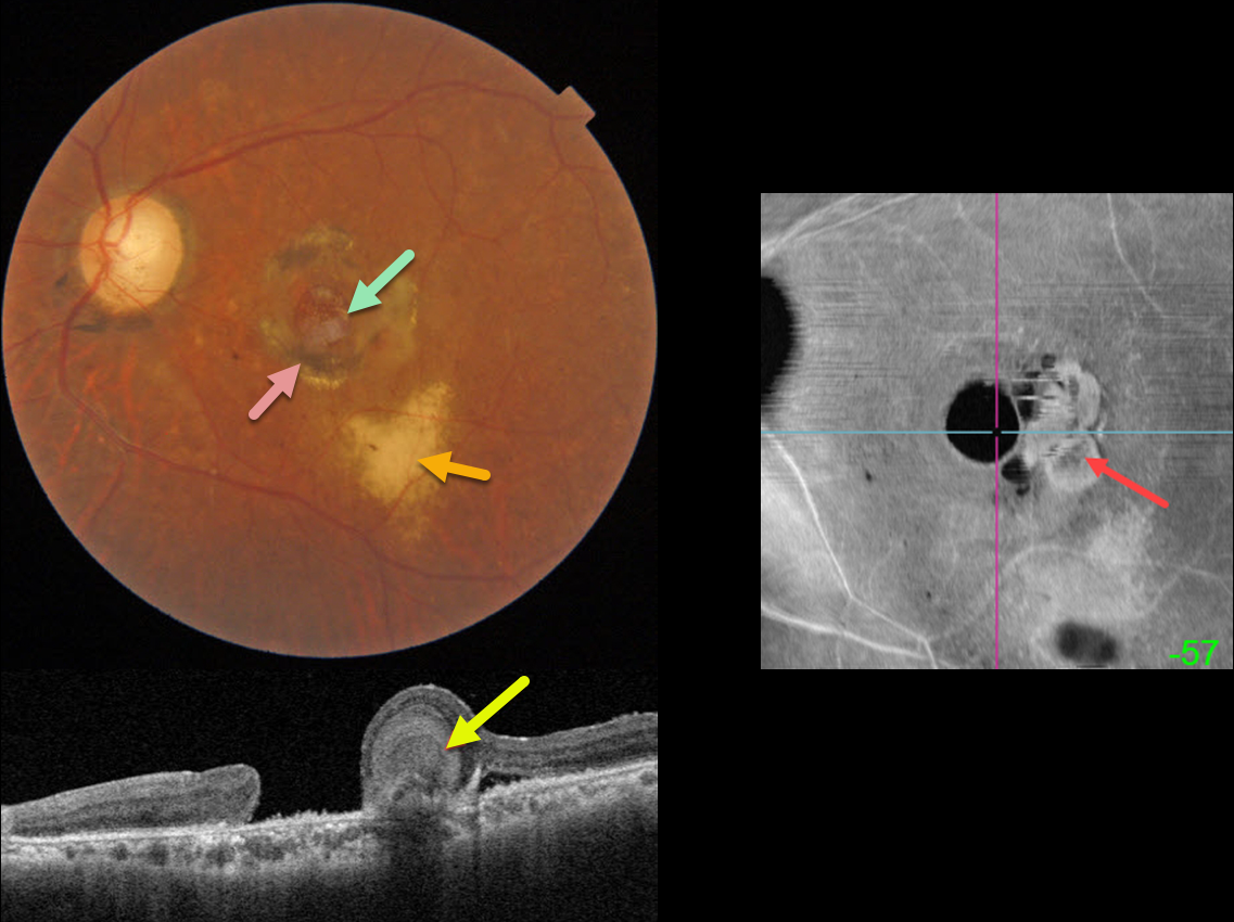
CHRONIC MACULAR HOLE
Originally posted on @retina.rocks September 19, 2022
This 59YO male has a complicated ocular and medical history, including HIV/AIDS, prior cytomegalovirus (CMV) retinitis that was treated with a ganciclovir implant in the 1990s, multiple retinal detachment surgeries in his right eye, eventually leading to evisceration for a blind and painful eye, and a chronic macular hole in his left eye.
Color imaging shows a large macular hole surrounded by a rim of variable subretinal hyperpigmentation. Yellowish subretinal scarring from prior CMV retinitis is noted more inferiorly.
Swept-source OCT shows an unusual ball of deep retinal hyperreflectivity. En face OCT shows a unique perspective for this macular hole, including the hyperreflective deep retinal material.
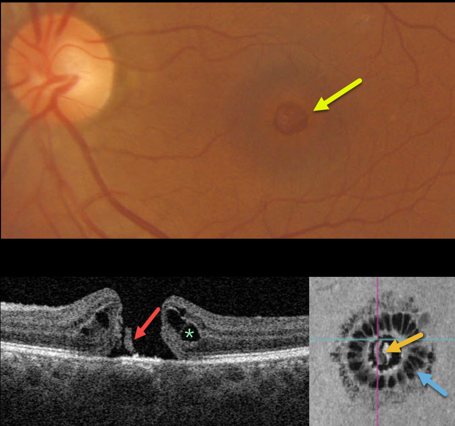
MACULAR HOLE
Originally posted on @retina.rocks August 4, 2022
This 51YO presented with counting-fingers vision secondary to a large (600-micron), full-thickness macular hole.
Swept-source OCT shows the classic appearance of a hole from a foveal dehiscence, with cystic thickening. A small remnant of the outer retina is adherent to the underlying RPE.
Vitrectomy surgery was recommended.
Learning Points:
En face imaging shows a unique and beautiful perspective of the hole. As described by Govetto et al (AJO 2020;212:43-56), idiopathic macular holes have a sunflower appearance showing features of both exudative and tractional cystoid spaces.
The macular hole is hyporeflective centrally with multiple hyporeflective cystoid spaces radiating from the center.
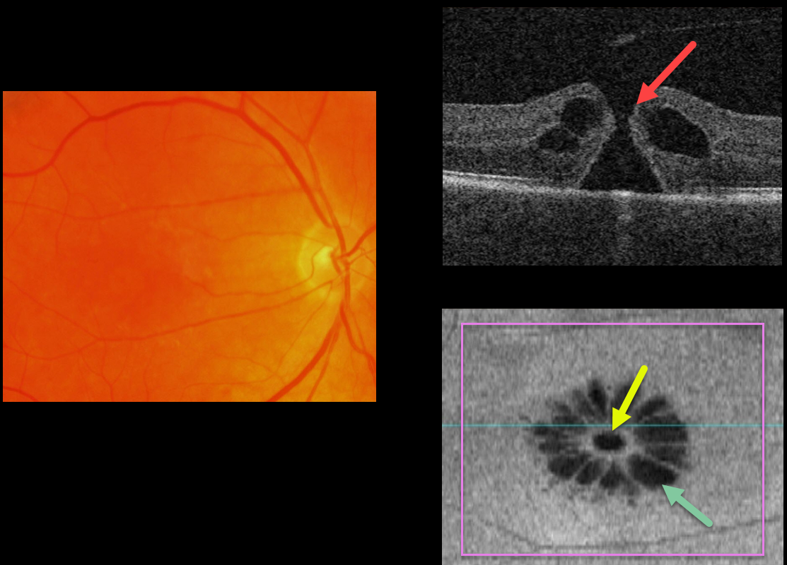
MACULAR HOLE
Originally posted on @retina.rocks May 5, 2022
This 63YO male patient has a full-thickness macular hole (MH) with 20/80 vision.
The hole is not well imaged in the color photograph.
OCT B-scan shows a small MH without traction.
The en face image beautifully depicts a unique perspective on an otherwise unremarkable case. The macular hole is hyporeflective centrally with multiple hyporeflective cystoid spaces radiating from the center.
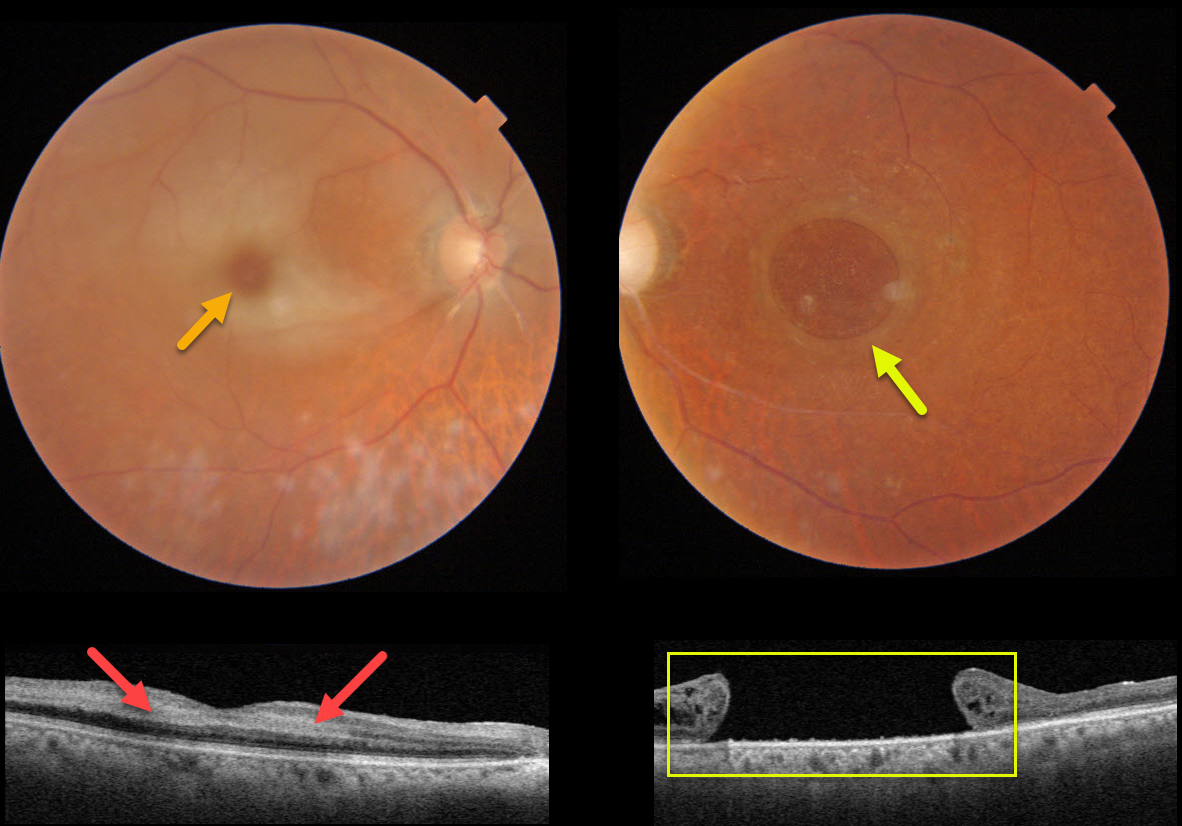
CRAO + MACULAR HOLE
Originally posted on @retina.rocks February 21, 2022
This 82YO female presented with counting fingers vision in her previously healthy right eye. Vision in her left eye had been counting fingers for many years due to a large chronic macular hole measuring 2,870 microns.
An acute central retinal artery occlusion (CRAO) was noted in her right eye. The foveal retina is still perfused by the underlying choroid, resulting in the classic cherry red spot.
Swept-source OCT shows hyperreflectivity from the opaque, ischemic inner retinal layers.
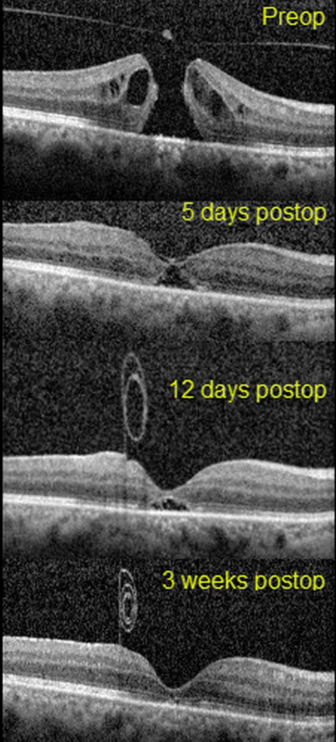
MACULAR HOLE
Erdem Dinç
Originally posted on @retina.rocks February 15, 2022
This 51yo presented with 20/1250 vision in her right eye from a primary full-thickness macular hole without traction measuring 407 microns. There was also a moderately severe nuclear sclerotic cataract.
Combined cataract surgery with 23-gauge pars plana vitrectomy, temporal half-moon inverted ILM flap, and fluid-air-SF6 exchange was performed, followed by prone positioning.
On the 5th postoperative day, the flap extended over the closed hole, but one week later, the ILM flap retracted and was rolled inward, suspended anterior to the macula.
Three weeks following vitrectomy, the hole was completely flattened with partial restoration of the outer retinal layers. The ILM flap continued to contract and scroll onto itself. Vision was 20/40.
Learning Points:
Introduced by Kelly and Wendel (Arch Ophthalmol 1991;109:654-659), macular hole surgery is one of the modern success stories in ophthalmology. ILM flaps appear to improve the surgical success for large (>400 microns) macular holes.
In this case, the ILM flap inadvertently scrolled onto itself and away from the macular hole by post-op day 12. Some authors have suggested that the ILM flap provides a scaffold for cells to grow over and help heal the hole.
However, most macular holes are actually foveal dehiscences without tissue loss, where the foveal retina splits and elevates like a drawbridge (Tornambe, Retina 2003;23:421-424). For successful surgery, there is thus no need or significant evidence for glial or neurosensory retina regrowth, just for the “drawbridge” to close.
As this case exemplifies, the ILM flap most likely serves the same purpose as the gas bubble, helping to sequester the macular hole from the overlying vitreous. This allows the RPE to actively pump (and the choroid to passively absorb) the subretinal fluid, allowing the foveal tissue to flatten into its normal position. Once the hole closes, as long as the vitreous and epimacular traction are relieved, there is likely no need for the ILM flap.
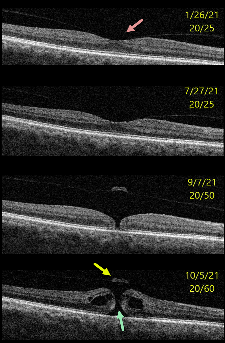
MACULAR HOLE
Originally posted on @retina.rocks November 9, 2021
This 64YO male presented with 20/25 vision and asymptomatic vitreomacular adhesion (VMA) in his right eye.
Over the next 8 months, this progressed to subtle focal vitreomacular traction (VMT) and eventually a small full-thickness macular hole without traction. Vitrectomy surgery was recommended.
Learning Points:
In 1988, Dr. Gass described his concept for the pathogenesis of idiopathic macular holes (Arch Ophthalmol 1988;106:629-639).
His classification system, from stage 1 for an impending macular hole through stage 4 for a fully developed macular hole with complete posterior vitreous detachment, was based on his concept of localized, mostly tangential traction caused by shrinking foveal vitreous cortex. This classic paper heralded the advent of modern macular hole surgery for a previously untreatable condition.
The advent of OCT technology, however, showed that macular hole development is driven more by focal vitreomacular traction, as our case beautifully illustrates.
The current classification system, described by the International Vitreomacular Traction Study Group, emphasizes the various characteristics that determine choice of treatment and visual prognosis (Ophthalmology 2013;120:2611-2619). This includes size (small <= 250 microns, medium >250 microns and <= 400 microns, large >400 microns), presence or absence of VMT, and cause (primary from VMT or secondary).
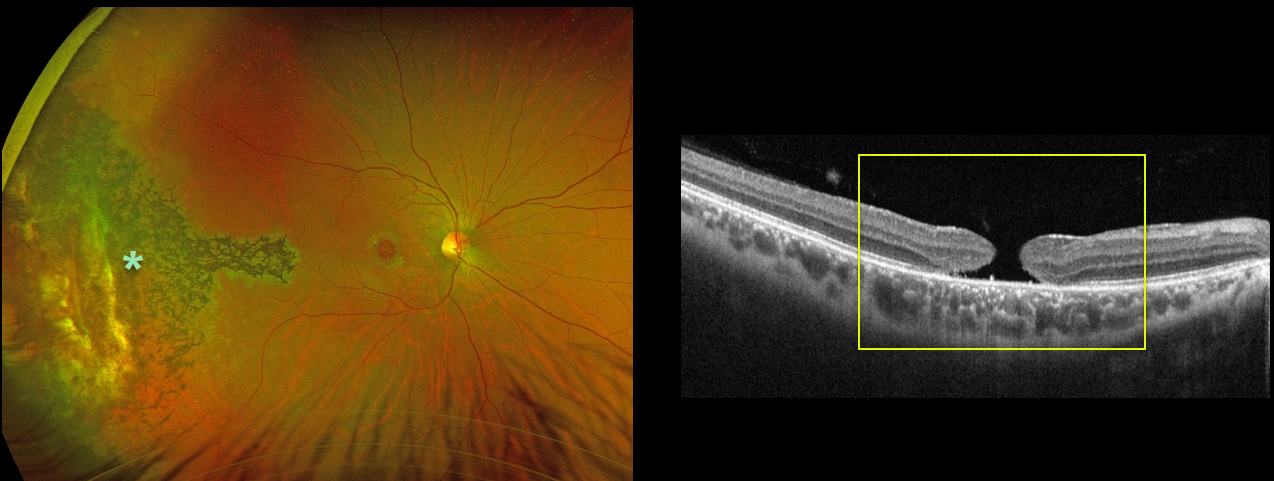
MACULAR HOLE
Originally posted on @retina.rocks August 24, 2021
This 31YO male was hit in his right eye by a paintball when he was 12 years old. His vision was 20/200 due to a large, chronic 1100-micron full-thickness macular hole.
Optos imaging shows chorioretinal scarring with intraretinal pigment migration extending from the distal macula into the temporal periphery, underlying the likely impact site.
Learning Points:
Traumatic macular holes are more common in younger patients, often from recreational or sports injury, and tend to be more eccentric in shape.
The mechanism could be sudden vitreous separation or stretching of the posterior pole resulting from anteroposterior ocular compression. The holes can spontaneously close.
Unfortunately for our patient, the hole remained open, and at this point, we saw little possible surgical benefit given its chronicity, size, and secondary pigmentary changes.
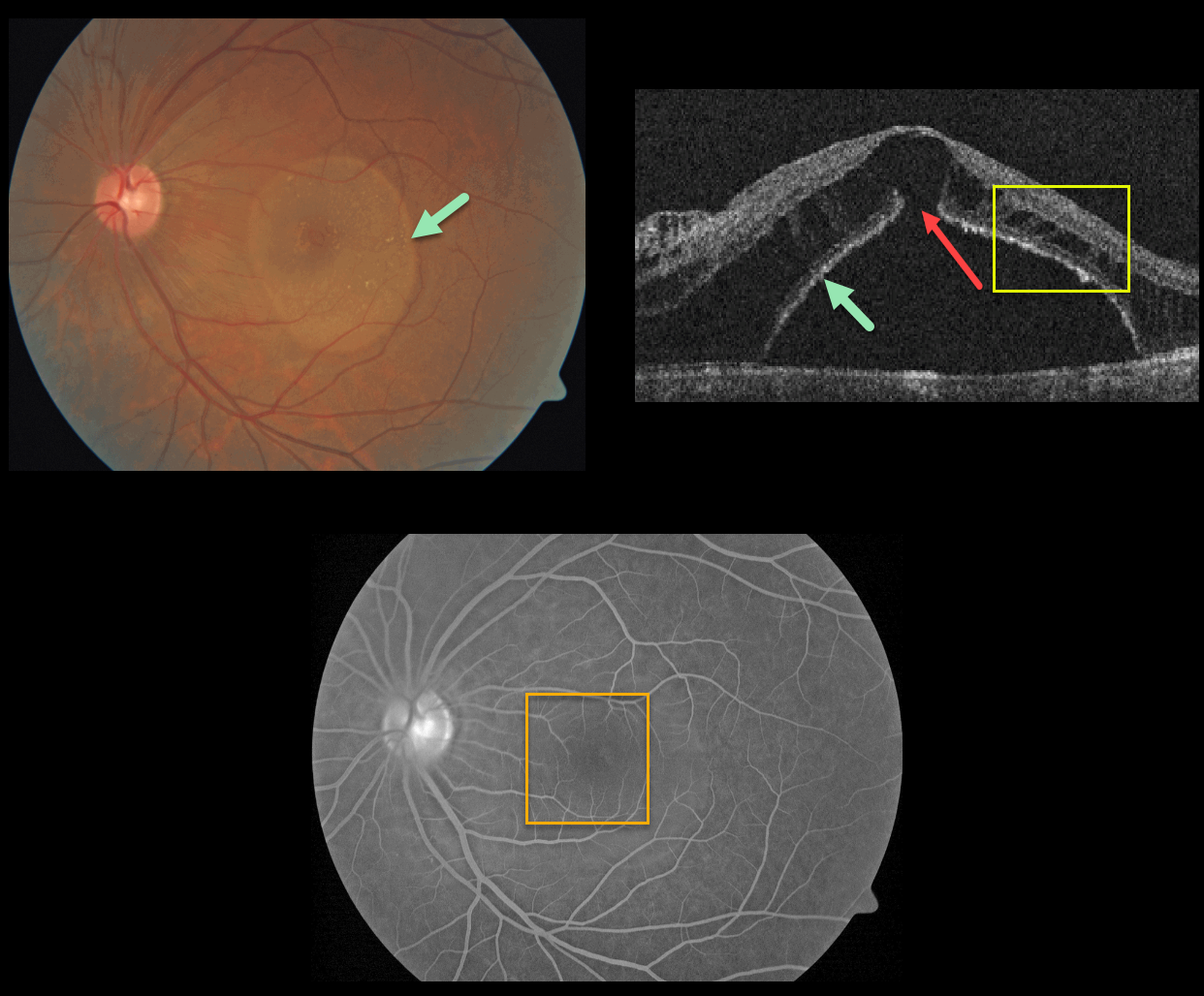
EXUDATIVE RETINAL DETACHMENT WITH OUTER MACULAR HOLE
Originally posted on @retina.rocks June 25, 2021
This patient presented with a sharply-circumscribed macular serous detachment, cystic outer retinal edema, and an outer macular hole. Fluorescein angiography is essentially normal without leakage.
An optic pit was absent clinically and on OCT imaging. Serum protein electrophoresis was normal, and a systemic cancer workup was negative.
Learning Points:
Serous detachment can be an uncommon ocular manifestation of paraproteinemia (see Mansour et al, Ophthalmology 2014;121;1925-1932). These detachments, which resemble those seen in optic pit maculopathy, are characterized by a well-defined serous detachment without angiographic leakage. A central outer macular hole is often present.
The fluid is likely due to an osmotic gradient generated by subretinal immunoglobulins that passively migrate into the subretinal space.
We do not know the cause of our patient’s findings, given his lack of an optic pit and a negative systemic workup.
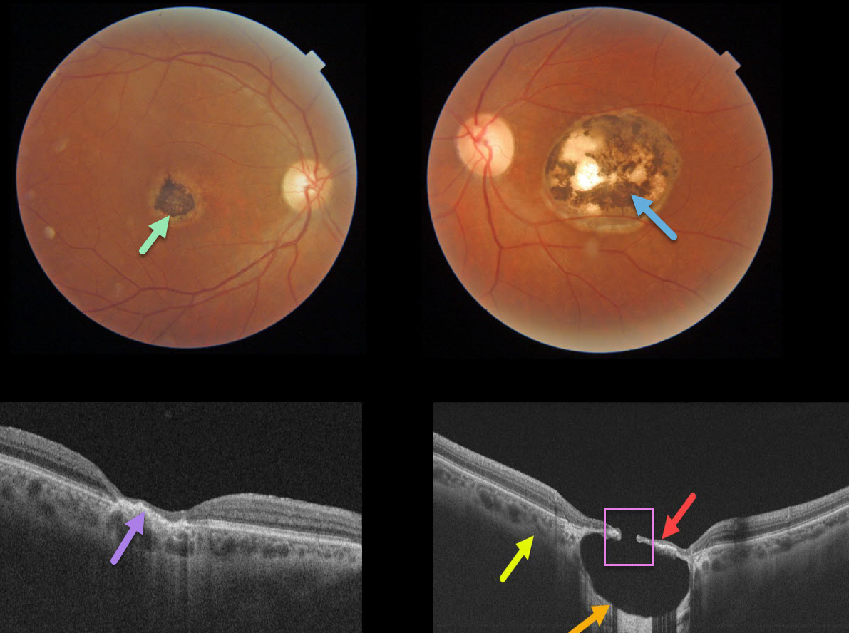
TOXOPLASMOSIS
Originally posted on @retina.rocks March 26, 2021
This 57YO male gave a history of lifelong poor vision in his left eye. Vision was 20/40 OD and 20/200 OS.
A small hyperpigmented foveal scar is noted in his right eye with neurosensory retinal atrophy and irregular elevation of the RPE.
A larger, round, variably pigmented, excavated macular scar is noted in his left eye. Triton swept-source OCT reveals a thinned atrophic retina and intact RPE suspended over an optically empty hyporeflective excavated colobomatous type lesion. The plane of the intact surrounding sclera is indicated by the yellow arrow. A small full-thickness retinal and RPE defect is noted centrally.
Learning Points:
For a great discussion on how to differentiate these infectious macular colobomatous lesions (congenital toxoplasmosis, Zika, and cytomegalovirus) from inherited dystrophies like North Carolina Macular Dystrophy (NCMD), see Kumar and Mahalingam AJO 2019;200;47-56.
In case you’re thinking our patient has NCMD, also see the first OCT description of congenital toxoplasmosis macular lesions (Garg et al, Retina 2009;29;631-637). Their Figures 4 and 6 look eerily similar to our patient’s right and left eyes!
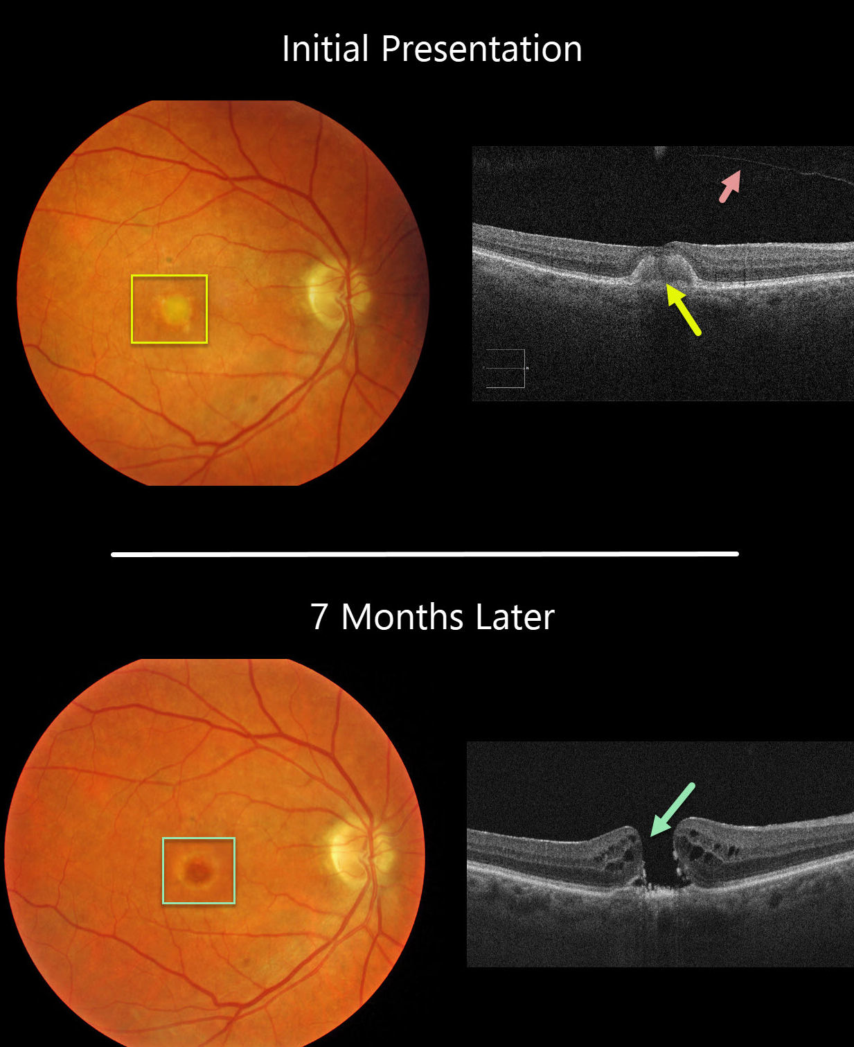
ADULT-ONSET FOVEOMACULAR VITELLIFORM DYSTROPHY
Originally posted on @retina.rocks November 18, 2020
This 77YO female was initially examined with 20/60 vision OD, 20/30 vision OS, and bilateral vitelliform lesions (only right eye pictured) consistent with Adult Best (adult-onset foveomacular vitelliform dystrophy). A complete posterior vitreous detachment (PVD) was noted in her right eye.
Seven months later, vision decreased to counting fingers OD due to a new full-thickness macular hole. This patient was asymptomatic due to good vision in her left eye and has opted against surgery for now.
Learning Points:
Macular holes are most commonly caused by a partial PVD with active vitreomacular traction. However, they can occasionally develop without traction following a prior PVD or vitrectomy surgery. Macular holes have also been reported as a rare complication of adult Best’s disease.
Adult-onset foveomacular vitelliform dystrophy is associated with a mutation in the BEST1 gene, which encodes the bestrophin-1 protein.
Bestrophin-1, a calcium-activated chloride channel, is primarily found in the basolateral plasma membrane of the RPE. BEST1 mutations cause a variety of phenotypes, including autosomal recessive bestrophinopathy, best vitelliform macular dystrophy, and autosomal dominant vitreoretinochoroidopathy.
MACULAR HOLE
Originally posted on @retina.rocks November 6, 2020
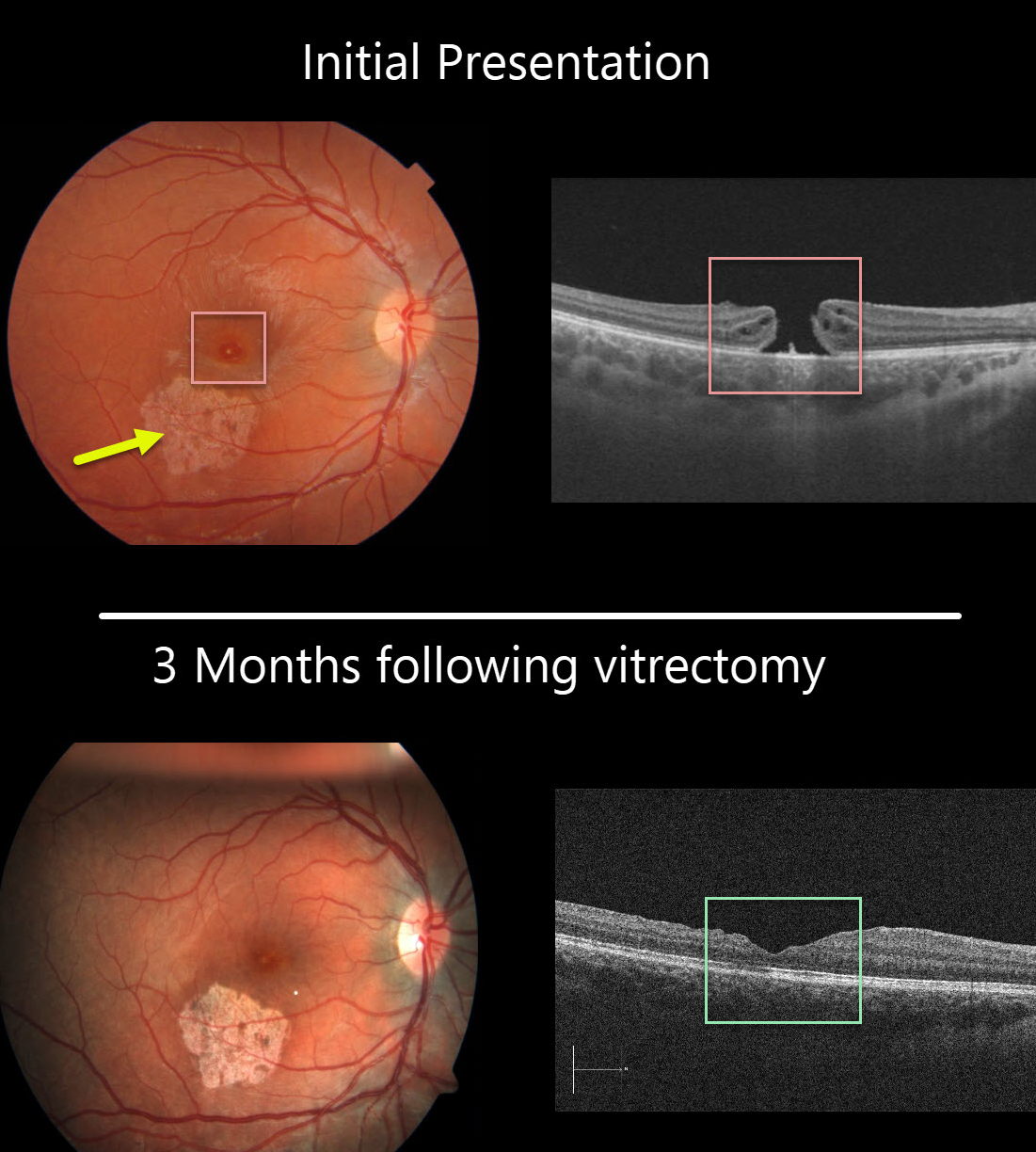
This 13YO girl was hit in her right eye while playing volleyball. She presented to us several months later with 20/200 vision, an inferotemporal macular scar from resolved commotio retina, and a full-thickness macular hole.
The macular hole was closed following successful vitrectomy, and she was 20/100 one month postoperatively. Vision improved to 20/40 three months following surgery. Since the scotoma from the macular scar was superior, she is now virtually completely visually asymptomatic and extremely lucky!
Learning Points:
Traumatic macular holes are more common in younger patients, often from recreational or sports injury, and tend to be more eccentric in shape.
The mechanism could be due to sudden vitreous separation or to stretching of the posterior pole resulting from anteroposterior ocular compression. The holes can spontaneously close and have a high surgical success rate, as in our patient (see Liu et al AJO 2020;210:174-183).
ADULT-ONSET FOVEOMACULAR VITELLIFORM DYSTROPHY WITH SECONDARY MACULAR HOLE
Originally posted on @retina.rocks May 28, 2020
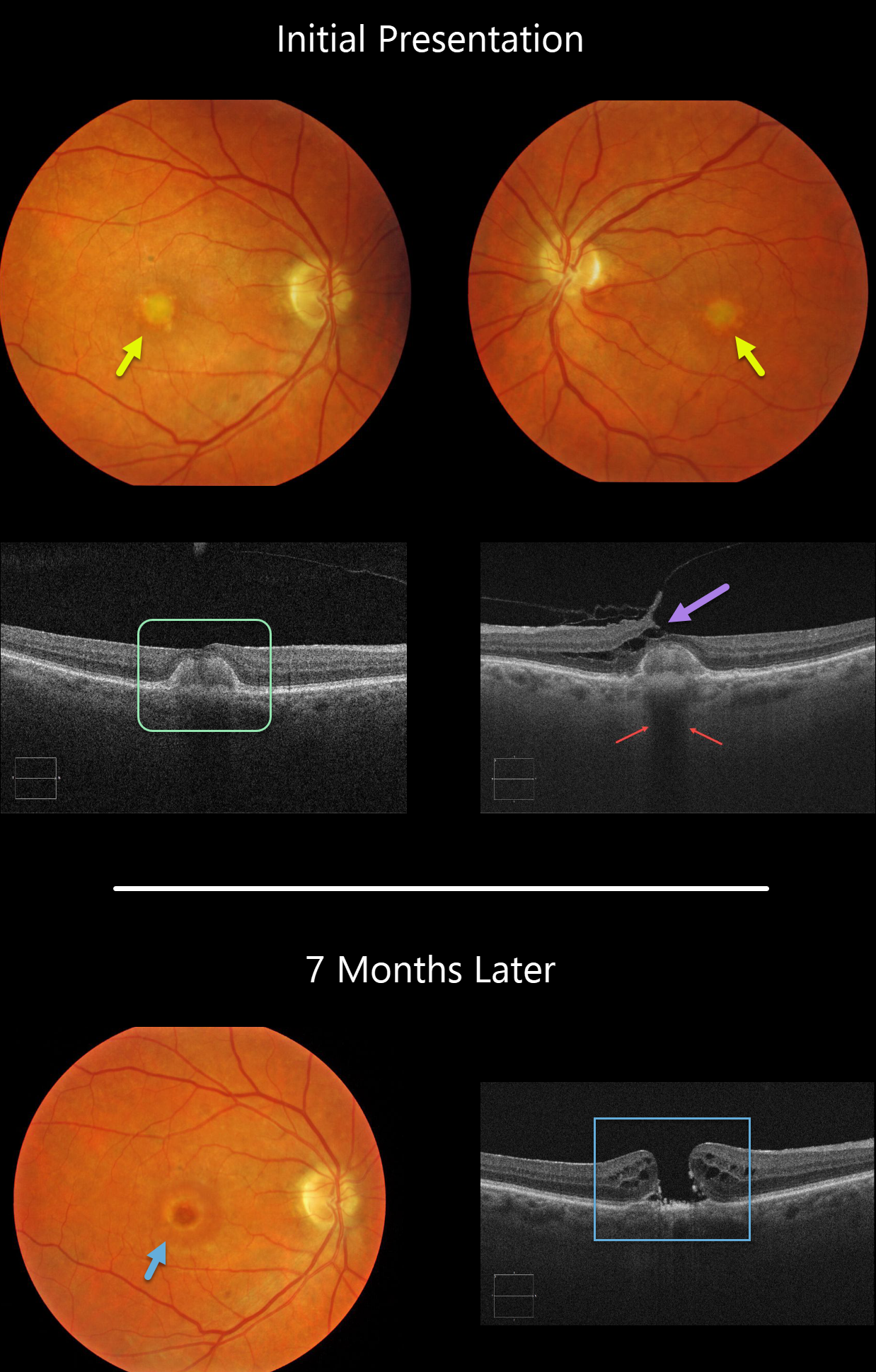
Our patient shows classic findings for adult-onset foveomacular vitelliform dystrophy (Adult Best’s) including small, round, symmetrical subfoveal yellow spots, which is seen on OCT as subretinal hyperreflective material between the RPE and outer retina.
Additionally, the vitelliform lesions provide a great example of shadowing of the underlying choroid. Shadowing occurs when a hyperreflective structure prevents the OCT laser from penetrating to illuminate deeper structures.
A bonus finding in the left eye is focal nasal vitreofoveal traction lifting the retina, splitting the outer plexiform layer and forming a partial tractional lamellar macular hole.
Learning Points:
There are numerous causes for the subfoveal accumulation of yellow material (acquired vitelliform lesions. Adult-onset foveomacular vitelliform dystrophy (Adult Best’s) is associated with a mutation in the BEST1 gene, which encodes for the bestrophin-1 protein.
Bestrophin-1, a calcium-activated chloride channel, is primarily found in the basolateral plasma membrane of the RPE. BEST1 mutations cause a variety of varied phenotypes, also including autosomal recessive bestrophinopathy, best vitelliform macular dystrophy, and autosomal dominant vitreoretinochoroidopathy.
These patients tend to maintain fairly good vision. Outer retinal and RPE atrophy can cause central vision loss, as can the rare development of macular neovascularization. Patients should therefore regularly monitor their vision with an Amsler grid.
MACULAR HOLE
Originally posted on @retina.rocks May 8, 2020
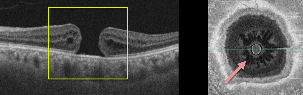
This patient has a full thickness macular hole without traction. The en face image offers a unique perspective on an otherwise unremarkable case.
Learning Points:
The radiating cysts, as described in Govetto et al AJO 2020;212;43-56, have a sunflower appearance showing features of both exudative and tractional cystoid spaces.
The macular hole should be hyporeflective centrally. It is hyperreflective in this image due to the OCT software mis-identifying the RPE as being within the mid-retina.
MACULAR HOLE
Originally posted on @retina.rocks April 3, 2020
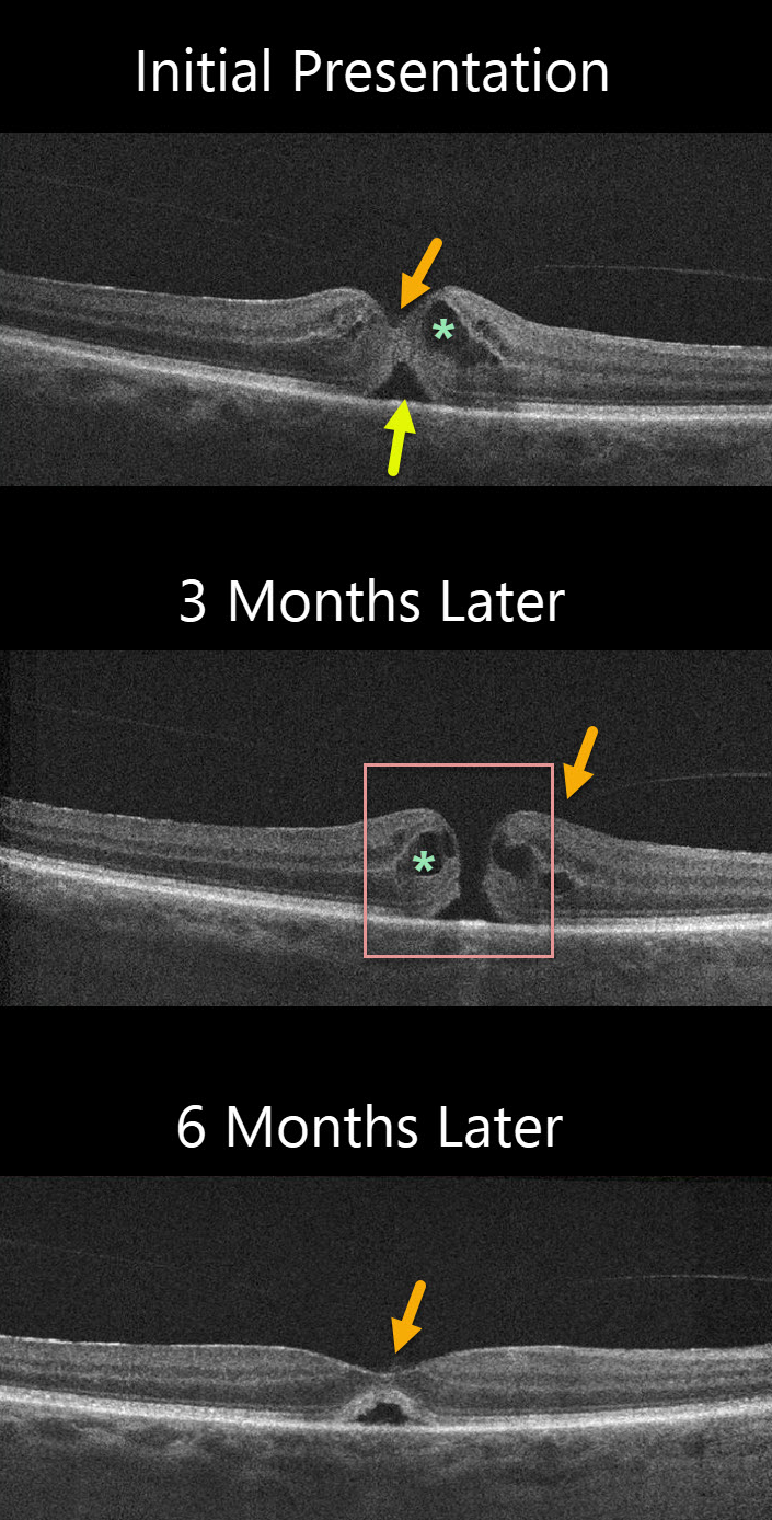
The patient was initially diagnosed with idiopathic cystoid macular edema (CME) and a small foveal detachment with vitreomacular adhesion (VMA). Vision was 20/50. Although the vitreous was attached centrally, a foveal depression remained which would be unusual if vitreomacular traction (VMT) were the cause.
The patient was observed without treatment, and 3 months later vision decreased to 20/60. There was a small full-thickness macular hole with an attached hyaloid. CME was still present. The patient elected to observe the hole.
Another three months later vision improved to 20/40 and the hole spontaneously closed with a normal foveal contour and resolved CME.
The central vitreous remained attached.
Learning Points:
The differential diagnosis for a localized serous foveal detachment include central serous chorioretinopathy, VMT, prior macular hole surgery, and resolved acquired vitelliform lesions.
This patient has a faintly visualized posterior hyaloid that is still attached centrally making VMT unlikely. The choroid probably has a normal thickness which makes central serous less likely, and moreover, the fellow eye is normal.
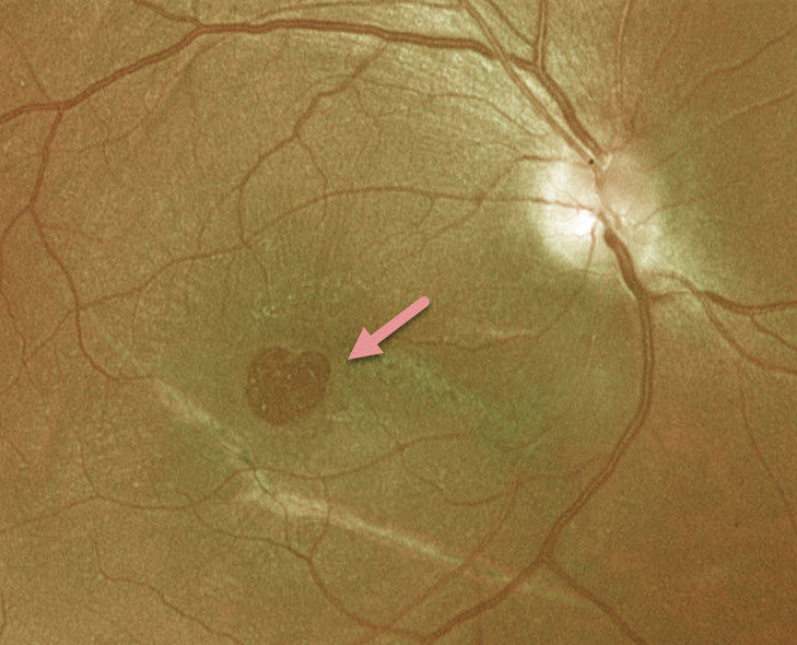
VALENTINE RETINOPATHY
Originally posted on @retina.rocks February 14, 2020
This patient has a heart-shaped macular hole just in time for Valentine’s Day!
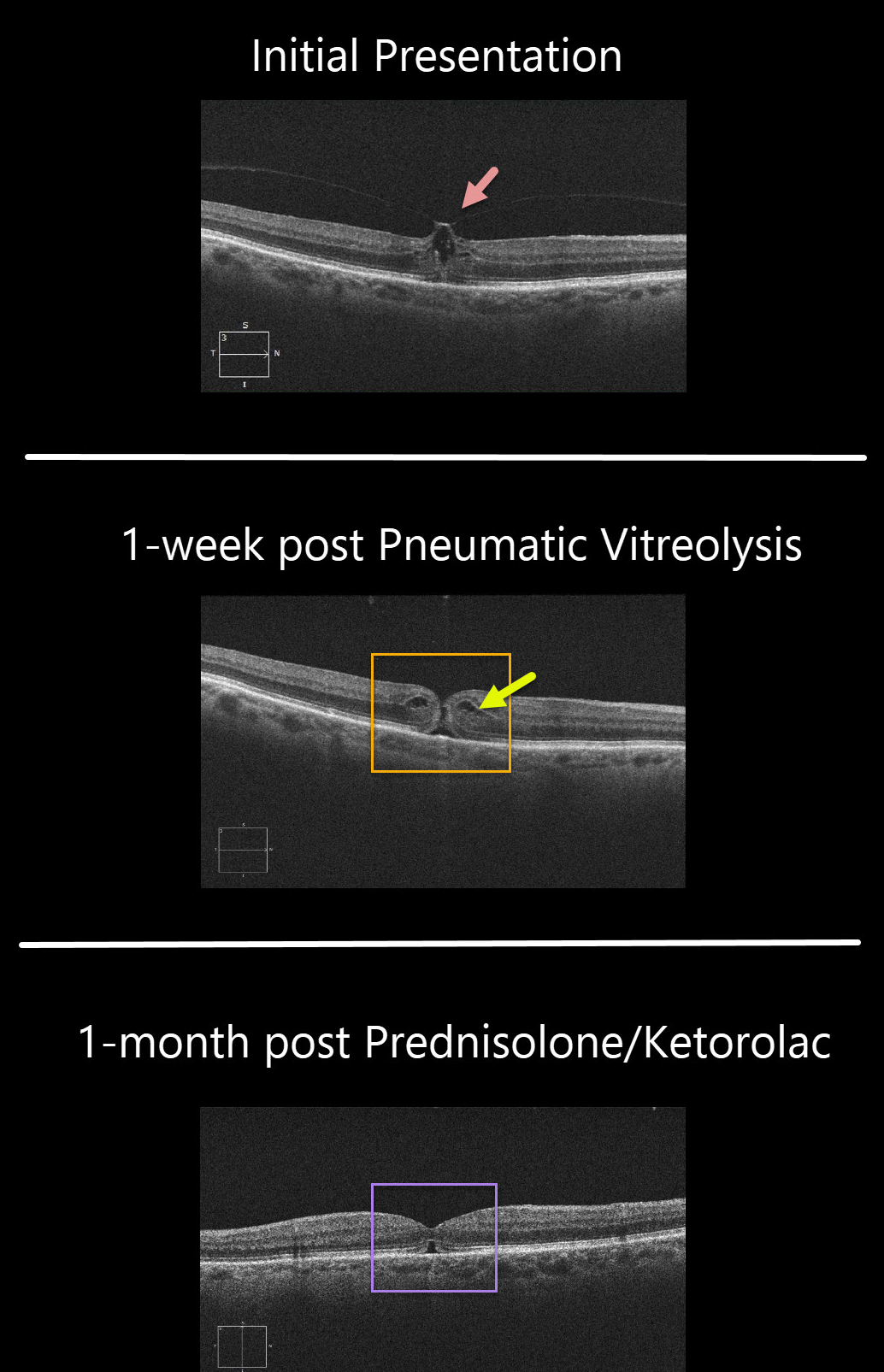
VITREOMACULAR TRACTION + MACULAR HOLE
Originally posted on @retina.rocks February 11, 2020
This patient’s symptomatic 20/80 vitreomacular traction was treated with pneumatic vitreolysis, where a small gas bubble was injected into the vitreous cavity to induce a posterior vitreous detachment. The patient wished to try this less invasive option instead of vitrectomy.
Although the traction successfully released 1 week postop, the patient developed an immediate macular hole with 20/100 vision.
Since the hole was tiny with some cystoid edema (CME), a trial of topical prednisolone acetate 1% and ketorolac was given.
One month later, the CME resolved and the hole closed with improvement to 20/60.
Learning Points:
Topical prednisolone acetate 1% and ketorolac is a newly described treatment that can occasionally be tried for macular holes without traction.


