

Macular Hole Lamellar
<< Back to Cases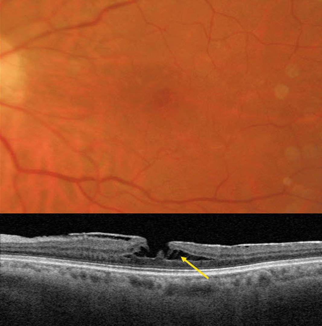
TRACTIONAL LAMELLAR MACULAR HOLE
Originally posted on @retina.rocks February 10, 2026
This 62YO female has a known history of an asymptomatic macular pucker. Vision was 20/70.
Triton color imaging shows a faint, irregular macular pseudohole. Swept-source OCT shows a variably adherent hyperreflective macular pucker. A tractional lamellar macular hole (LMH) is noted centrally, with pillars of Müller cells spanning the split outer plexiform layer (OPL).
Learning Points:
LMH can be defined as tractional or degenerative (Govetto et al, AJO 2016;164:99-109). Tractional LMH shows OPL splitting, an intact ellipsoid zone, and is associated with vitreomacular traction and tractional epiretinal membranes. OCT features of a degenerative LMH include loss of outer retinal tissue below the OPL and an outer retinal ‘bump’. Non-tractional epimacular proliferation is often present and will often extend around the posterior edge of the degenerative LMH.
FOCAL LASER FOR DIABETIC MACULAR EDEMA (DME) WITH SUBSEQUENT DEGENERATIVE LAMELLAR MACULAR HOLE
Originally posted on @retina.rocks August 15, 2024
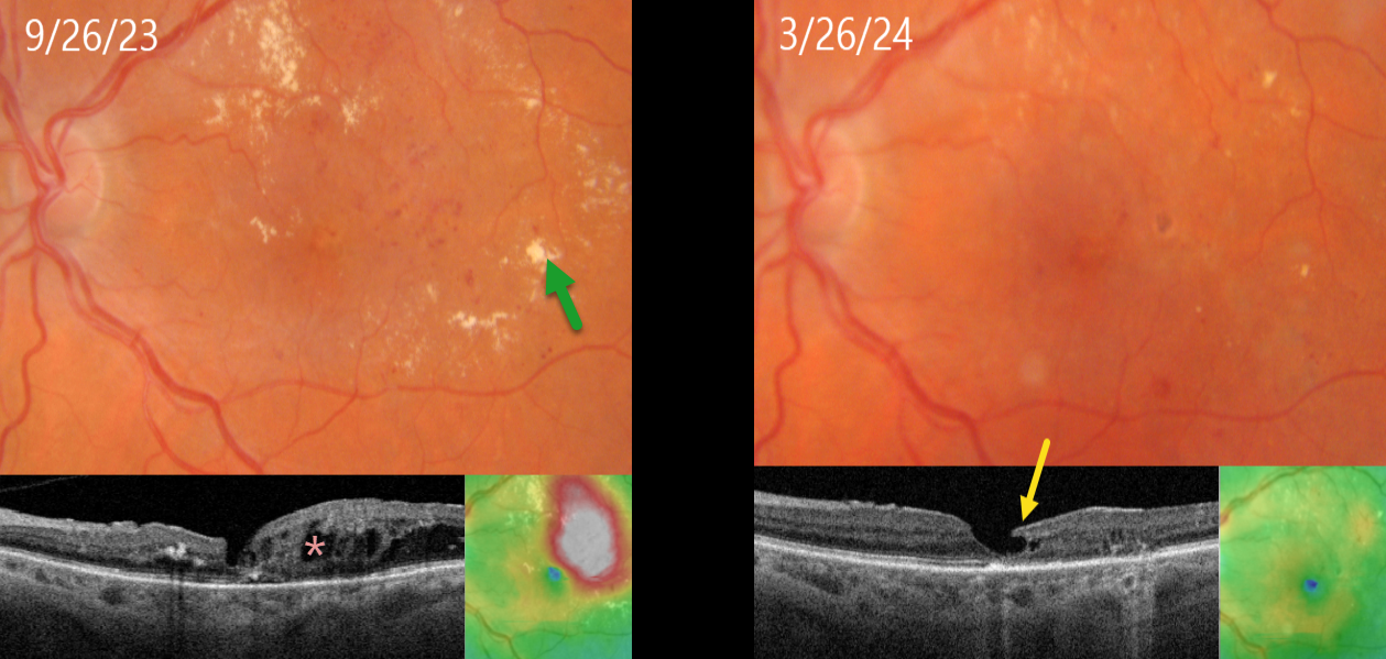
This 71YO female with type 2 diabetes presented diabetic macular edema (DME) and lipid exudates. Triton swept-source OCT shows central foveal thinning with surrounding cystic edema. Vision was 20/60. Thermal macular laser was applied.
Six months later, the edema completely resolved following this single treatment, with near total resolution of the macular lipid. A partial degenerative lamellar macular hole (LMH) is noted. Vision improved to 20/50.
Learning Points:
Our patient exemplifies why macular laser still has a place in any retinal surgeon’s toolbox and should be considered a viable option for treating non-central involved diabetic macular edema (NCI-DME) and even center-involved edema (CI-DME) when the leaking microaneurysms are outside the foveal avascular zone (FAZ). In our practice, macular laser remains the initial treatment for eyes with NCI-DME. When applied gently with small, low-power, and short-duration applications well outside the FAZ, patients rarely, if ever, experience scotoma. Both the treatment burden and the financial costs to society are dramatically less. And there is no risk for endophthalmitis.
Although the role of thermal laser for treating DME is currently a source of debate (see Eye 2022;36:485-486 and Eye 2022;36:483-484 for great pro-laser and anti-laser editorials) we are not sure what all the fuss is about. Old-fashioned lasers work and, in our opinion, are incredibly under-utilized.
LMH can be defined as tractional or degenerative (Govetto et al, AJO 2016;164:99-109). Our patient developed a partial degenerative LMH following likely degeneration or rupture of a foveal cyst following closure of the leaking microaneurysms. There is some debate regarding the benefits of surgery for symptomatic LMH, with some suggesting visual improvement (Morescalchi et al, Retina, 2020;40:1087-1093) and others noting little visual benefit (Mohammed and Thompson, J Vitreoretinal Diseases 2024;8:125-130). Since our patient was visually happy, observation was recommended.
LAMELLAR HOLE
Originally posted on @retina.rocks February 23, 2023
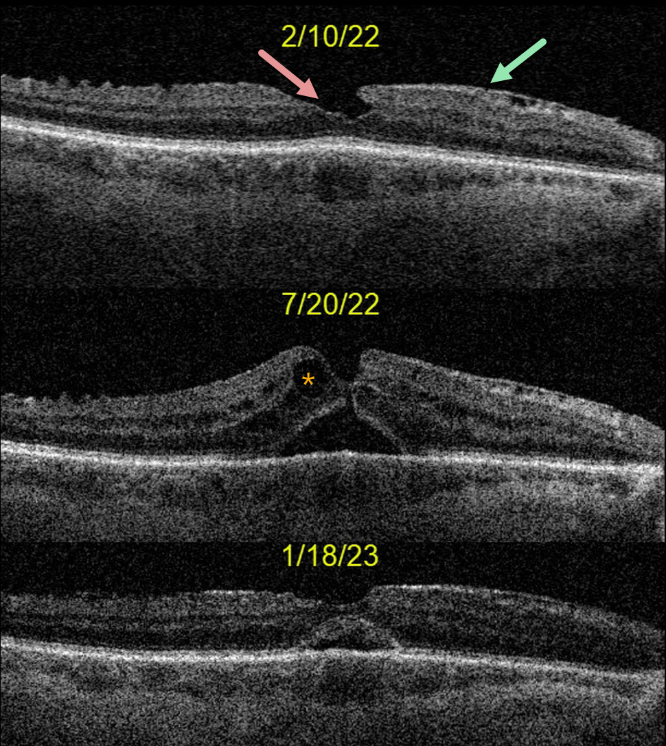
This 73YO male was seen on 2/10/22 with 20/40 vision and an asymptomatic partial lamellar macular hole from an epiretinal membrane. Cataract surgery was performed on 5/25/22, and vision improved to 20/50 postoperatively.
However, on 7/20/22, vision decreased to 20/200 due to a new, extremely tiny full-thickness macular hole and pseudophakic cystoid macular edema (CME). Topical prednisolone acetate 1% and diclofenac QID were prescribed.
The macular hole partially closed one month later with 20/80 vision, and he was tapered off of topical therapy. When last examined on 1/18/23, vision had improved to 20/70.
OCT shows a stable appearance with a persistent outer retinal defect. We recommended continued observation.
Learning Points:
Topical steroid and non-steroidal therapy can sometimes close macular holes without traction, especially those that are small and associated with significant CME (see Ophthalmology Retina 2020;4:695-699). We therefore usually recommend topical therapy for selected symptomatic patients before proceeding to surgery.
ACQUIRED VITELLIFORM LESION
Originally posted on @retina.rocks January 30, 2023
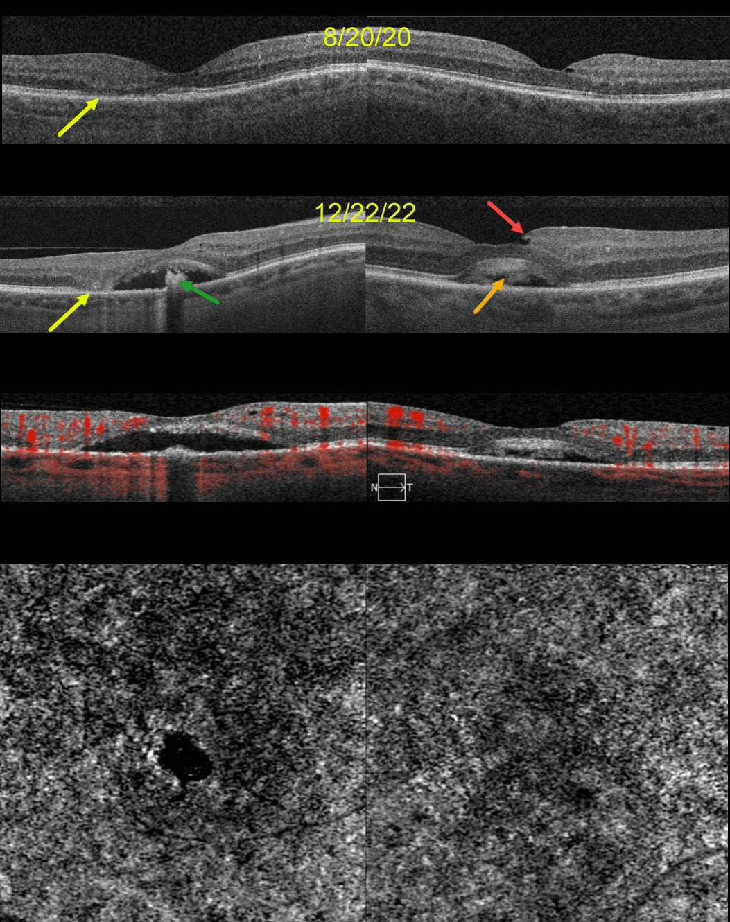
This 68YO female was seen on 8/20/20. Vision was 20/70 OD and 20/40 OS. Although there were no visual complaints, OCT scanning showed bilateral defects in the EZ bands, along with some temporal outer retinal atrophy OD.
She returned two years later, complaining of gradual vision loss OU, although vision was stable at 20/70 OD and 20/30 OS. OCT scanning shows bilateral acquired vitelliform lesions (AVL). The lesion OD is mostly scrambled with a hyporeflective space between the RPE and outer retina, along with a central hyperreflective clump on top of the RPE. There is now complete outer retinal atrophy temporally.
The AVL in her left macula shows variable hyperreflective material. An unrelated small partial lamellar macular hole is noted temporally. OCT angiography (OCTA) shows no flow signals from either AVL.
Learning Points:
Vision loss from AVLs most commonly results from lesions collapsing due to secondary atrophy or macular neovascularization, which can develop in about 8% of eyes (Balaratnasingam et al., AJO 2016;172:28-38).
The absence of OCTA flow signals in either eye confirmed that these lesions were avascular, and we continue to follow her.
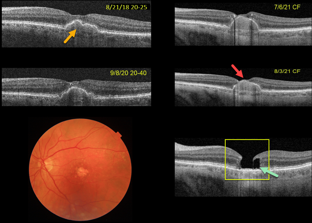
LAMELLAR HOLE AFTER AMD PIGMENT EPITHELIAL DETACHMENT (PED) COLLAPSE
Originally posted on @retina.rocks December 30, 2022
This 75YOF was seen in 2018 with non-neovascular AMD and a central pigment epithelial detachment (PED). Vision was 20/25. She returned on 9/8/20 with 20/40 vision with mild enlargement of the PED.
She complained of decreased vision when seen nearly one year later. Vision had dropped to 20/80. The PED had enlarged to involve full-thickness retina. She elected for an Eylea injection in hopes of possibly treating an occult macular neovascularization vs an avascular PED.
On 7/6/21, vision decreased to counting fingers although the OCT looked fairly stable. An additional Eylea injection followed by half-fluence PDT was given. On 8/3/21, the dome of the PED now extended through the inner retina and vision remained at counting fingers.
On 1/4/22, the PED had completely collapsed, with secondary retinal atrophy and a degenerative lamellar macular hole (LMH).
The progression of her PED through full-thickness retina and subsequent collapse with secondary retinal/RPE atrophy and a LMH are all quite unusual. Fortunately her right eye is 20/20 with intermediate dry AMD.
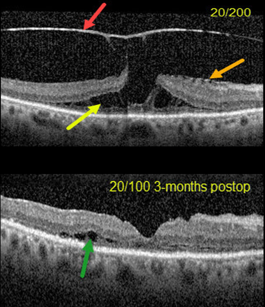
VITREOMACULAR TRACTION + LAMELLAR MACULAR HOLE
Erdem Dinç
Originally posted on @retina.rocks March 29, 2022
This 70YO female presented with 20/200 vision in her right eye from severe vitreomacular traction with a tractional lamellar macular hole (LMH).
OCT scanning shows a highly elevated posterior hyaloid adherent to the underlying fovea, with tractional splitting of the outer plexiform layer (OPL). There is also a faint nasal epiretinal membrane.
Combined cataract surgery, 23-gauge pars plana vitrectomy, membrane peeling, temporal half-moon inverted ILM flap, and fluid-air-C3F8 exchange was performed.
Three months postoperatively, the tractional LMH was closed with 20/100 acuity. There is still some temporal OPL splitting along with disorganization and thinning of the outer macular layers.
Learning Points:
LMH can be defined as tractional or degenerative (see Govetto et al, AJO 2016;164:99-109). Tractional LMH shows OPL splitting, an intact ellipsoid zone, and is associated with vitreomacular traction and tractional epiretinal membranes.
OCT features of a degenerative LMH include loss of outer retinal tissue below the OPL and an outer retinal ‘bump’. Non-tractional epimacular proliferation is often present and will often extend around the posterior edge of the degenerative LMH.
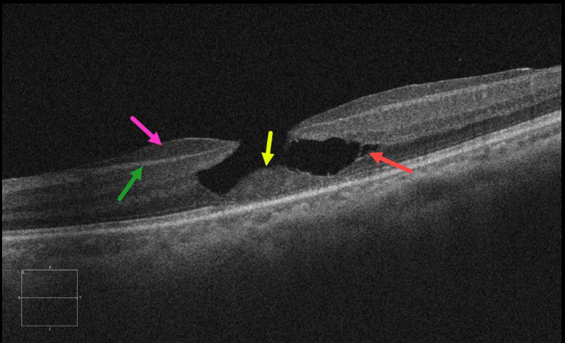
LAMELLAR HOLE
Originally posted on @retina.rocks December 15, 2021
This 62YO female presented with 20/80 vision in her left eye from an asymptomatic lamellar macular hole (LMH). Epiretinal proliferation is adherent to the underlying nerve fiber layer. A localized splitting of the outer plexiform layer (OPL) is seen temporally. There is a loss of tissue below the OPL with a central mound of remaining featureless outer retina.
Learning Points:
LMH can be defined as tractional or degenerative (see Govetto et al, AJO 2016;164:99-109).
Our case has all the classic findings of a degenerative LMH, which Govetto et al. described as resembling the brim of a top hat. The epimacular proliferation often extends around the posterior edge of the LMH, but this was not seen in our case.
Although surgery is usually observed, it can close these holes with some visual improvement (see Morescalchi et al, Retina, 2020;40:1087-1093).


