

Macular Hole Outer
<< Back to Cases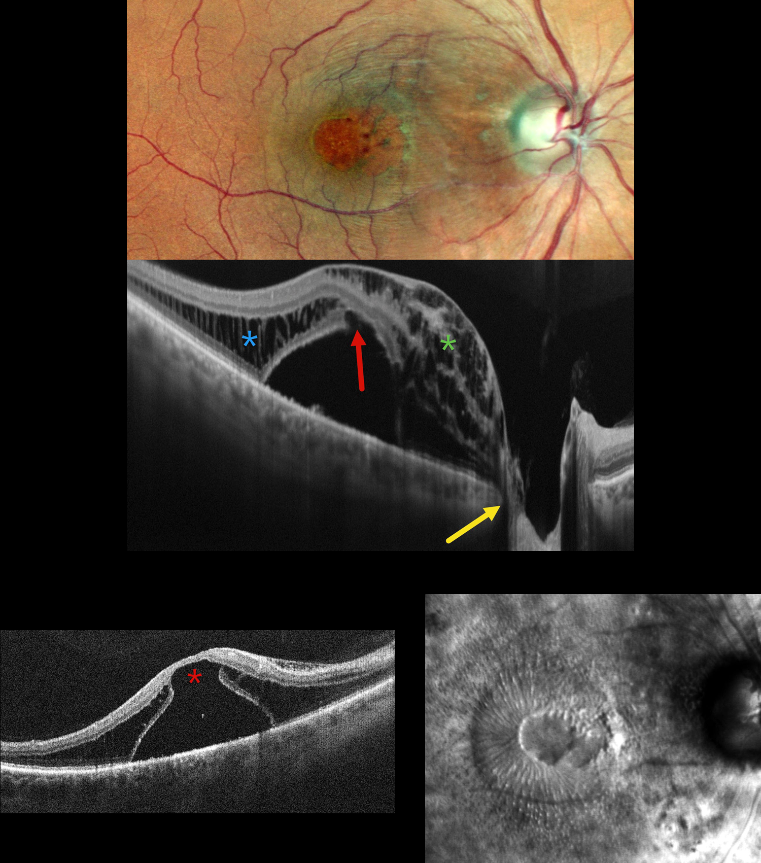
OPTIC NERVE PIT
Shraddha Raj Shrivastava and Manish Nagpal
Originally posted on @retina.rocks July 24, 2026
This 50YO male presented with 1 year of decreased vision in his right eye. He had been treated elsewhere with intravitreal injections for cystoid macular edema. Vision was 20/200 OD and 20/20 in his normal OS.
Pseudocolor SLO imaging shows a possible serous macular detachment with a large macular pseudohole. A possible pit is noted along the temporal aspect of the optic nerve. OCT scanning shows an elevated serous detachment. An outer-layer macular hole is noted along the posterior aspect of the fovea, along with full-thickness cystic retinal edema nasally and outer nuclear layer cystic edema temporally. The subretinal fluid directly communicates with the optic nerve pit. Retro mode reveals a unique view of the macular pathology, with the central outer macular hole and radiating fluid.
Learning Points:
An optic nerve pit is a rare (about 0.1% prevalence) congenital anomaly thought to be caused by incomplete closure of the optic fissure during gestation. Pits are most often located within or along the inferior-temporal disc margin and usually appear as a gray depression. While most optic pits are asymptomatic, they can cause vision loss by forcing fluid into the inner retina, outer retina, and, ultimately, the subretinal space (optic pit maculopathy). The source of the fluid remains a subject of ongoing debate, with opinions ranging from the vitreous to the subarachnoid space.
Vitrectomy surgery was discussed, and the patient elected for observation unless his symptoms worsened.
DIABETIC MACULAR EDEMA (DME)
Originally posted on @retina.rocks January 26, 2023
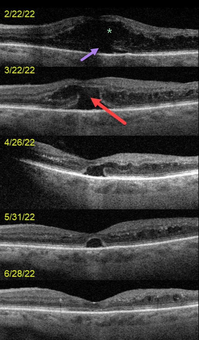
This 72YO female presented on 2/22/22 with counting-finger vision in her left eye.
OCT scanning at the initial visit shows severe center-involved diabetic macular edema (CI-DME) with shallow subretinal fluid. Anti-VEGF therapy was started.
One month later, vision remained at counting fingers. The retinal thickness had improved, but a new outer macular hole was noted.
Following 3 additional monthly injections, the edema continued to resolve with spontaneous closure of the outer macular hole. Vision was 20/70 at the 6/28/22 visit.
Learning Points:
Traditional primary full-thickness macular holes are caused by vitreomacular traction. Outer macular holes, however, have a much broader range of etiologies.
In our practice, we most commonly see these lesions in optic pit maculopathy and paraproteinemia (see Mansour et al, Ophthalmology 2014;121:1925-1932).
Our patient’s outer macular hole was likely caused by inner retinal exudation that extended through the outer retina. The return of central vision and closure of the defect, with relatively intact outer macular architecture, likely indicates that this was an outer macular dehiscence rather than a true loss of tissue. See Kumawat et al for a great review of atypical macular holes (Retina 2019;39:1236-1264).
EXUDATIVE RETINAL DETACHMENT WITH OUTER MACULAR HOLE
Originally posted on @retina.rocks June 25, 2021
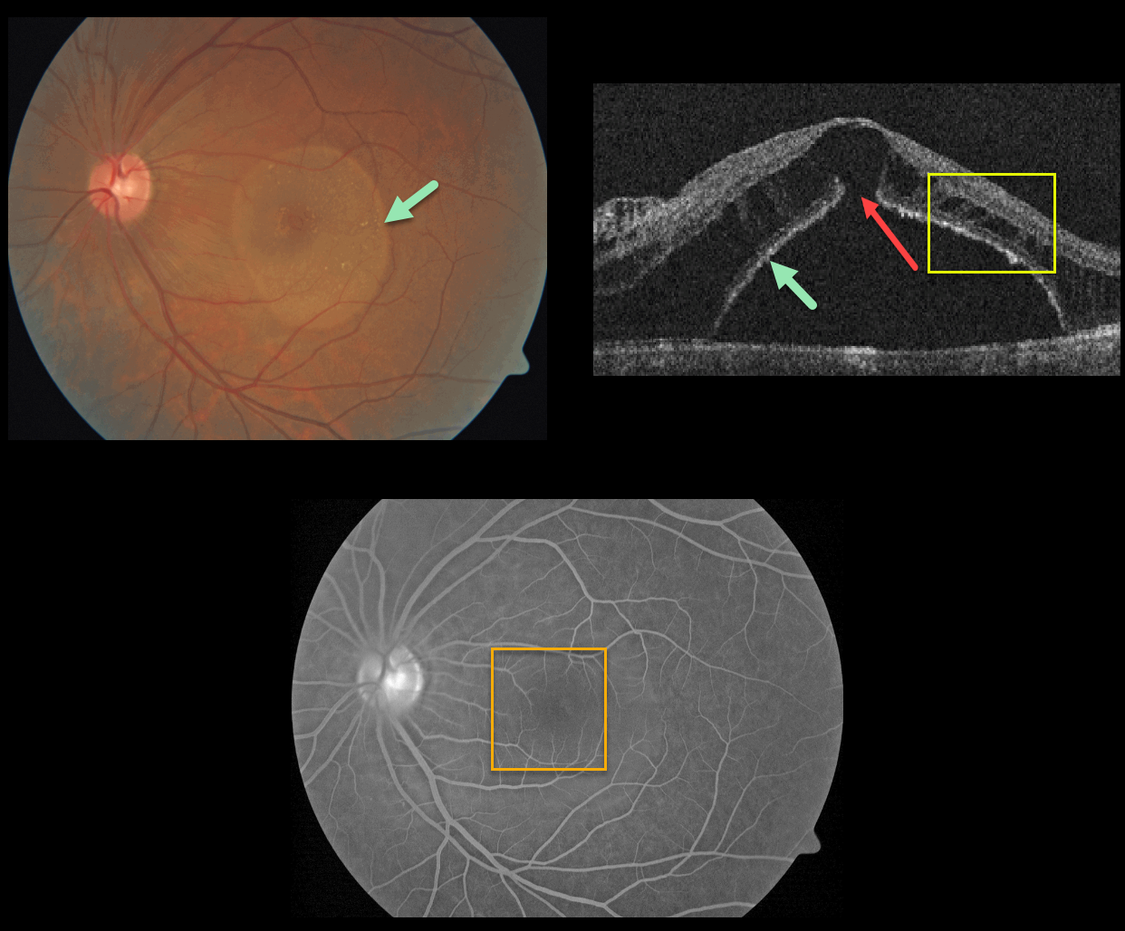
This patient presented with a sharply-circumscribed macular serous detachment, cystic outer retinal edema, and an outer macular hole. Fluorescein angiography is essentially normal without leakage.
An optic pit was absent clinically and on OCT imaging. Serum protein electrophoresis was normal, and a systemic cancer workup was negative.
Learning Points:
Serous detachment can be an uncommon ocular manifestation of paraproteinemia (see Mansour et al, Ophthalmology 2014;121;1925-1932). These detachments, which resemble those seen in optic pit maculopathy, are characterized by a well-defined serous detachment without angiographic leakage. A central outer macular hole is often present.
The fluid is likely due to an osmotic gradient generated by subretinal immunoglobulins that passively migrate into the subretinal space.
We do not know the cause of our patient’s findings, given his lack of an optic pit and a negative systemic workup.


