

Macular Pucker
<< Back to Cases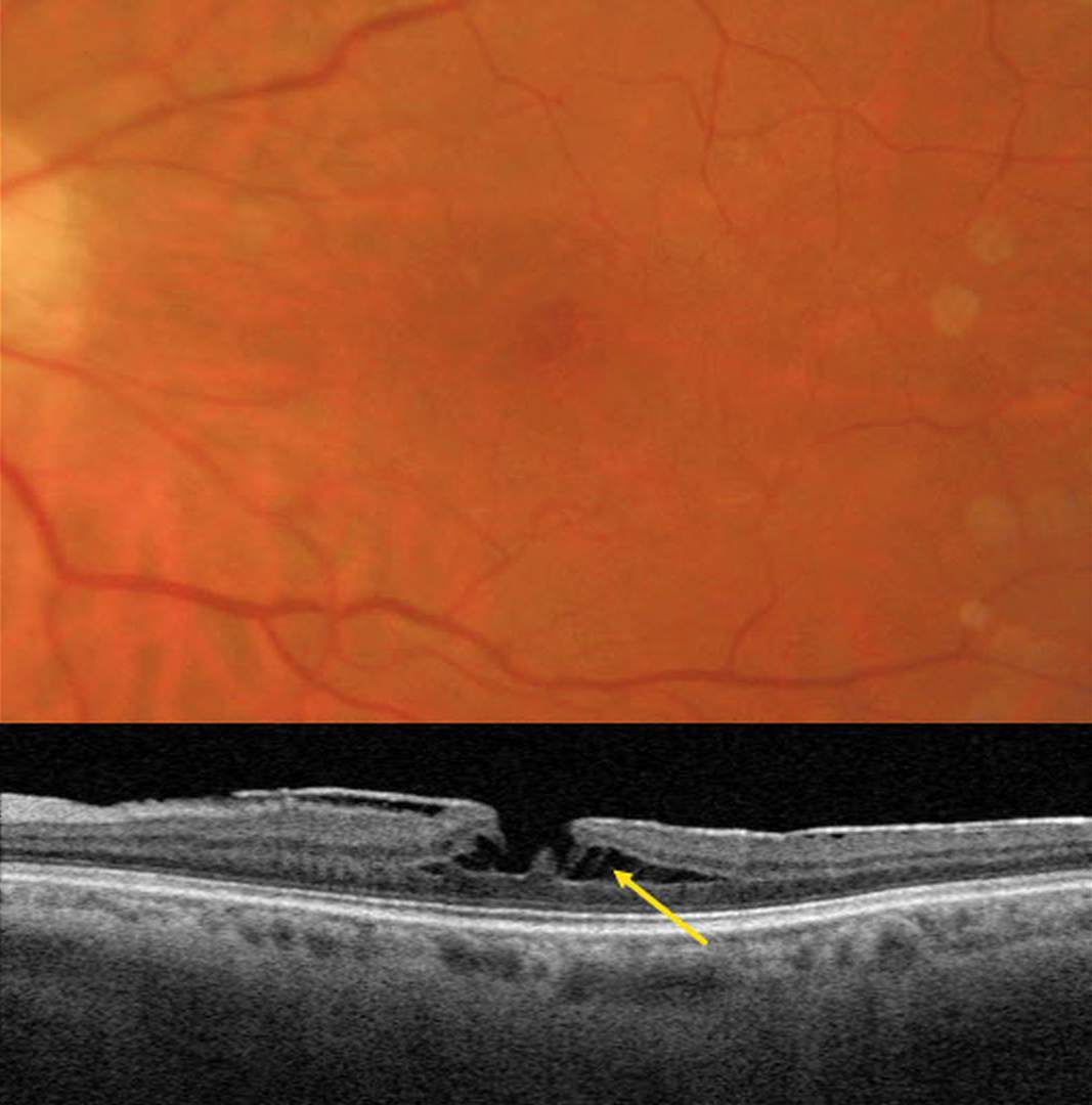
TRACTIONAL LAMELLAR MACULAR HOLE
Originally posted on @retina.rocks February 10, 2026
This 62YO female has a known history of an asymptomatic macular pucker. Vision was 20/70.
Triton color imaging shows a faint, irregular macular pseudohole. Swept-source OCT shows a variably adherent hyperreflective macular pucker. A tractional lamellar macular hole (LMH) is noted centrally, with pillars of Müller cells spanning the split outer plexiform layer (OPL).
Learning Points:
LMH can be defined as tractional or degenerative (Govetto et al, AJO 2016;164:99-109). Tractional LMH shows OPL splitting, an intact ellipsoid zone, and is associated with vitreomacular traction and tractional epiretinal membranes. OCT features of a degenerative LMH include loss of outer retinal tissue below the OPL and an outer retinal ‘bump’. Non-tractional epimacular proliferation is often present and will often extend around the posterior edge of the degenerative LMH.
MACULAR PUCKER WITH RNFL SCHISIS
Originally posted on @retina.rocks January 5, 2026
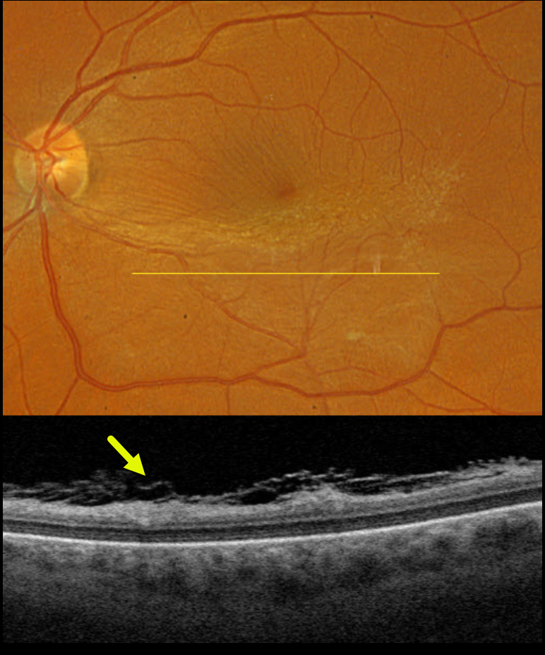
This 58YO female was referred for an asymptomatic macular pucker in her left eye. Vision was 20/25.
Optos color RGB imaging shows a moderate macular pucker centered in the inferior macula. Fine inner retinal striae are noted superiorly. Triton swept-source OCT shows a shredded appearance of multi-layered hyperreflective tissue overlying the retinal surface (the ‘spaghetti sign’).
Learning Points:
Fibrillary changes overlying an epiretinal membrane (ERM) were initially noted by Kim et al (AJO 2012;153:692-697), and termed schisis of the retinal nerve fiber layer by Hussnain et al (AJO 2019;207:304-3120). Despite rarely being described in the medical literature, it is present in over 50% of ERMs (Russell et al, Ophthalmology Retina 2023;7:325-332). Felt to arise from tangential tractional forces from the overlying epimacular tissue, it can appear clinically as whitish retinal areas overlying a pucker. During intraoperative membrane peeling, this tissue can be mistaken for residual epimacular proliferation and should not be removed to avoid iatrogenic retinal damage.
MACULAR PUCKER
The European VitreoRetina Society (EVRS), Mohamed Tawfik and Norhan Lotfy
Originally posted on @retina.rocks November 14, 2025
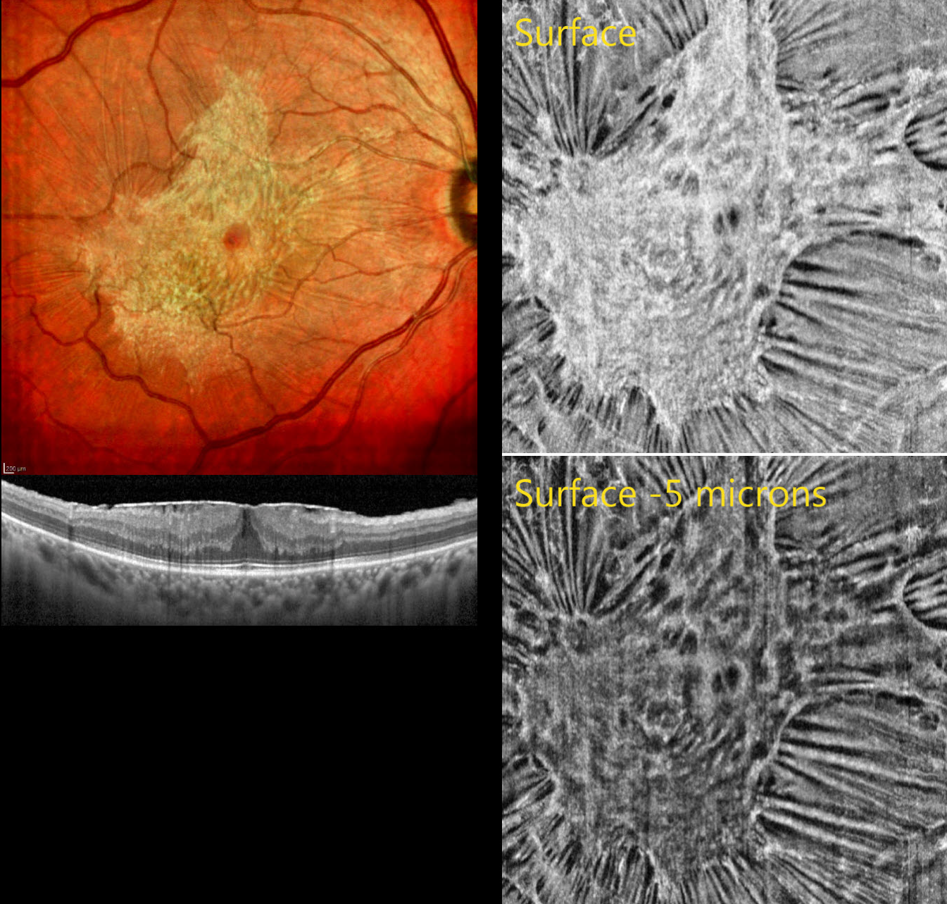
This 60YO male presented with gradual blurred vision in his right eye. Vision was 20/30.
Color SLO imaging shows a variably dense and glistening macular pucker with radiating retinal striae extending outwards from multiple areas of tangential traction. OCT shows the hyperreflective epiretinal membrane causing diffuse retinal thickening. The retinal bands are fairly well-preserved. En face imaging at the membrane’s surface reveals better detail for the irregular pucker and surface traction. Imaging 5 microns below the surface reveals dimpling of the underlying tissue.
Surgery will be recommended if our patient develops increased symptoms.
MACULAR PUCKER
Originally posted on @retina.rocks September 8, 2025
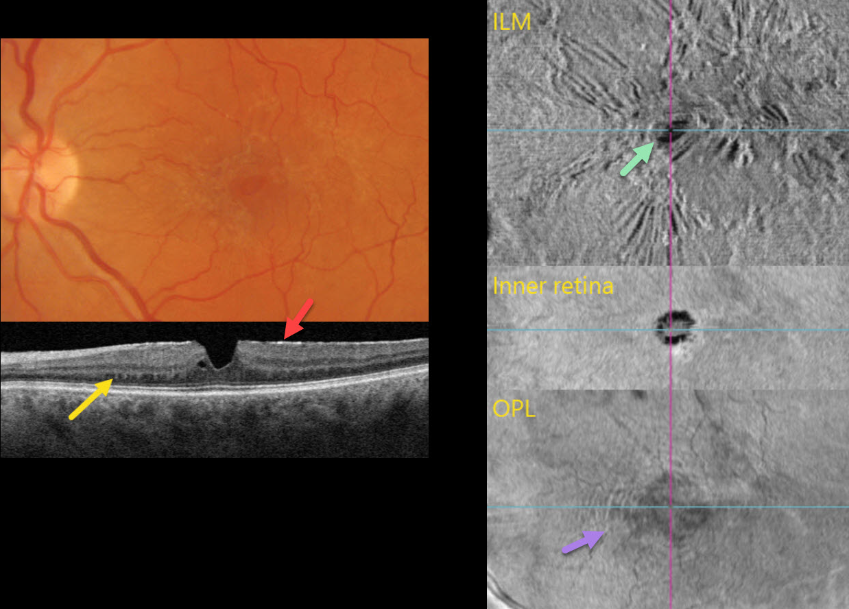
This 56YO female was referred for asymptomatic retinal findings. Vision was 20/20.
Multimodal imaging shows numerous pucker-related findings.
Triton color photography shows a diffuse macular pucker with a central teardrop-shaped pseudohole.
Triton swept-source OCT B-scanning shows an adherent hyperreflective epiretinal membrane, a vertical foveal contour, and trace adjacent outer plexiform splitting. The nasal outer plexiform layer (OPL) contains numerous peg-like hyperreflective lesions that extend into the outer nuclear layer.
En face OCT of the retinal surface shows variable striae and distortion from the pucker, along with the hyporeflective pseudohole. En face OCT through the inner retina shows the irregular pseudohole. En face of the OPL, through the peg-like lesions noted on the B-scan, shows a hyperreflective fingerprint.
Learning Points:
The ‘fingerprint sign’ is a unique OCT en face finding, described by Griffin et al (Retina 2021;41:381-386), appearing as concentric, fingerprint-like waves of Henle’s layer underlying epiretinal membranes. The authors felt that the undulating OPL creates hyperreflective lines when oriented perpendicular to the incident OCT beam and hyporeflective lines when oriented parallel to it.
MACULAR PUCKER WITH LAMELLAR DEFECTS
The European VitreoRetina Society (EVRS) and Shishir Verghese
Originally posted on @retina.rocks July 11, 2025
This 58YO female presented with 6 months of vision loss in her left eye. Vision was 20/60 OS.
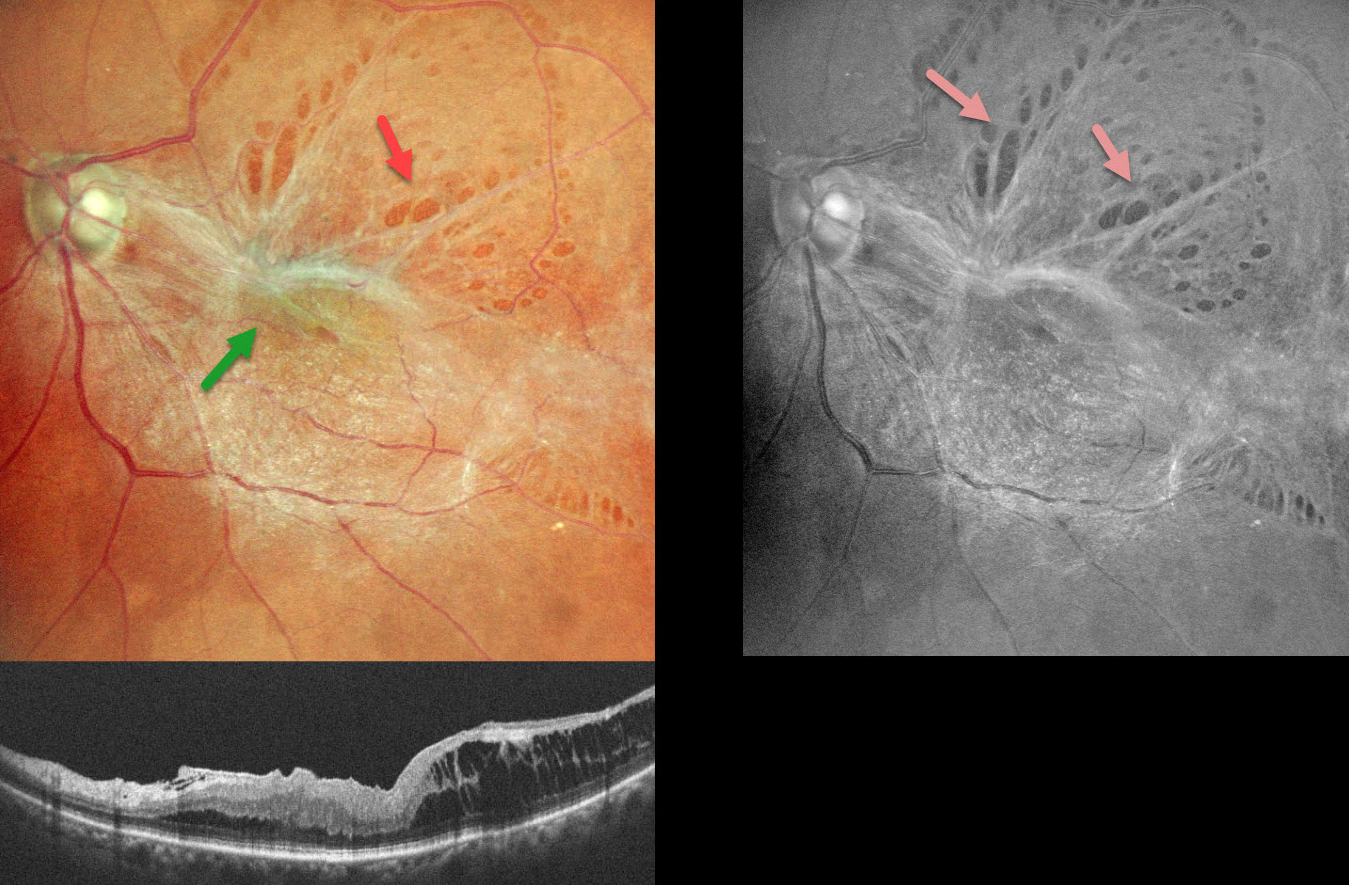
Color SLO imaging shows a taut epiretinal membrane (ERM) with multiple paravascular lamellar defects, which are better visualized on red-free imaging. OCT scanning shows a stage 3 macular pucker with ectopic inner foveal layer (EIFL) and temporal inner layer schisis.
Learning Points:
An ERM is a thin, fibrous membrane that forms on the retinal surface, often causing central vision loss with metamorphopsia. It can be classified into 4 OCT-based stages (Gonzalez-Saldivar et al, Retina 2020;40:710-717): Stage 1 ERM with preserved foveal contour, Stage 2 Loss of foveal depression with thickened outer nuclear layer, Stage 3 EIFL, and Stage 4 EIFL with complete loss of inner retinal anatomy.
ERMs can cause paravascular lamellar defects due to shearing forces at the vessel borders, and these defects can extend towards the fovea. Our patient’s highly contractile membrane caused multiple striking lamellar defects visualized on fundus photography.
ECTOPIC MACULAR PUCKER
Shelby Culver
Originally posted on @retina.rocks May 15, 2025
This 51YO male was referred for a minimally symptomatic, unusual-appearing macular pucker. Vision was 20/20.
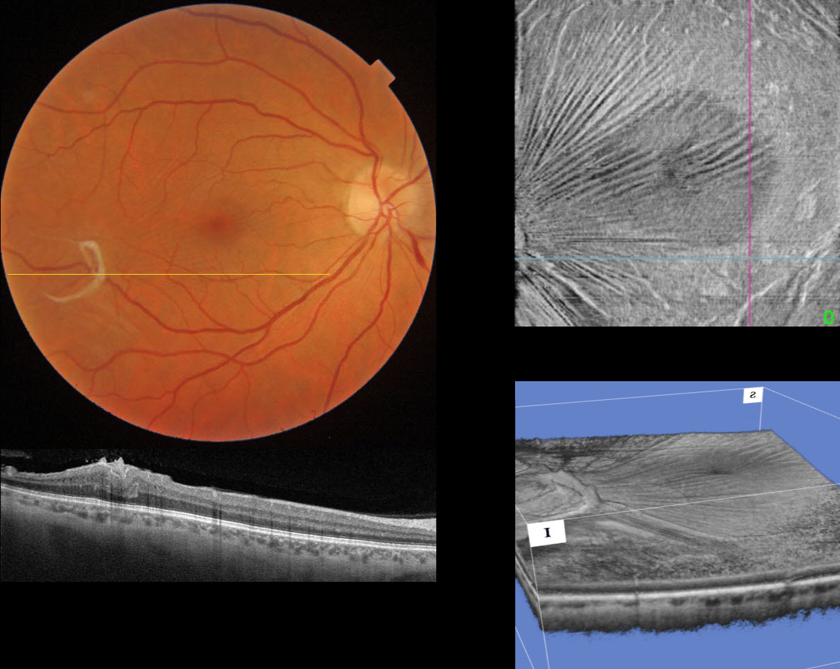
Triton color imaging and swept-source OCT show a semicircular area of epiretinal proliferation and traction in the distal inferotemporal macula. Radiating inner retinal striae are drawn towards this area of traction, which are best seen on en face OCT of the inner retina. A 3D reconstruction shows another view of this traction.
Since our patient was virtually asymptomatic, observation was recommended.
MACULAR PUCKER WITH FOVEAL HERNIATION
Originally posted on @retina.rocks February 27, 2025
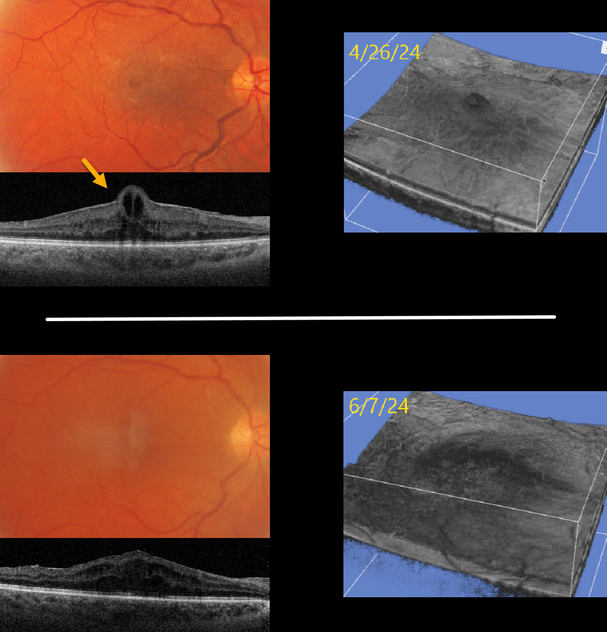
This 53YO male presented with counting-fingers vision in his right eye due to a symptomatic macular pucker.
Optos color RG imaging shows a macular pucker with a foveal cyst. Triton swept-source OCT shows an adherent pucker with central foveal herniation. Vitrectomy surgery was performed, and one month later, vision improved to 20/100. OCT scanning shows no pucker, moderate diffuse cystic retinal thickening, and resolution of the foveal herniation. The pre- and postoperative appearances are also well visualized with 3D OCT reconstruction.
Learning Points:
Foveal herniation is found in about 1% of eyes with idiopathic epiretinal membranes (Uzel et al, Eye 2023;37:1357-1360). Epiretinal membranes not uncommonly surround the foveal center, and central contraction can result in uninvolved inner foveal tissue being squeezed through the central membrane defect, resulting in this unique appearance. Vitrectomy surgery with membrane peeling usually results in si
MACULAR PUCKER
Originally posted on @retina.rocks February 19, 2025
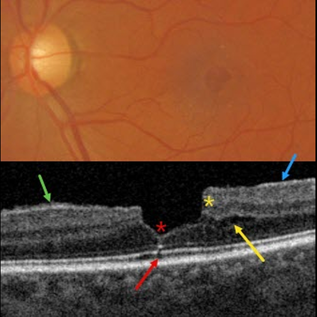
This patient presented with 20/30 vision due to a mildly symptomatic clinical macular pucker with a pseudohole; observation was recommended. Multimodal imaging reveals extensive pucker-related findings.
Triton color imaging shows an irregular macular pseudohole with a faint surrounding epiretinal membrane (ERM). True full-thickness macular holes are virtually always perfectly round, and these pucker-related pseudoholes are often irregularly round, as in this eye.
Swept-source OCT shows an adherent ERM with some shaggy-appearing adherent vitreous cortex, favoring epimacular proliferation. The fovea has a vertical contour, often present with ERM, due to the encircling membrane contracting towards the foveal center, dragging the inner retina centrally.
There are features of both a tractional lamellar macular hole (LMH) with splitting of the outer plexiform layer, along with a degenerative LMH with descending loss of tissue into the outer nuclear layer (Govetto et al, AJO 2016;164:99-109).
Finally, a tiny area of shadowing vs an outer foveal microdefect (Cohen et al, Ophthalmology Retina 2021;5:553-561) is seen.
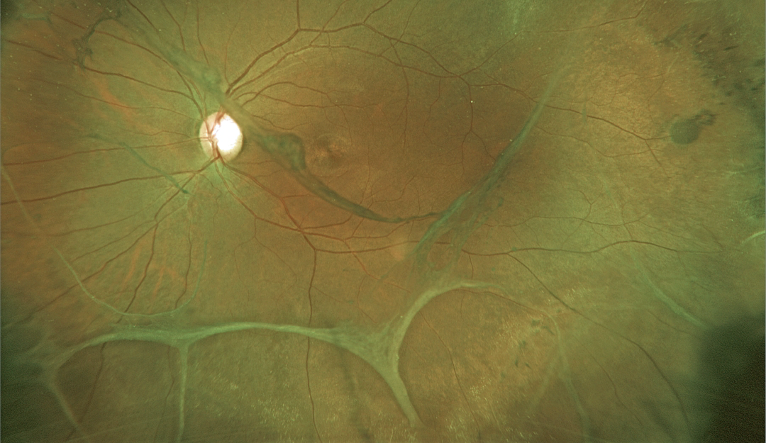
VITREOSCHISIS WITH MACULAR PUCKER
Originally posted on @retina.rocks February 11, 2025
This 28YO 8-diopter myopic female presented with recent floaters in her left eye. Vision was 20/30 OD and 20/40 OS.
Optos color RG imaging shows extensive bands of variably adherent vitreoschisis and epiretinal membranes. A prominent vitreous membrane vs pucker is noted in the inferonasal macula. There was bilateral variably pigmented paravenous lattice (not shown).
We recommended twice-yearly examinations, but unfortunately, the patient was immediately lost to follow-up.
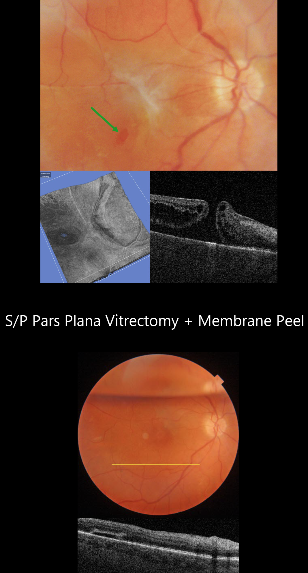
MACULAR PUCKER WITH ECTOPIC MACULAR HOLE
Originally posted on @retina.rocks July 25, 2024
This 52YO female underwent vitrectomy for a hand motion, macula-off rhegmatogenous retinal detachment on 1/25/21. Despite successful reattachment, vision was counting fingers postoperatively, in part due to a severe macular pucker identified on 8/4/21. An ectopic secondary macular hole was present in the inferotemporal macula.
Vitrectomy surgery with membrane peeling was performed on 10/14/21. One week postoperatively, the ectopic hole was closed, and the macula was completely free of epimacular tissue. When last examined on 3/24/23, vision was 20/70.
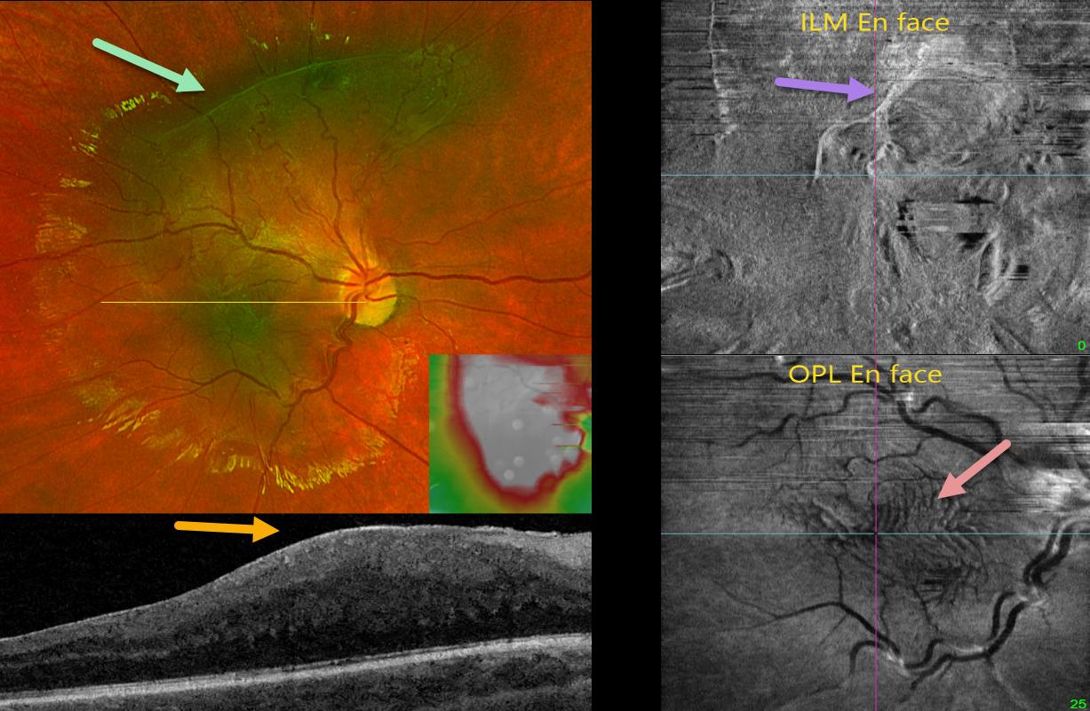
MACULAR PUCKER
Originally posted on @retina.rocks May 29, 2024
This healthy 9YO boy was referred for asymptomatic retinal changes in his right eye. Vision was 20/25 OU. The left eye was normal.
Optos color RG imaging shows a greenish hyperpigmented lesion in the macula extending into the superior midperiphery. Diffuse macular thickening is noted on swept-source OCT. En face imaging offers another multimodal perspective. En face of the internal limiting membrane/vitreous interface shows the epimacular fibrosis, while en face of the outer plexiform layer shows variable fingerprint-like whorls centrally.
Learning Points:
In 2017, Govetto et al described an OCT finding of ectopic inner foveal layers (EIFL) associated with epiretinal membranes (AJO 2017;175:99-113). The findings range from mild underlying retinal changes (stage 1, thin ERM with foveal depression), widening of the ONL and loss of foveal depression (stage 2), and ERM continuous with EIFL crossing the entire foveal area (stage 3). All retinal layers are clearly visualized in stages 1 through 3. In stage 4, the ERM is thick, and all retinal layers are disrupted. Vision is worse with increasing stage severity. It is also associated with worse postoperative visual recovery (Yang et al, Retina 2022;42:1472-1478).
The ‘fingerprint sign’ is a unique en face OCT finding described by Griffin et al (Retina 2021;41:381-386) that appears as concentric, fingerprint-like waves of Henle’s layer underlying epiretinal membranes. The authors found that the undulating OPL creates hyperreflective lines when oriented perpendicular to the incident OCT beam and hyporeflective lines when parallel to it.
Combined hamartomas are classically described as benign congenital lesions composed of glial cells, vascular tissue, and pigmented retinal pigment epithelial cells. However, more recent data indicate that these lesions primarily arise from the inner retinal layers (Chawla et al, AJO 2017;181:88-96).
Hamartomas and severe macular puckers can look very similar, even to some of the most seasoned retina specialists. OCT may be helpful in differentiating these entities (Ophthalmic Surg Lasers Imaging Retina 2017;48;122-125). In our experience, we feel that many ‘combined hamartomas’ described in the literature are most likely severe macular pucker with ectopic inner retina layers as in our case.
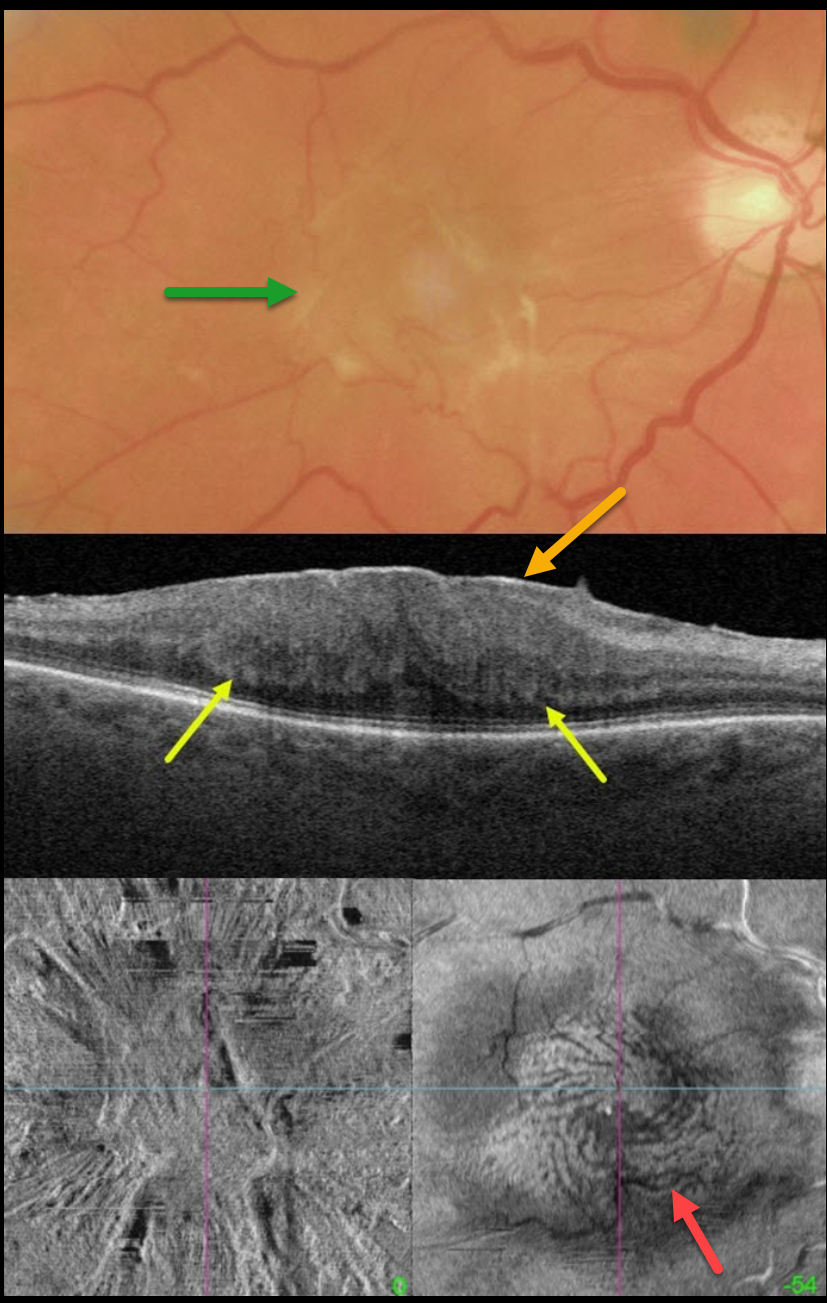
MACULAR PUCKER WITH FINGERPRINT SIGN
Matt Meredith
Originally posted on @retina.rocks December 4, 2023
This 67YO male presented with 20/400 vision in his right eye from a symptomatic macular pucker.
Color imaging shows a severe central macular pucker. Triton swept-source OCT shows a variably adherent epiretinal membrane (ERM) with inner retinal hyperreflectivity and thickening, and complete loss of the normal inner retinal structures. The outer plexiform layer (OPL) is scalloped along its margin with the outer nuclear layer.
En face OCT of the inner retina shows variable surface distortion. En face imaging through the OPL shows a fingerprint-like whorl of hyperreflective lines.
Vitrectomy surgery was scheduled.
Learning Points:
The ‘fingerprint sign’ is a unique OCT en face finding, described by Griffin et al (Retina 2021;41:381-386), appearing as concentric, fingerprint-like waves of Henle’s layer underlying epiretinal membranes.
The authors found that the undulating OPL creates hyperreflective lines when oriented perpendicular to the incident OCT beam and hyporeflective lines when parallel to it.
In 2017, Govetto et al described an OCT finding of ectopic inner foveal layers (EIFL) associated with epiretinal membranes (AJO 2017;175:99-113). The findings range from mild underlying retinal changes (stage 1, thin ERM with foveal depression), widening of the ONL and loss of foveal depression (stage 2), and ERM continuous with EIFL crossing the entire foveal area (stage 3). All retinal layers are clearly visualized in stages 1 through 3. In stage 4, the ERM is thick, and all retinal layers are disrupted. Vision is worse with increasing stage severity. It is also associated with worse postoperative visual recovery (Yang et al, Retina 2022;42:1472-1478).
MACULAR PUCKER
Originally posted on @retina.rocks October 23, 2023
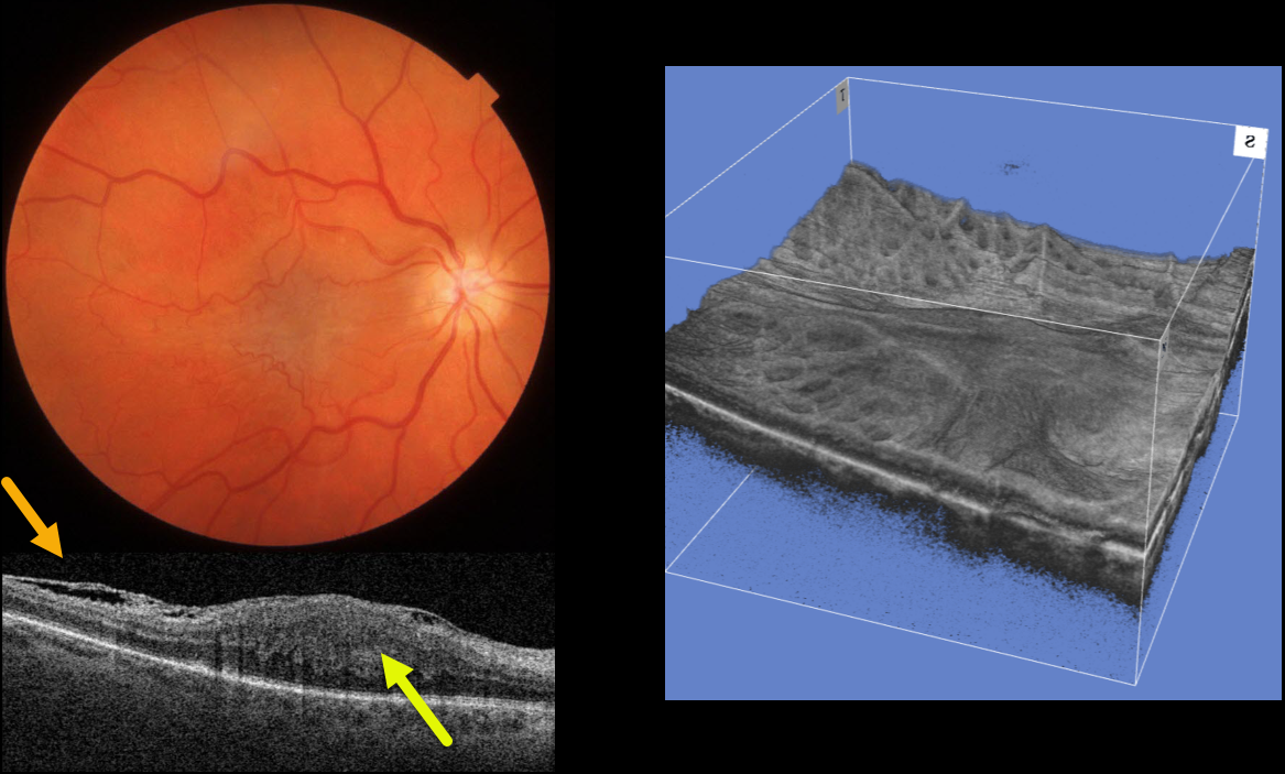
This 64YO female was referred for an asymptomatic macular pucker in her right eye. Vision was 20/200 OD and 20/20 OS.
Color imaging shows a severe central macular pucker. Triton swept-source OCT shows a variably adherent epiretinal membrane (ERM) with inner retinal hyperreflectivity and thickening, and complete loss of the normal inner retinal structures.
A 3D OCT reconstruction offers another view of the inner macular appearance, with unusual radiating dimples more peripherally.
Learning Points:
In 2017, Govetto et al described an OCT finding of ectopic inner foveal layers (EIFL) associated with epiretinal membranes (AJO 2017;175:99-113). The findings range from mild underlying retinal changes (stage 1, thin ERM with foveal depression), widening of the ONL and loss of foveal depression (stage 2), and ERM continuous with EIFL crossing the entire foveal area (stage 3). All retinal layers are clearly visualized in stages 1 through 3.
In stage 4, the ERM is thick, and all retinal layers are disrupted. Vision is worse with increasing stage severity. It is also associated with worse postoperative visual recovery (Yang et al, Retina 2022;42:1472-1478).
Combined hamartomas are classically described as benign congenital lesions composed of glial cells, vascular tissue, and pigmented retinal pigment epithelial cells. However, more recent data indicate that these lesions primarily arise from the inner retinal layers (see Chawla et al AJO 2017;181:88-96).
Combined hamartomas and severe macular puckers can look very similar, even to some of the most seasoned retina specialists. In our opinion, many cases of combined hamartomas in the literature most likely represent severe ERMs with EIFL.
Our patient has stage 4 EIFL. We recommended observation since she is binocularly asymptomatic.
MACULAR PUCKER
Originally posted on @retina.rocks July 4, 2023
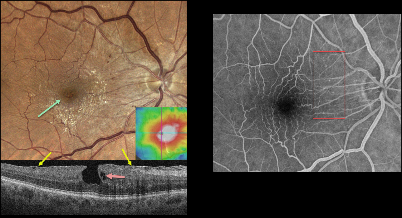
This 79YO male was referred for asymptomatic macular findings in his right eye. Vision was 20/20.
Color imaging shows a shiny, mostly nasal and inferior, macular pucker with a small, irregular macular pseudohole. Small dots of retinal blood vs telangiectasia are seen nasally.
OCT scanning shows an adherent macular pucker, a vertical foveal contour, and a small area of outer plexiform layer (OPL) schisis. The macula was diffusely thickened.
Fluorescein angiography shows no macular leakage, with filling of the nasal telangiectatic vessels.
Learning Points:
This case nicely illustrates many clinical and multimodal imaging findings for macular pucker. Clinically, true macular holes are virtually always perfectly round, whereas these pseudoholes are usually somewhat oval. The adherent epimacular tissue usually spares the foveal center. The adherent surrounding tissue contracts, drawing the surrounding tissue towards the macular center, giving the fovea a more vertical contour.
The Müller cell spans nearly the full thickness of the retina and helps mechanically hold the 3 layers of retinal cells together. Henle’s layer (the macular OPL) is inherently weakened since the photoreceptor axons and Muller cells are splayed laterally. Vitreomacular traction or traction from an overlying epimacular membrane will often therefore split Henle’s causing non-exudative schisis or cysts.
Finally, the secondary telangiectatic vascular changes are caused by mechanical damage from the overlying membrane. In severe cases, a pucker with the vascular changes can simulate a combined hamartoma.
MACULAR PUCKER
Originally posted on @retina.rocks March 27, 2023
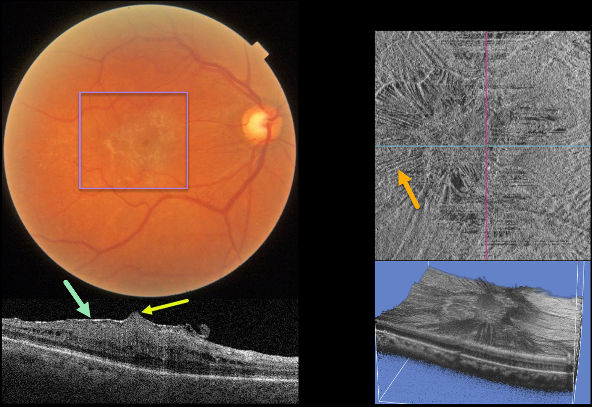
This 61YO female presented with 20/40 vision in her right eye from a mildly symptomatic macular pucker.
Color imaging shows a moderate macular pucker. Swept source OCT shows a variably adherent hyperreflective pucker, moderate retinal thickening with loss of the inner retinal layer detail, and a small foveal prolapse. En face imaging shows variable inner retinal striae from the pucker, and 3D reconstruction shows another unique perspective.
Learning Points:
Herniation of the central fovea is a rare finding with macular pucker. A recent case series (Shah et al, Retina 2023;43:182-190) found that larger foveal herniation height was associated with greater preoperative retinal thickness and worse pre- and postoperative vision.
However, vitrectomy with membrane peeling still results in significant anatomic and visual improvement. We have been observing our patient since she has been completely asymptomatic.
MACULAR PUCKER
Originally posted on @retina.rocks February 13, 2023
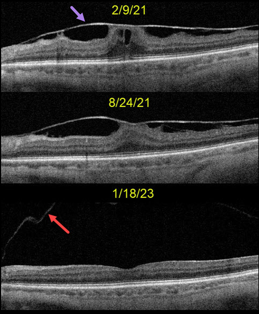
This 71YO female was seen on 2/9/21 with a completely asymptomatic variably adherent macular pucker. Vision was 20/80, and observation was recommended. On 8/24/21, vision improved to 20/40, and the macular traction had decreased.
When last examined on 1/18/23, the membrane had completely and spontaneously peeled itself as part of a macular posterior vitreous detachment. The back surface of the posterior hyaloid is seen nasally. Vision was 20/50.
Learning Points:
Spontaneous macular pucker separation is a known but rare event, occurring in about 2% of eyes with pre-existing posterior vitreous detachment (PVD) and 13% of eyes without PVD (Yang et al., Retina 2014;34:2079-2087).
We generally avoid macular pucker, vitreomacular traction, or macular hole surgery in visually happy patients. This patient also reminds us that these conditions can sometimes resolve on their own.
MACULAR PUCKER
Vaidehi Sathaye and Manish Nagpal
Originally posted on @retina.rocks December 9, 2022
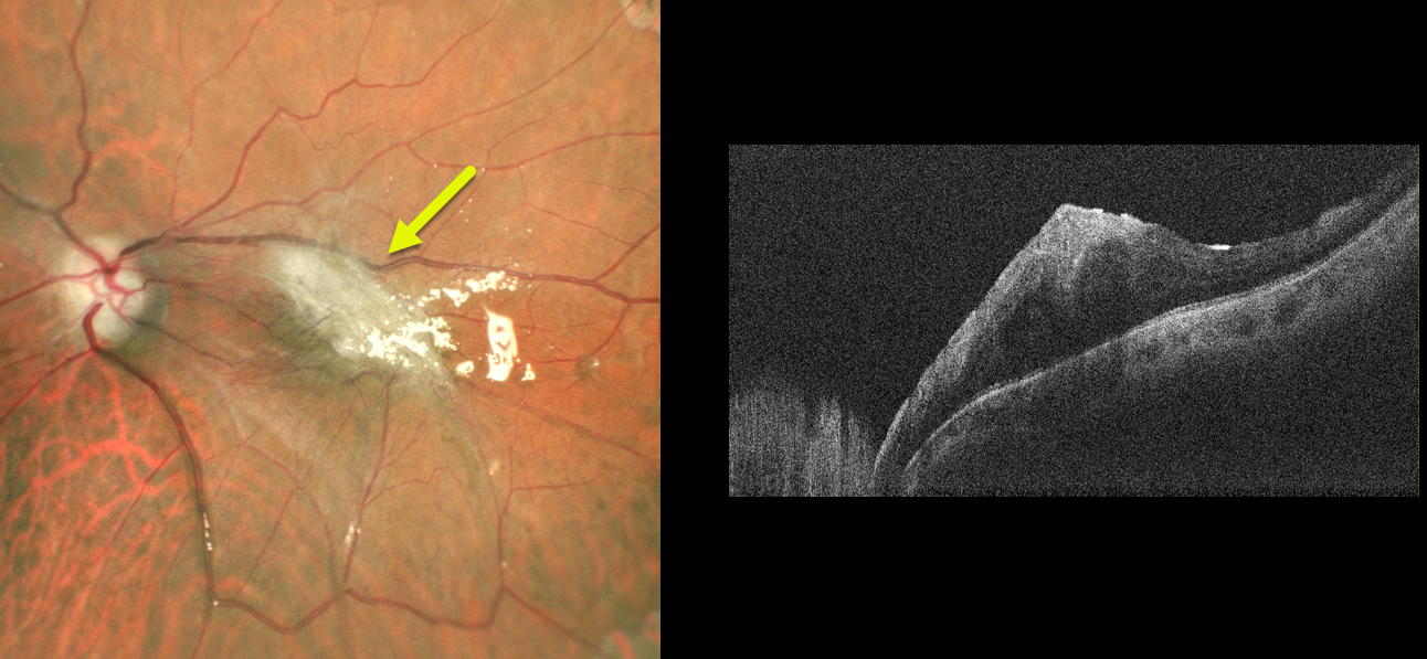
This 45YO male underwent vitrectomy with silicone oil for a recurrent rhegmatogenous retinal detachment (RRD).
Although the retina remained attached, he developed a severe macular pucker postoperatively. Removal of the silicone oil with membrane peeling is planned in the near future.
Learning Points:
Macular pucker following vitrectomy for RRD develops clinically in about 10% of patients (Katira et al, Retina 2008;28:744-748), with the incidence likely being higher than this using OCT.
Half of the eyes developing macular pucker can occur after the first postoperative year (Szigiato et al, International Journal of Retina and Vitreous 2022;8:70).
MACULAR PUCKER FOLLOWING ENDOPHTHALMITIS
Originally posted on @retina.rocks March 31, 2022
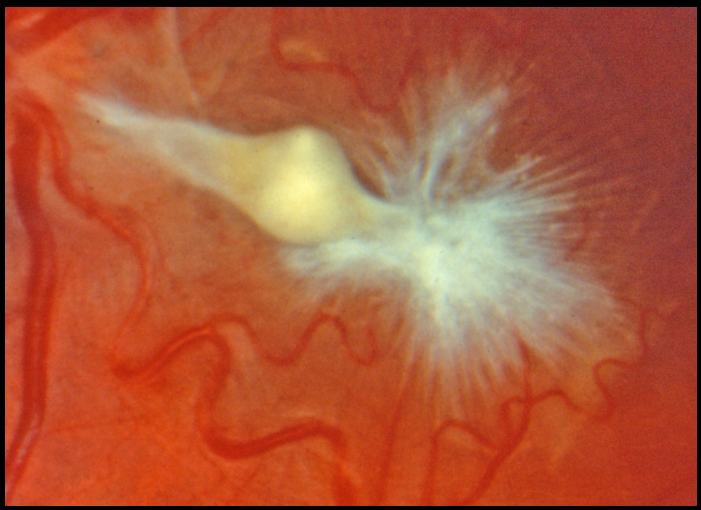
This eye shows a severely contracted and complex epimacular membrane in a patient who recovered from prior endophthalmitis. Unfortunately, we don’t have any further clinical information.
MACULAR PUCKER
Seif Anwar
Originally posted on @retina.rocks January 4, 2022
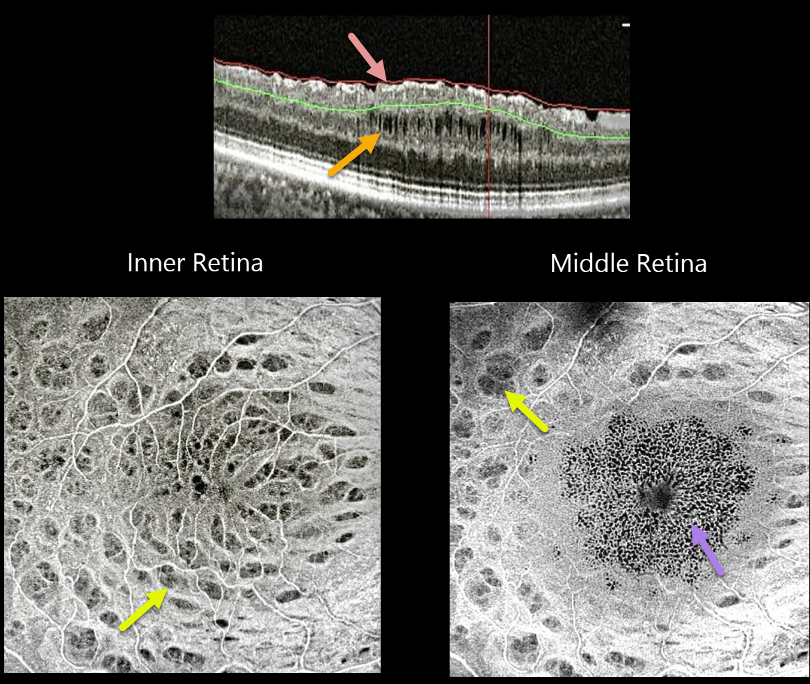
This 52YO pseudophakic female with an idiopathic macular pucker and 20/60 vision underwent pars plana vitrectomy (PPV) with internal limiting membrane (ILM) peeling. Vision improved to 20/30 after surgery, followed 6 months later by a gradual decrease to 20/50.
SOLIX OCT B-scan imaging shows multiple inner surface irregularities and thickening, with middle-layer cavitations.
En face OCT of the inner retina shows multiple concentric dark spots (dimples), characteristic of dissociated optic nerve fiber layer (DONFL). En face imaging through the mid-retinal region shows a central pattern of tiny hyporeflective cysts, with still-visible dimples more peripherally.
Learning Points:
PPV with membrane peeling is frequently performed to manage many vitreomacular interface disorders, including vitreomacular traction, macular hole, and epiretinal membrane.
While ILM peeling decreases epiretinal membrane recurrence and enhances macular hole closure, it also causes mechanical trauma to the inner retina, as evidenced by DONFL.
Many mechanisms have been proposed, including Müller cell damage from direct contact with surgical instruments, damage to the deep inner retinal layer, damage from the visualization dye, or tractional thinning of the temporal retina.
CYSTOID MACULAR EDEMA
Alex Hynes
Originally posted on @retina.rocks November 23, 2021
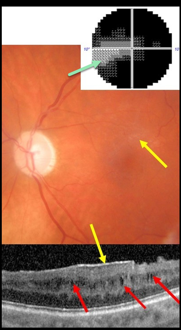
This 61YO male presented with end-stage glaucoma OS, 20/30 vision, and an IOP of 8 mmHg following successful trabeculectomy ten years earlier. He had prior cataract surgery in this eye 15 years ago with no documented history of pseudophakic cystoid macular edema.
There was a moderate macular pucker noted clinically and on OCT, along with multiple inner nuclear layer (INL) microcysts on OCT scanning. He has a tiny island remaining in his visual field.
Learning Points:
Microcystic macular changes have been associated with both advanced primary open-angle glaucoma and idiopathic macular pucker (see Govetto et al, AJO 2017;181:156-165).
Macular pucker-induced mechanical stress, coupled with retrograde trans-synaptic degeneration of bipolar cells secondary to retinal ganglion cell loss, likely predisposes towards the accumulation of fluid within the inner retinas of these patients.
Relative to non-glaucomatous eyes, microcystic macular changes in glaucomatous eyes tend to be more persistent following membrane peeling. This is likely due to retrograde trans-synaptic degeneration.
It is important that these microcystic spaces in eyes with optic neuropathy are not mistaken for inflammatory edema following membrane peeling, which could lead to unnecessary treatment.
Finally, disproportionate preservation of overall retinal thickness despite severe thinning of the ganglion cell and/or nerve fiber layers should not be surprising in glaucomatous eyes.
MACULAR PUCKER
Originally posted on @retina.rocks August 5, 2021
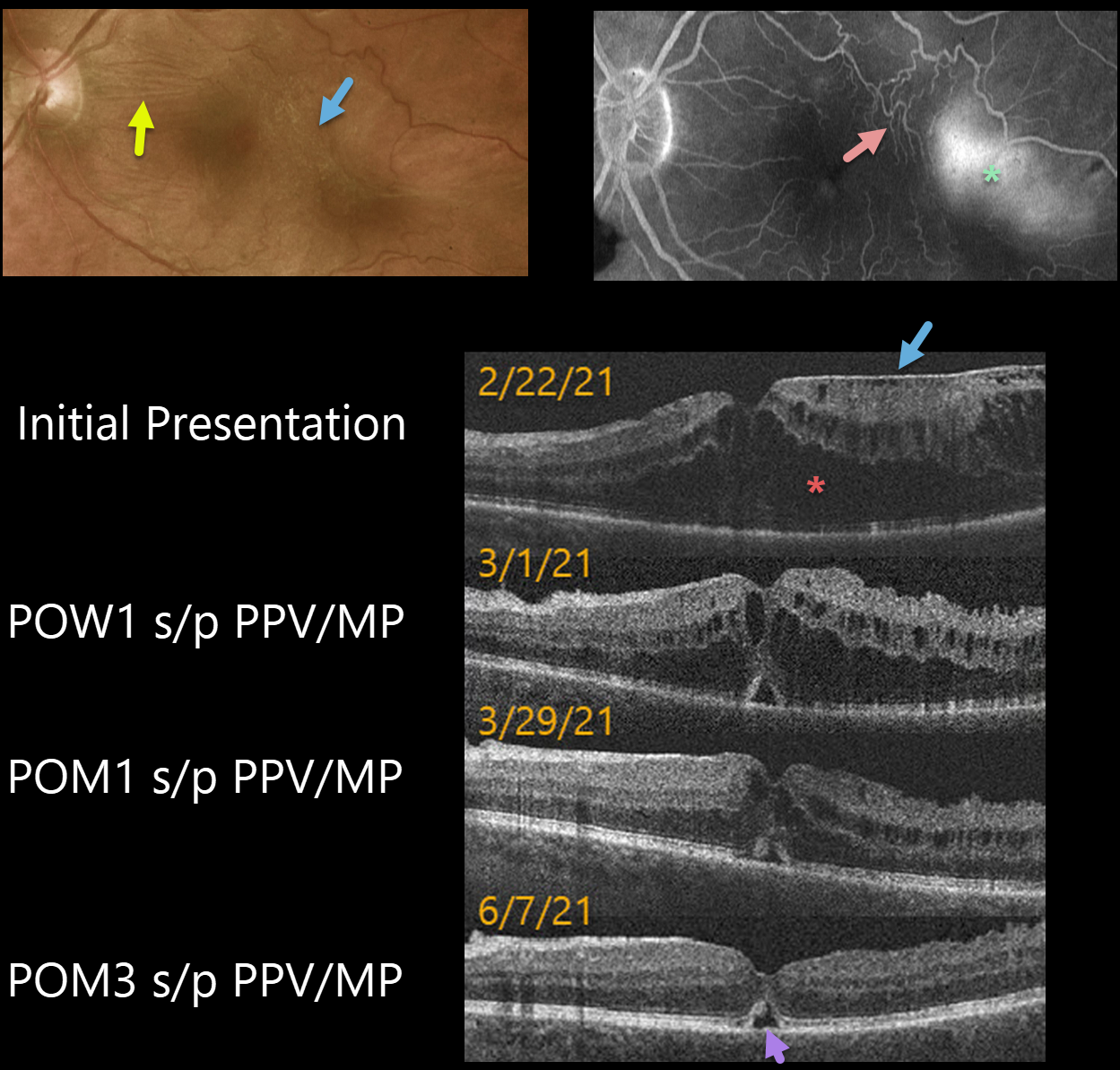
This 62YO male presented with 20/200 vision in his left eye. An epiretinal membrane (ERM) was noted in the temporal macula, associated with more nasal horizontal retinal striae.
Optos fluorescein angiography shows distortion of the temporal macular vessels with late leakage.
Vitrectomy with membrane peeling was performed the following day, and one week later, on 3/1/21, vision improved to 20/70 with a significant decrease in macular thickening.
When last examined on 6/7/21, about 3 months post-op, vision was still 20/70, but the macular thickening and contour were almost normal except for a small blister of foveal subretinal fluid.
Learning Points:
OCT scanning confirmed the temporal ERM, but the severe cystic macular thickening with possible tractional schisis that extended into the nasal macula seemed disproportionate to the imaged temporal ERM. Fortunately, both the vision and macular thickening significantly improved following vitrectomy with membrane peeling.
COMBINED HAMARTOMA OF THE RETINA AND RPE
Originally posted on @retina.rocks August 25, 2020
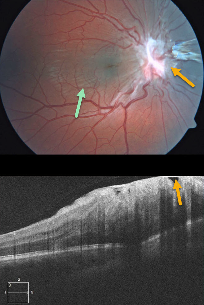
This 7YO boy presented without visual symptoms and 20/50 vision OD. The left eye was normal.
Contracted fibrotic tissue overlying the optic nerve is causing distortion of the surrounding retina, with striae visible through the macula. Some of this fibrosis may also be pigmented nasal to the nerve.
This case represents either a combined hamartoma of the retina and RPE or an unusual pucker centered over the optic nerve.
Learning Points:
Combined hamartomas are classically described as benign congenital lesions composed of glial cells, vascular tissue, and pigmented retinal pigment epithelial cells. However, more recent data indicate that these lesions primarily arise from the inner retinal layers.
MACULAR PUCKER
Originally posted on @retina.rocks July 3, 2020
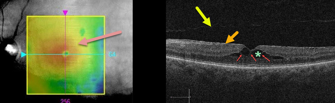
This patient was sent in for treatment for possible diabetic macular edema. The retinal thickness map shows central macular thickening.
The OCT B-scan shows an adherent, hyperreflective epiretinal membrane (ERM), a faint posterior vitreous detachment overlying the macula, and central cystic-like inner retinal spaces.
Muller cells, which extend vertically from the internal limiting membrane to the outer retina (external limiting membrane), essentially hold the 3 layers of retinal cells together.
In the central retina, the photoreceptor axons and Muller cells (Henle’s layer, which is the central OPL) are splayed outwards. This creates an intrinsically vertically weakened central macula. The central thin hyperreflective columns are Muller cells that span the region of split OPL.
Unlike exudative intraretinal cysts caused by leaking retinal blood vessels, these hyporeflective spaces are caused by tractional splitting of the outer plexiform layer (OPL) from either aborted vitreomacular traction and/or the ERM.
Learning Points:
When interpreting OCT scans, it is important to consider all the information in the OCT report, ideally by scrolling through the entire macular cube data in the review software.
The macula here is not truly edematous. It is thickened due to a tractional splitting of the retina. For a great discussion of tractional vs exudative cystoid spaces, see Govetto et al AJO 2020;212;43-56.
MACULAR PUCKER (PSEUDO-COMBINED HAMARTOMA)
Originally posted on @retina.rocks June 25, 2020
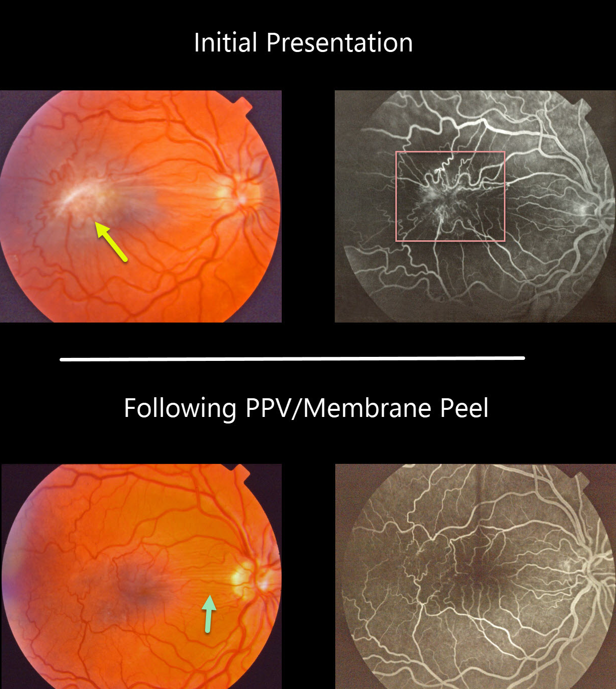
This patient had an isolated severe macular pucker. The fluorescein angiogram shows a vascular hamartomatous component due to severe retinal vascular distortion and leakage. This vascular component, along with the pigmented appearance of the pucker, helps give the false appearance that this was a combined hamartoma of the RPE.
The macular pucker was treated with vitrectomy and membrane peeling. Postoperatively, the retina appears nearly normal, with some residual peripapillary retinal folds and no vascular leakage.
Learning Points:
Hamartomas and severe macular puckers can look very similar, even to some of the most seasoned retina specialists. OCT may be helpful in differentiating these entities (see Ophthalmic Surg Lasers Imaging Retina 2017;48;122-125).
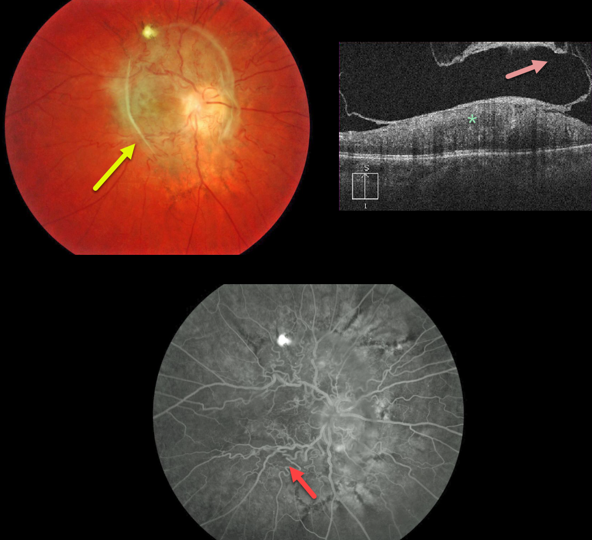
MACULAR PUCKER VS COMBINED HAMARTOMA
Originally posted on @retina.rocks May 25, 2020
The fundus photo shows an unusual peripapillary pigmented epiretinal membrane distorting the underlying retina.
The OCT shows a partially detached epiretinal membrane and marked hyperreflectivity of the inner retina, with loss of normal retinal layer detail.
Fluorescein angiography shows a distorted retinal vasculature with late leakage.
Occasionally, vitrectomy with membrane peeling can be offered, but we are electing to observe this eye since vision has been poor since birth.
Learning Points:
A combined hamartoma of the RPE is a benign congenital lesion composed of glial cells, vascular tissue, and pigmented epithelial cells.
Even though we’re calling this a combined hamartoma, we’re not positive that this may not just be an unusual peripapillary/macular epiretinal membrane.
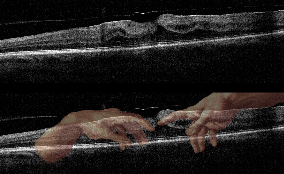
MICHELANGELO MACULOPATHY
Originally posted on @retina.rocks May 1, 2020
This patient has an epiretinal membrane with some vitreomacular traction.
This case was submitted by Dr. Jennifer Reynolds. The “artistic” mind of the submitting doctor, noticed the hand of the Creator in this scan!
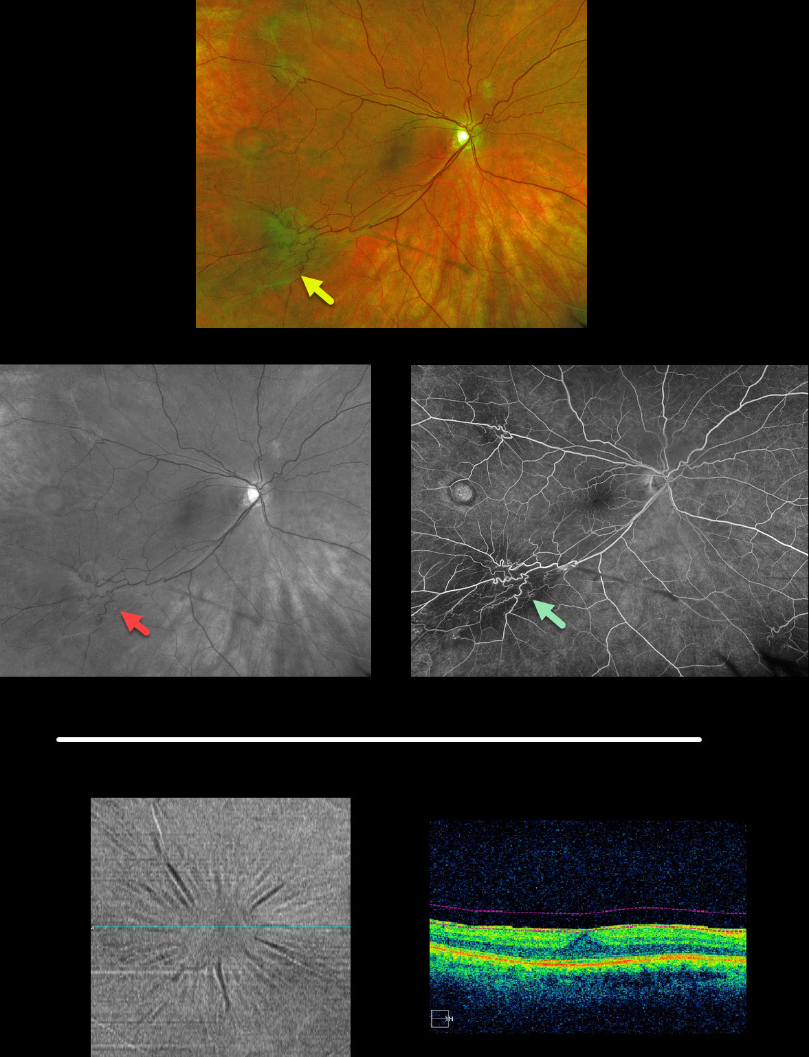
MACULAR PUCKER
Originally posted on @retina.rocks February 12, 2020
Multifocal images of an extramacular epiretinal membranes with an unrelated temporal retinal pigmented epithelial detachment are displayed here (Optos, red-free, and fluoroscein angiography).
BONUS: The second set of images is a different patient with a macular pucker that is well-visualized using OCT en face analysis, which provides transverse views of the retinal and choroidal layers using user-specified depths and thickness. This is an underutilized yet fantastic tool that gives another perspective for many chorioretinal disorders.


