

Macular Telangiectasia (MacTel)
<< Back to Cases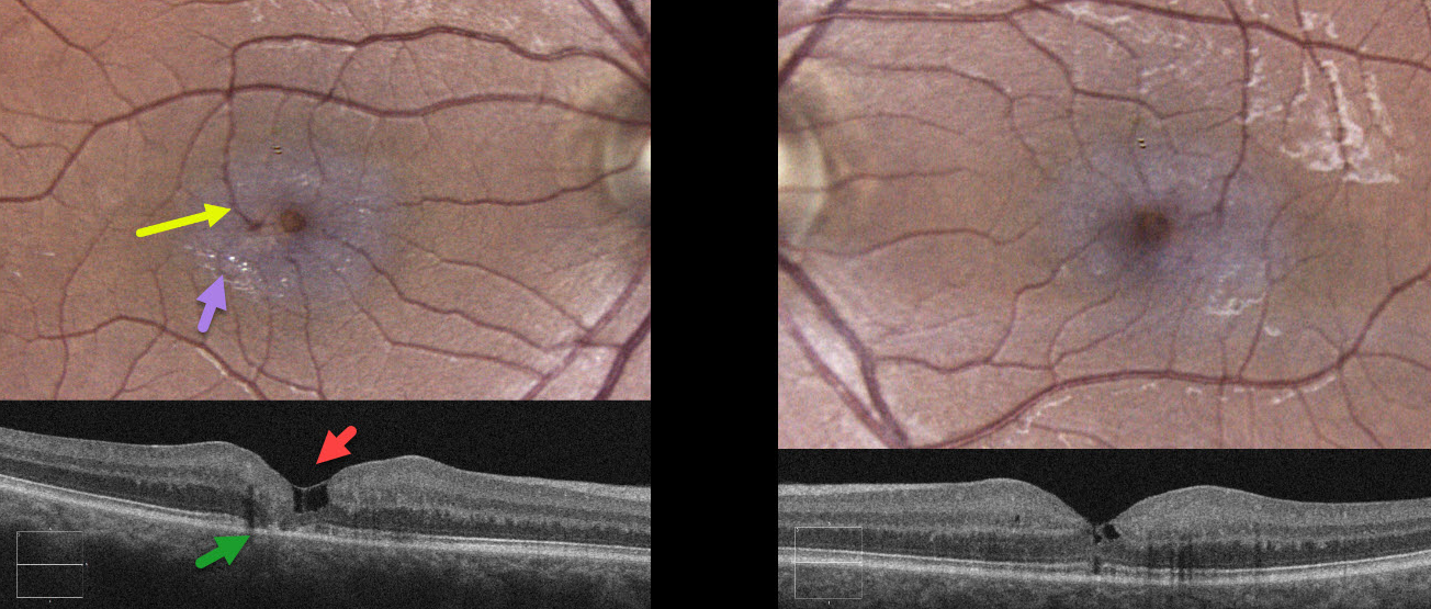
MACULAR TELANGIECTASIA
Originally posted on @retina.rocks March 26, 2025
This healthy 43YO male was referred for asymptomatic retinal findings. Vision was 20/40 OD and 20/30 OS.
Color imaging of the right eye shows a foveal cyst with pericentral opaque whitened retina and numerous inner retinal crystals. A right-angle venule is noted superotemporally.
OCT scanning shows inner foveal cavitations with variable loss of the outer retinal bands. Temporal foveal outer retinal atrophy is also noted. The left macula shows an encircling, pericentral, opaque retina with mild crystals. OCT reveals milder inner cavitations with an outer foveal microdefect.
Learning Points:
Originally described by Gass (Arch Ophthalmology 1982;100:769-780), macular telangiectasia type 2 (MacTel2) is a neurodegenerative disorder, most likely originating from Muller cell dysfunction. The term telangiectasia is misleading, as the funduscopic findings are mostly non-vascular. The refractile inner retinal ‘crystals’ are thought to be Muller cell footplates. The foveal retina has a whitish discoloration, most likely due to retinal opacification from dysfunctional Muller cells. Photoreceptor loss allows RPE cells to migrate along intraretinal capillaries, causing the coarse clinical pigment clumping (not seen in our patient).
Most patients will also have subclinical chorioretinal anastomosis in association with right-angle venules (Spaide et al, Retina 2018;38:1920-1929). Secondary macular neovascularization can be successfully treated per age-related macular degeneration protocols.
MACULAR TELANGIECTASIA
Originally posted on @retina.rocks March 4, 2024
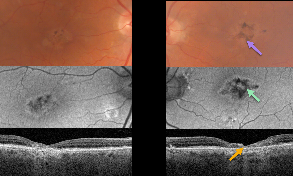
This 80YO male was referred for bilateral macular findings. He was without new visual complaints. Vision was 20/70 OD and 20/200 OS.
Optos color RG imaging shows bilateral foveal pigment loss with coarse intraretinal pigment migration. The changes are mostly hypo-autofluorescent.
Triton swept-source OCT shows atrophy and disorganization of all retinal layers in the central and temporal foveas.
Learning Points:
Originally described by Gass (Arch Ophthalmology 1982;100:769-780), macular telangiectasia type 2 (MacTel2) is a neurodegenerative disorder, most likely originating from Muller cell dysfunction.
The term telangiectasia is misleading, as the funduscopic findings are mostly non-vascular. The refractile inner retinal ‘crystals,’ not seen in our patient, are thought to be Muller cell footplates. The foveal retina shows a whitish discoloration, most likely due to retinal opacification caused by dysfunctional Muller cells. Photoreceptor loss allows RPE cells to migrate along intraretinal capillaries, causing the coarse clinical pigment clumping.
The majority of patients will also have subclinical chorioretinal anastomosis associated with right-angle venules (Spaide et al, Retina 2018;38:1920-1929). Secondary macular neovascularization can be successfully treated per age-related macular degeneration protocols.
MACULAR TELANGIECTASIA
Originally posted on @retina.rocks November 8, 2022
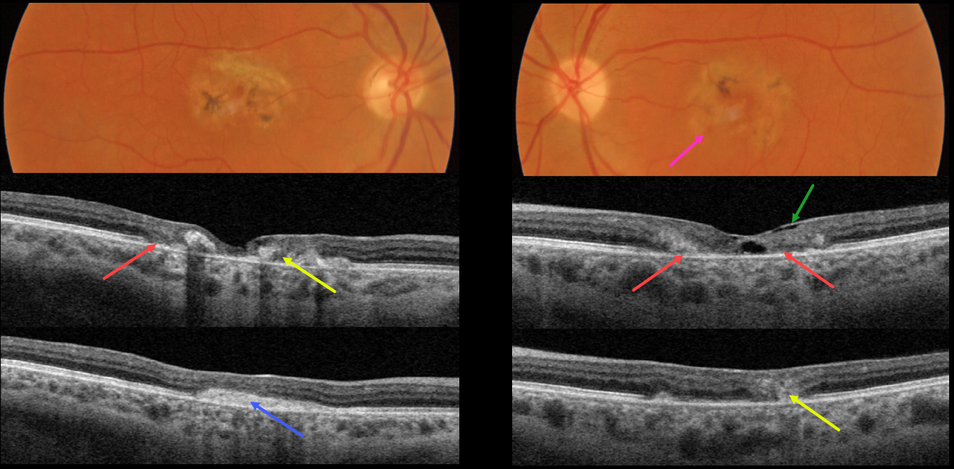
This 49YO female presented with several weeks of vision loss in her left eye. A macular neovascularization (MNV) in her right eye was treated 4 years earlier with intravitreal Avastin. Vision was 20/200 OD and 20/70 OS.
Both maculas had central pigment loss with intraretinal pigment migration. A small dot of blood was noted inferonasally in the left macula.
OCT scanning shows outer retinal atrophy, pericentral retinal disorganization, a fibrosed, inactive MNV in the right macula, and hyporeflective loss of retinal tissue (cavitation) in the left outer macula. Faint loss of the temporal inner retinal tissue is highlighted by intact internal limiting membrane (ILM drape sign).
Anti-VEGF therapy was started for a presumed MNV in her left eye.
Learning Points:
Originally described by Gass (Arch Ophthalmology 1982;100:769-780), MacTel2 is a neurodegenerative disorder, most likely originating from Muller cell dysfunction.
The term, telangiectasia, is misleading in that the funduscopic findings are mostly non-vascular. The refractile inner retinal ‘crystals’ are thought to be Muller cell footplates. The foveal retina shows a whitish discoloration, most likely due to retinal opacification caused by dysfunctional Muller cells.
Photoreceptor loss allows RPE cells to migrate along intraretinal capillaries, causing the coarse clinical pigment clumping.
All of these changes are usually in the temporal fovea, so our patient is atypical in that they involve the entire central macular regions.
MACULAR TELANGIECTASIA
Originally posted on @retina.rocks June 7, 2022
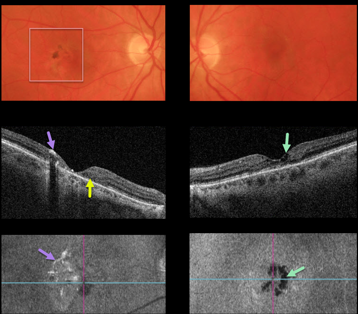
This 60YO female presented with 20/70 vision OD and 20/50 vision OS, along with classic findings of macular telangiectasia type 2 (MacTel2).
The perifoveal retina is somewhat opaque with significant pigmentary changes, particularly in the right eye.
OCT scanning of the right eye shows significant central outer retinal atrophy with hyper-reflective pigment migration. The left OCT shows milder atrophy, central inner cavitations, but no pigment migration.
En face imaging helps highlight some of the findings described above. The right en face image shows scattered patchy hyperreflective areas corresponding to the pigment migration, while the left en face image highlights the hyporeflective cavitations.
Learning Points:
Originally described by Gass (Arch Ophthalmology 1982;100:769-780), MacTel2 is a neurodegenerative disorder, most likely originating from Müller cell dysfunction. The term telangiectasia is misleading because the funduscopic findings are mostly nonvascular.
The refractile inner retinal ‘crystals’ are thought to be Müller cell footplates. The foveal retina shows a whitish discoloration, most likely due to retinal opacification resulting from Müller cell dysfunction.
Photoreceptor loss allows RPE cells to migrate along intraretinal capillaries, causing the coarse clinical pigment clumping (not seen in our patient).
The majority of patients will also have subclinical chorioretinal anastomosis associated with right-angle venules (Spaide et al, Retina 2018;38:1920-1929).
Secondary macular neovascularization can be successfully treated according to age-related macular degeneration protocols.
MACULAR TELANGIECTASIA
Originally posted on @retina.rocks December 27, 2021
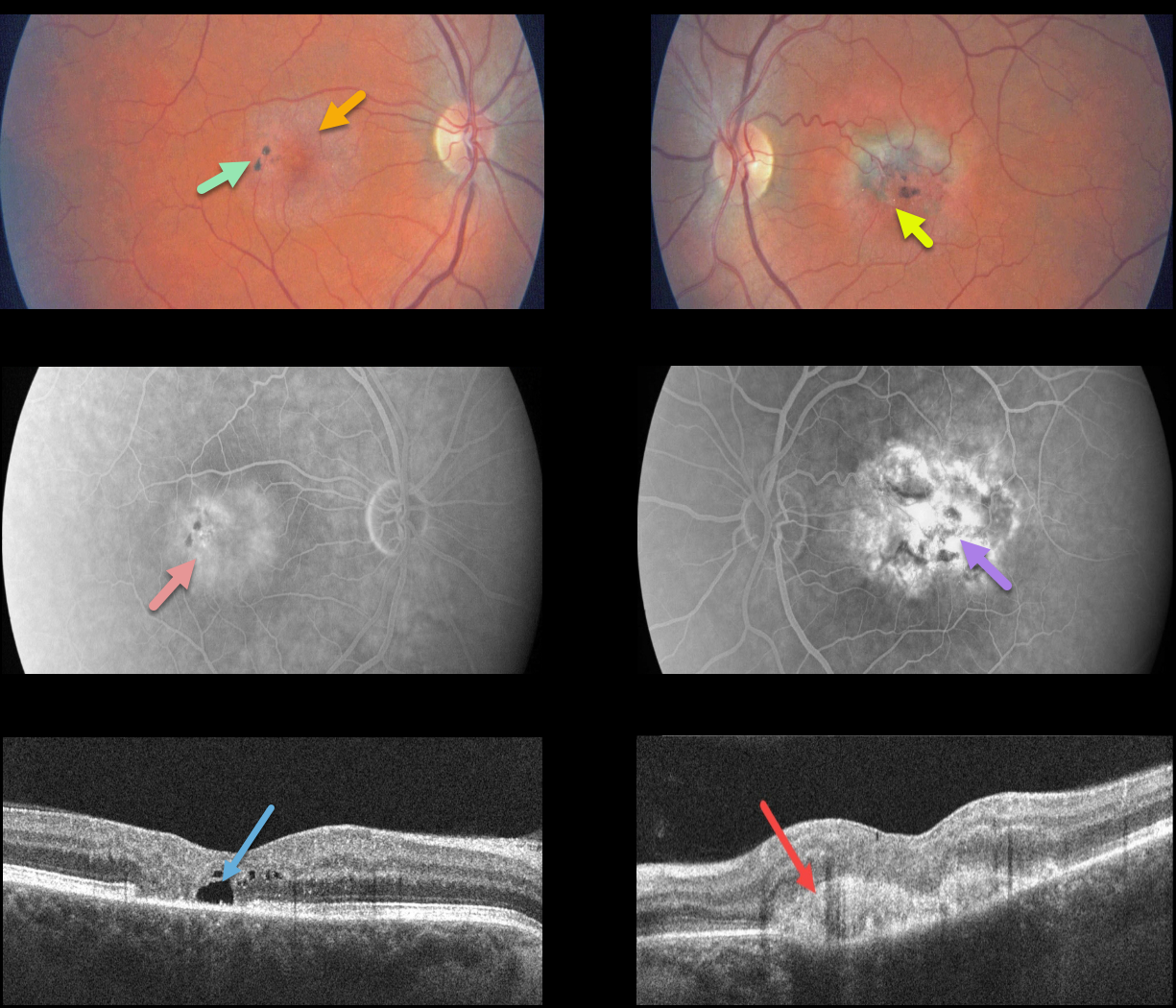
This patient presented with classic findings of macular telangiectasia type 2 (MacTel2), including an opaque perifoveal retina, inner retinal crystal, and black clumps of intraretinal pigment migration.
Deep retinal angiographic leakage is noted in the right eye with more diffuse staining in the left eye. OCT scanning shows an outer retinal cavitation OD and type 2 macular neovascularization (MNV) OS.
Learning Points:
Originally described by Gass (Arch Ophthalmology 1982;100:769-780), MacTel2 is a neurodegenerative disorder, most likely originating from Müller cell dysfunction.
The term telangiectasia is misleading because the funduscopic findings are mostly nonvascular.
The refractile inner retinal ‘crystals’ are thought to be Müller cell footplates. The foveal retina shows a whitish discoloration, most likely due to retinal opacification resulting from Müller cell dysfunction.
Photoreceptor loss allows RPE cells to migrate along intraretinal capillaries, causing the coarse clinical pigment clumping.
The majority of patients will also have subclinical chorioretinal anastomosis associated with right-angle venules (Spaide et al., Retina 2018;38:1920-1929).
Secondary MNV can be successfully treated per age-related macular degeneration protocols.
MACULAR TELANGIECTASIA
Originally posted on @retina.rocks July 30, 2021
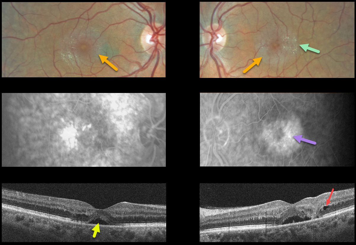
This 52YO male presented with 20/70 vision OD, 20/100 vision OS, and many classic findings for macular telangiectasia type 2 (MacTel2).
The perifoveal retina is somewhat opaque with inner retinal crystals and deep retinal angiographic leakage.
OCT scanning of the right eye shows some temporal outer retinal atrophy with an atypical small serous detachment. OCT of the left eye shows somewhat similar findings, along with a small temporal outer nuclear layer cyst vs cavitation. The serous detachment in the left fovea may represent an atypical outer macular cavitation.
Learning Points:
Originally described by Gass (Arch Ophthalmology 1982;100:769-780), MacTel2 is a neurodegenerative disorder, most likely originating from Muller cell dysfunction.
The term telangiectasia is misleading because the funduscopic findings are mostly nonvascular. The refractile inner retinal ‘crystals’ are thought to be Muller cell footplates. The foveal retina shows a whitish discoloration, most likely due to retinal opacification resulting from Müller cell dysfunction. Photoreceptor loss allows RPE cells to migrate along intraretinal capillaries, causing the coarse clinical pigment clumping (not seen in our patient).
The majority of patients will also have subclinical chorioretinal anastomosis associated with right-angle venules (Spaide et al., Retina 2018;38:1920-1929). Secondary macular neovascularization can be successfully treated according to age-related macular degeneration protocols.
MACULAR TELANGIECTASIA
Originally posted on @retina.rocks May 14, 2021
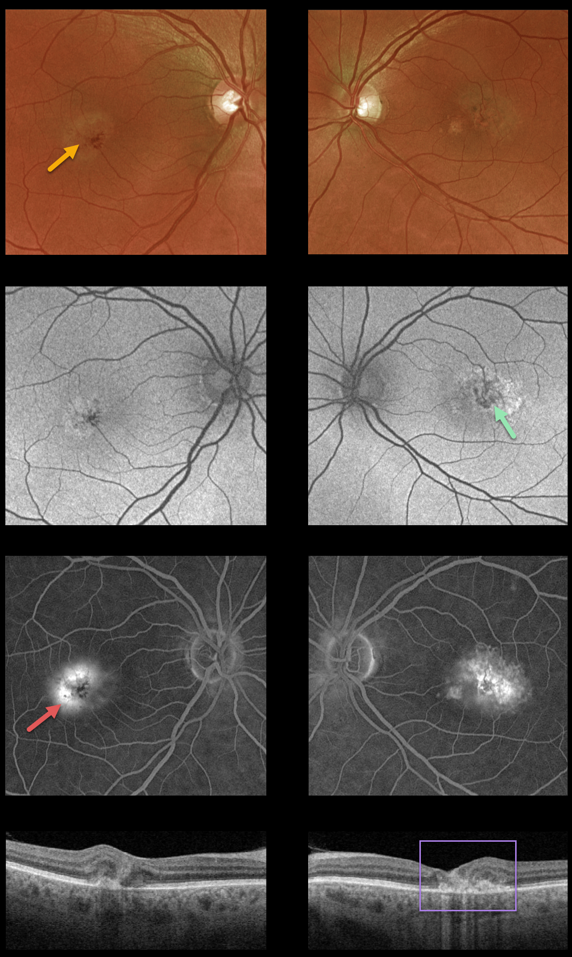
This 55YO male presented with macular telangiectasia type 2 (MacTel2). He was visually asymptomatic, and vision was 20/40 OD and 20/50 OS.
This case highlights various multimodal imaging findings. The foveal retina shows a whitish discoloration, most likely due to retinal opacification resulting from Müller cell dysfunction.
There are variable macular hyper- and hypofAF abnormalities, with angiographic staining and leakage. OCT scanning shows mostly temporal foveal outer retinal atrophy and disorganization.
Characteristic MacTel2 features absent in our patient include refractile inner retinal ‘crystals’ (thought to be Muller cell footplates, similar to the refractile dots found in retinoschisis) and coarse black clumps of intraretinal pigment migration.
Learning Points:
Originally described by Gass (Arch Ophthalmology 1982;100:769-780), MacTel2 is a neurodegenerative disorder, most likely originating from Muller cell dysfunction. The term telangiectasia is misleading because the funduscopic findings are mostly nonvascular.
Although much remains to be learned about its pathophysiology and there is no treatment for the underlying disease, secondary macular neovascularization can be successfully treated according to age-related macular degeneration protocols.
MACULAR TELANGIECTASIA
Originally posted on @retina.rocks January 22, 2021
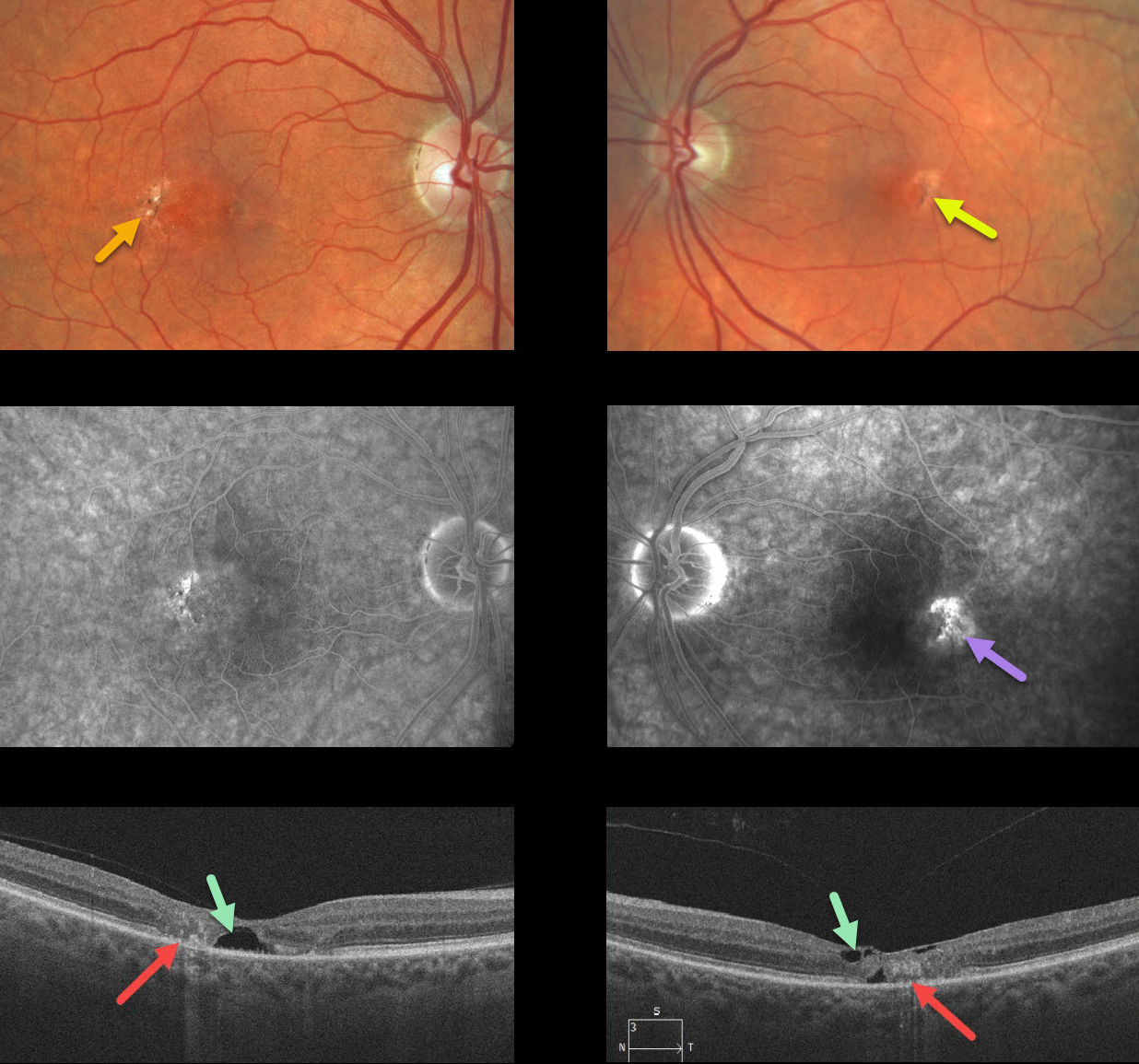
Our patient shows classic bilateral findings of macular telangiectasia type 2 (MacTel2) with temporal foveal intraretinal pigment migration and inner retinal crystals, as well as angiographic staining and leakage.
Our patient’s optical coherence tomography scans also show classic findings of mostly temporal foveal retinal atrophy, disorganization, and hyporeflective tissue loss (cavitations).
Learning Points:
MacTel type 2 is a neurodegenerative disorder, most likely originating from Müller cell dysfunction. The term telangiectasia is misleading because the funduscopic findings are mostly nonvascular.
It is thought that photoreceptor loss allows RPE cells to migrate along intraretinal capillaries, creating the pigment clumping. This is a similar mechanism to that found in other causes of intraretinal pigment migration, including photoreceptor loss or damage, such as retinitis pigmentosa, chronic retinal detachment, and blunt trauma.
The refractile inner retinal ‘crystals’ are thought to be Muller cell footplates, similar to the refractile dots found in retinoschisis.
Although much remains to be learned about its pathophysiology and there is no treatment for the underlying disease, secondary macular neovascularization can be successfully treated according to age-related macular degeneration protocols.
MACULAR TELANGIECTASIA
Originally posted on @retina.rocks April 9, 2020
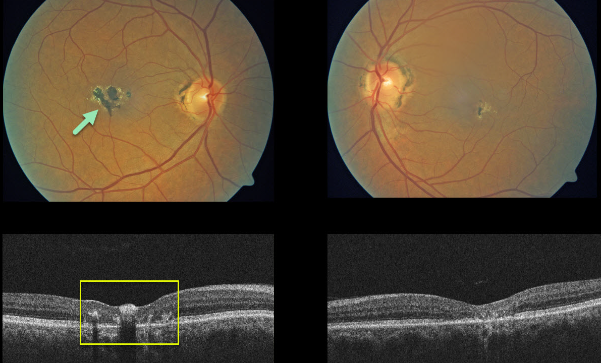
Our patient shows classic bilateral findings for macular telangiecasia (MacTel) type 2 with coarse pigment clumps.
The OCTs show central/temporal retinal atrophy with disorganized retinal layers. Absent in our patient, the OCT will also often display hyporeflective cavitations of tissue loss.
The majority of patients will also have subclinical chorioretinal anastomosis in association with right-angle venules.
Learning Points:
MacTel type 2 is a neurodegenerative disorder, most likely originating from Muller cell dysfunction. The term, telangiectasia, is misleading in that the funduscopic findings are mostly non-vascular.
It is thought that photoreceptor loss allows RPE cells to migrate along intraretinal capillaries causing the clinical pigment clumping. There are also numerous refractile inner retinal ‘crystals,’ thought to be Muller cell footplates.
Although there is much to be learned about its pathophysiology and there is no treatment for the underlying disease, secondary macular neovascularization can be successfully treated per AMD protocols.


