

Maternally Inherited Diabetes and Deafness (MIDD)
<< Back to Cases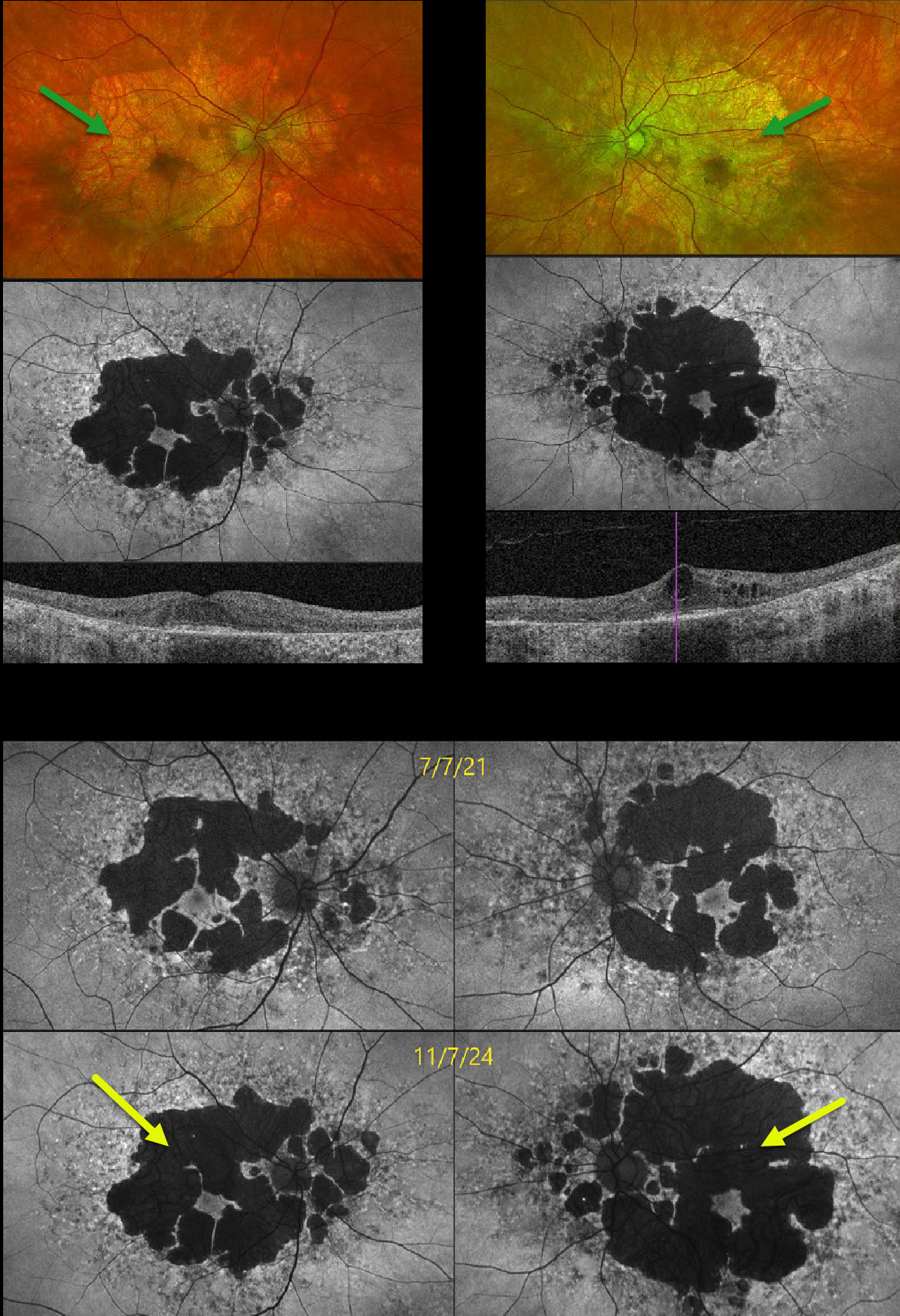
MATERNALLY INHERITED DIABETES AND DEAFNESS (MIDD)
Originally posted on @retina.rocks April 17, 2025
This 58YO male presented with several years of progressive vision loss. He has a history of well-controlled type 2 diabetes. Vision was 20/30 OD and 20/60 OS.
Optos color RG imaging shows fairly symmetric areas of variable, confluent, non-central macular atrophy. Fundus autofluorescence (FAF) shows hypo-FAF from the areas of atrophy, along with areas of hyper-FAF around each nerve and macula. Swept-source OCT shows outer retinal and RPE atrophy bilaterally with central cystic edema OS. FAF shows significant progression of atrophy compared with 3 years earlier. The subclinical edema OS was felt to be diabetic and has been observed since it was spontaneously fluctuating with unchanged vision and symptoms.
Learning Points:
Maternally inherited diabetes and deafness (MIDD) accounts for up to 3% of all cases of diabetes and results from the A3243 G mutation in mitochondrial DNA. MIDD often masquerades as a pattern macular dystrophy. In our experience, fundus autofluorescence (FAF) is the best way to visualize these changes. The FAF appearance somewhat resembles that seen with Elmiron toxicity. Peripapillary hypoautofluorescence, more densely packed macular autofluorescent changes, and earlier central macular involvement suggest Elmiron toxicity over other causes (Barnes et al Ophthalmology Retina 2020;4:1196-1201).
Despite our patient’s multimodal findings being classical for MIDD, mitochondrial genetic testing revealed variants of uncertain significance (homozygous AMPD1 and heterozygous ACAD5). Invitae Inherited Retinal Disorders Panel revealed homozygous variants of u
MATERNALLY INHERITED DIABETES AND DEAFNESS (MIDD)
Originally posted on @retina.rocks July 20, 2023
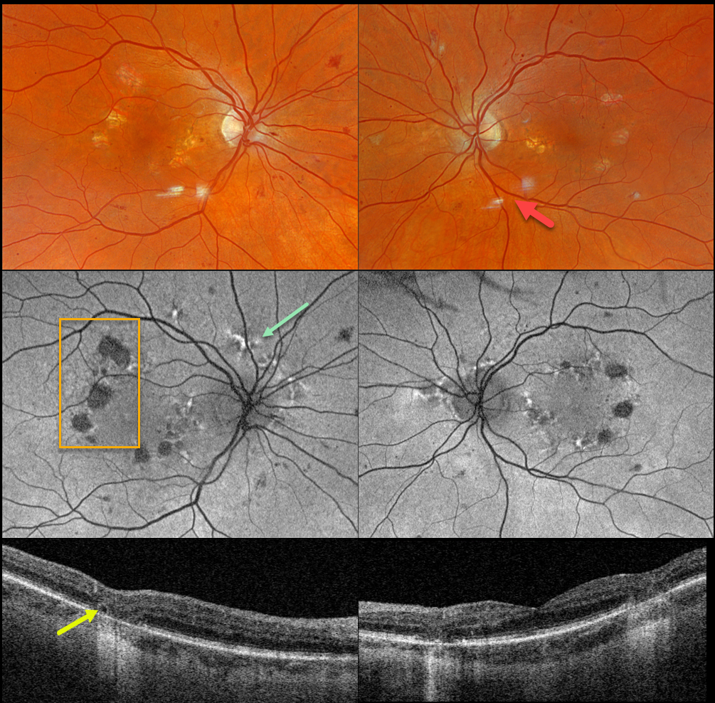
This 66YO female was seen for a diabetic retinopathy screening. She had a prior history of age-related macular degeneration (AMD). There were no visual symptoms. Vision was 20/40 OD and 20/30 OS.
Optos color RGB imaging shows moderate non-proliferative diabetic retinopathy (NPDR) with some scattered retinal hemorrhages and nerve fiber layer infarcts. Some focal areas of non-central macular atrophy are also seen.
Fundus autofluorescence (FAF) shows hypo-FAF from the areas of atrophy, along with areas of hyper-FAF around each nerve and macula.
Swept-source OCT shows outer retinal and RPE atrophy. A small area of outer retinal tubulation (ORT) is noted along the edge of an area of atrophy OD.
On further questioning, the patient’s mother had type 2 diabetes. Hearing was normal in our patient and her mother. Genetic testing of our patient showed a pathologic mitochondrial A3243G mutation.
Learning Points:
Maternally inherited diabetes and deafness (MIDD) accounts for up to 3% of all cases of diabetes and results from a mutation in mitochondrial DNA at position A3243G. MIDD often masquerades as a pattern macular dystrophy.
In our experience, fundus autofluorescence (FAF) is the best way to visualize these changes. The FAF appearance somewhat resembles that seen with Elmiron toxicity. Peripapillary hypoautofluorescence, more densely packed macular autofluorescent changes, and earlier central macular involvement suggest Elmiron toxicity over other causes (see Barnes et al Ophthalmology Retina 2020;4:1196-1201).
ORT is often noted overlying inactive MNV with ongoing anti-VEGF therapy and should not be confused with exudative fluid or cysts, which lack a hyperreflective border. The outer hyperreflective band likely represents inner segment mitochondria undergoing fission and translocation toward the nucleus (Litts et al, Retina 2018;38:445-461).
ORT, initially described by Zweifel et al (Arch Ophthalmol 2009;127:1596-1602), is a neurodegenerative condition of the photoreceptors and Muller cells associated with atrophy affecting the outer retina and retinal pigment epithelium, including advanced AMD and inherited retinal diseases.
MATERNALLY INHERITED DIABETES AND DEAFNESS (MIDD) + OUTER RETINAL TUBULATION (ORT)
Originally posted on @retina.rocks October 3, 2022
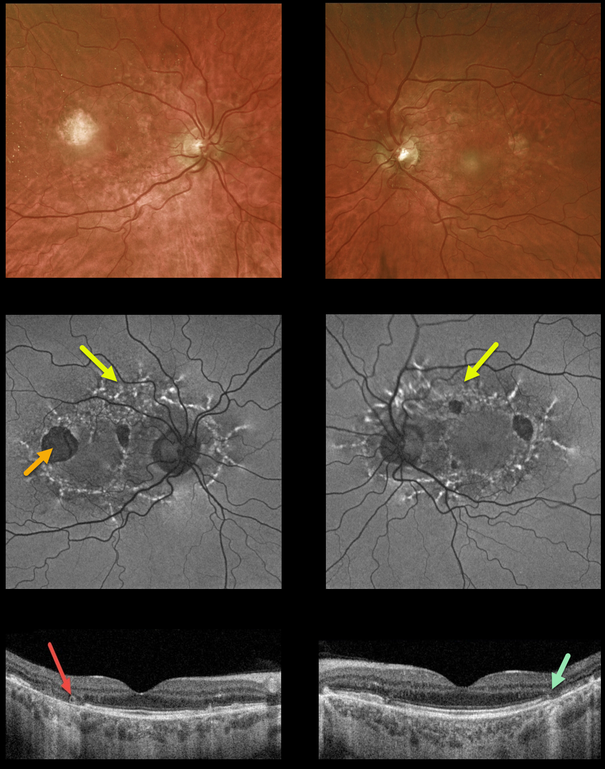
This 59YO male presented with vision of 20/400 OD and 20/30 OS. He has severe lifelong hearing loss, and both he and his mother have type 2 diabetes.
Optos ultrawidefield imaging shows temporal macular atrophy. Fundus autofluorescence shows the true extent of the pathology, with linear interconnected subretinal streaks of hyper-FAF associated with areas of hypo-FAF macular atrophy.
Swept-source OCT shows variable, mostly temporal outer retinal and RPE atrophy, along with a small area of temporal outer retinal tubulation (ORT) OD.
Learning Points: Maternally inherited diabetes and deafness (MIDD) is responsible for up to 3% of all cases of diabetes, and results from a mutation of mitochondrial DNA A3243G. MIDD often masquerades as a pattern macular dystrophy.
Fundus autofluorescence (FAF) in our experience is the best way to visualize these changes. The FAF appearance somewhat resembles that seen with Elmiron toxicity.
Peripapillary hypoautofluorescence, more densely-packed macular autofluorescent changes, and earlier central macular involvement suggest Elmiron toxicity over other causes (see Barnes et al Ophthalmology Retina 2020;4:1196-1201). Note that our case spares the central macula and peripapillary retina, which is more consistent with MIDD.
ORT is often noted overlying inactive MNV with ongoing anti-VEGF therapy and should not be confused with exudative fluid or cysts which lack a hyperreflective border. The outer hyperreflective band likely represents inner segment mitochondria undergoing fission and translocation toward the nucleus (Litts et al, Retina 2018;38:445-461).
ORT, initially described by Zweifel et al (Arch Ophthalmol 2009;127:1596-1602), is a neurodegenerative condition of the photoreceptors and Muller cells associated with atrophy affecting the outer retina and retinal pigment epithelium, including advanced AMD and inherited retinal diseases.
MATERNALLY INHERITED DIABETES AND DEAFNESS (MIDD)
Originally posted on @retina.rocks January 11, 2022
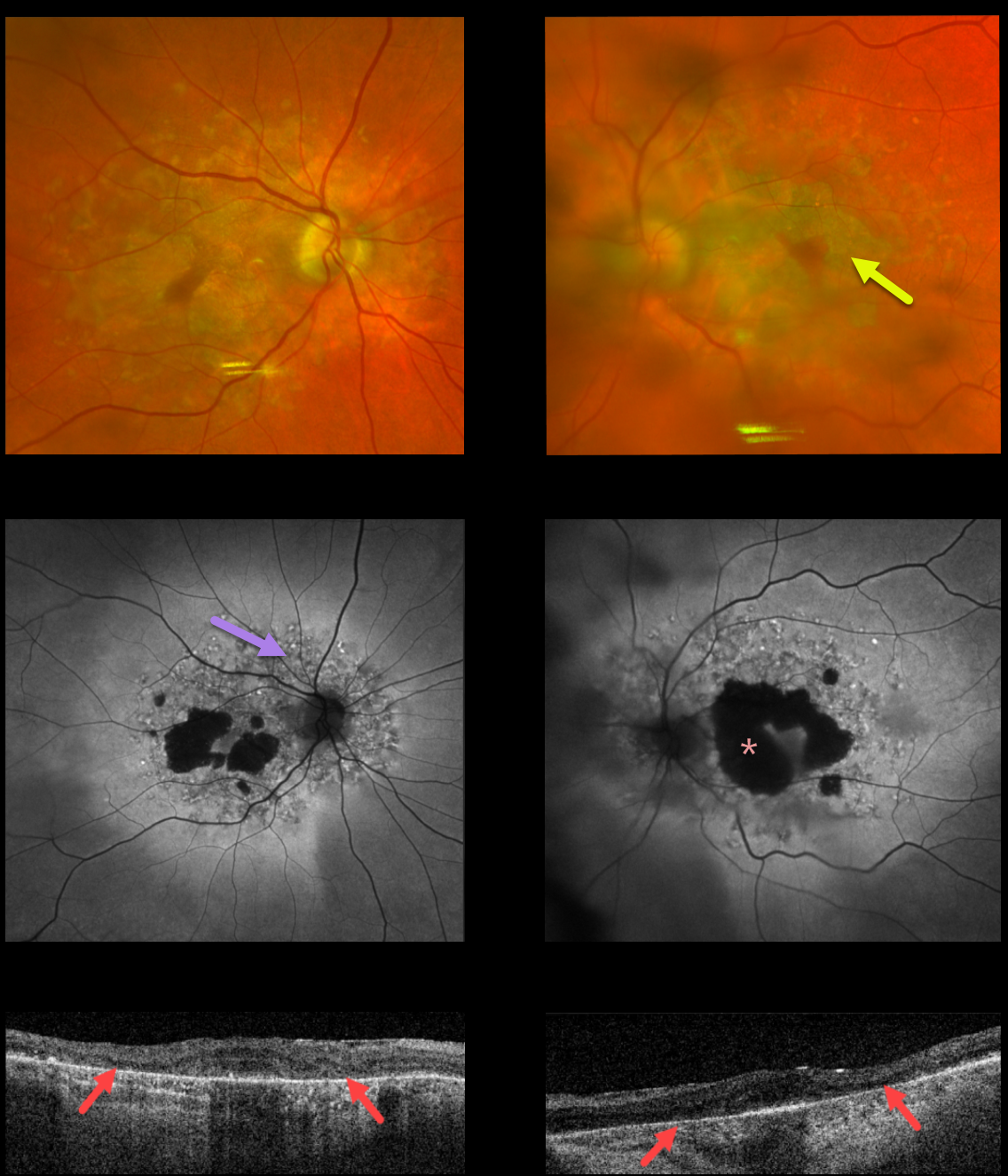
This 75YO male presented with 20/80 vision OU. Optos color imaging shows bilateral, relatively confluent patchy areas of macular atrophy.
Fundus autofluorescence (FAF) shows hypo-FAF within the areas of atrophy, with more extensive peripapillary and macular variably hyper-FAF spots.
Triton swept-source OCT shows abnormal macular architecture throughout, with the most pronounced outer retinal atrophy nasally and temporally within the areas of clinical atrophy.
Our patient has non-insulin-dependent type 2 diabetes, but there are no hearing abnormalities. MIDD patients need to be screened for other potential co-existing medical conditions, including cardiac arrhythmias, myopathy, and renal disease.
Learning Points:
Maternally inherited diabetes and deafness (MIDD) accounts for up to 3% of all cases of diabetes and results from a mutation in mitochondrial DNA at position A3243G.
MIDD often masquerades as a pattern macular dystrophy. In our experience, fundus autofluorescence (FAF) is the best way to visualize these changes. The FAF appearance somewhat resembles that seen with Elmiron toxicity.
Peripapillary hypoautofluorescence, more densely packed macular autofluorescent changes, and earlier central macular involvement suggest Elmiron toxicity over other causes (see Barnes et al., Ophthalmology Retina 2020;4:1196-1201). Note that our case spares the central macula and peripapillary retina, which is more consistent with MIDD.
MATERNALLY INHERITED DIABETES AND DEAFNESS (MIDD)
Originally posted on @retina.rocks October 19, 2021
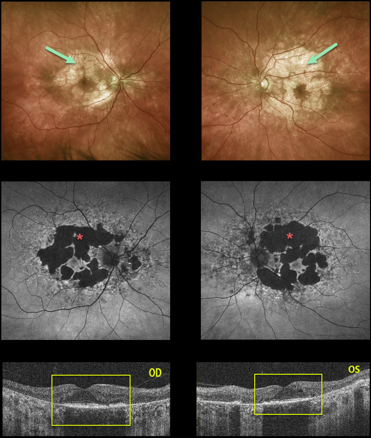
This 55YO male presented with 20/50 vision OD and 20/100 vision OS. Optos color imaging shows bilateral areas of relatively confluent patchy macular atrophy.
The RPE changes are much more pronounced on fundus autofluorescence. OCT scanning shows outer retinal and RPE loss within the areas of clinical atrophy.
Learning Points:
Maternally inherited diabetes and deafness (MIDD) accounts for up to 3% of all cases of diabetes and results from a mutation in mitochondrial DNA at position A3243G. MIDD often masquerades as a pattern macular dystrophy.
In our experience, fundus autofluorescence (FAF) is the best way to visualize these changes. The FAF appearance somewhat resembles that seen with Elmiron toxicity. Peripapillary hypoautofluorescence, more densely packed macular autofluorescent changes, and earlier central macular involvement suggest PPS toxicity over other causes (see Barnes et al Ophthalmology Retina 2020;4:1196-1201).
Note that our case spares the central macula and peripapillary retina, which is more consistent with MIDD.
MIDD patients need to be screened for other potential co-existing medical conditions, including cardiac arrhythmias, myopathy, and renal disease.
MATERNALLY INHERITED DIABETES AND DEAFNESS (MIDD)
Originally posted on @retina.rocks December 4, 2020
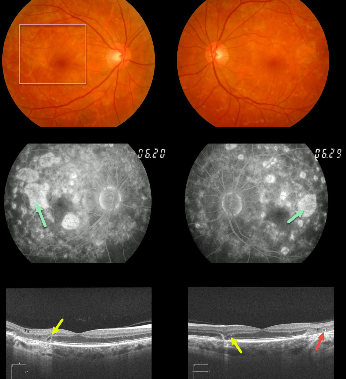
This 62yo female with a history of type 2 diabetes and mild hearing loss initially presented in 2012 with asymptomatic 20/30 vision OU and bilateral areas of focal macular atrophy and subretinal yellow fleck-like lesions.
The retinal pigment epithelial changes were much more pronounced on fluorescein angiography. OCT scanning shows classic wedge defects along the border of the macular atrophy as well as an area of outer retinal tubulation in the left eye.
Learning Points:
Maternally inherited diabetes and deafness (MIDD) accounts for up to 3% of all cases of diabetes and results from a mutation in mitochondrial DNA at position A3243G. MIDD often masquerades as a pattern macular dystrophy.
Fundus autofluorescence (FAF), in our experience, is the best way to visualize these changes, but it was not available at the office when we initially saw her. MIDD patients need to be screened for other potential co-existing medical conditions, including cardiac arrhythmias, myopathy, and renal disease.


