

Melanocytoma
<< Back to Cases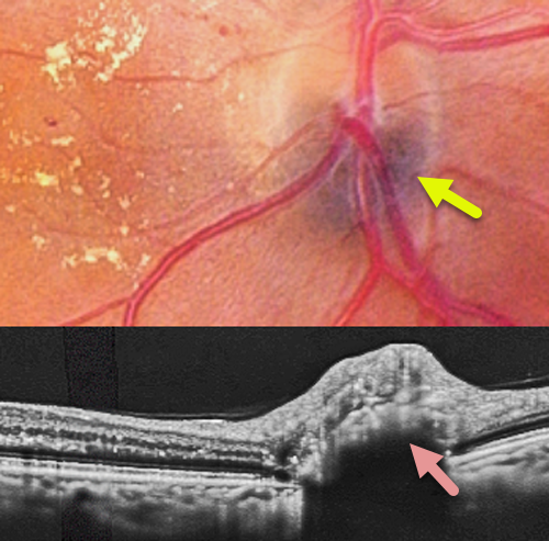
MELANOCYTOMA
Abhishek Karra and Ravindra Karra
Originally posted on @retina.rocks August 24, 2026
This 62YO female with type 2 diabetes was referred to us for diabetic retinopathy screening. Vision was 20/40 OD.
Color photography shows moderate non-proliferative diabetic retinopathy with nasal lipid exudation. The inferior portion of the optic disc has a black discoloration deep to the vessels. OCT scanning through this lesion shows a dome-shaped hyperreflective mass with posterior shadowing.
Learning Points:
Melanocytomas are benign melanocytic tumors intrinsic to the optic nerve. They are intensely pigmented with feathery margins, sometimes spreading into the adjacent retina and choroid. A retinal component, as seen in our patient, is found in about 30% of cases (Shields et al, Ophthalmology 2004;111:1739-1746). Although they may enlarge over time, malignant transformation is rare. Spontaneous necrosis may present with a similar appearance to vitreous seeding from melanoma. Vision and visual field loss can rarely occur from compressive optic neuropathy.

MELANOCYTOMA
Ayushi Gupta and Vishal Agrawal
Originally posted on @retina.rocks July 17, 2026
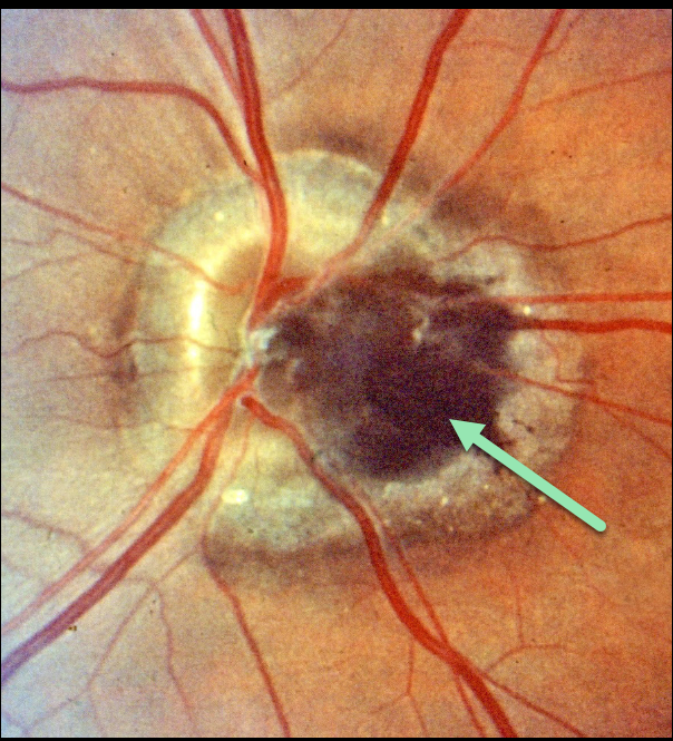
This 42YO female presented without complaint. Vision was 20/20 in her normal OD and 20/20 OS. Color fundus photography of the left eye reveals a well-defined, densely pigmented, jet-black lesion arising from the optic disc and extending slightly into the peripapillary retina. On OCT, the elevated lesion is hyperreflective with complete posterior shadowing.
Learning Points:
Melanocytomas are benign melanocytic tumors intrinsic to the optic nerve. They are intensely pigmented with feathery margins, sometimes spreading into the adjacent retina and choroid. A retinal component, as seen in our patient, is found in about 30% of cases (Shields et al, Ophthalmology 2004;111:1739-1746). Although they may enlarge over time, malignant transformation is rare. Spontaneous necrosis may present with a similar appearance to vitreous seeding from melanoma. Vision and visual field loss can rarely occur from compressive optic neuropathy.
MELANOCYTOMA
Originally posted on @retina.rocks June 22, 2022
This is just a great image of a melanocytoma! Unfortunately, we don’t have any clinical information for this patient.
Learning Points:
Melanocytomas are benign melanocytic tumors intrinsic to the optic nerve. They are intensely pigmented with feathery margins, sometimes spreading into the adjacent retina and choroid.
Although they may enlarge over time, malignant transformation is very rare. Spontaneous necrosis may present with a similar appearance to vitreous seeding from melanoma. Vision and visual field loss can rarely occur from compressive optic neuropathy.
MELANOCYTOMA
Originally posted on @retina.rocks October 15, 2021
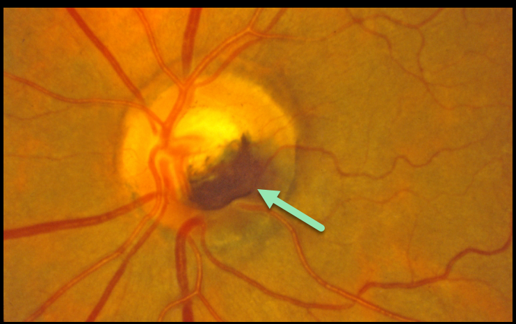
This patient has a melanocytoma, which is a benign melanocytic tumor intrinsic to the optic nerve.
They are intensely pigmented with feathery margins, sometimes spreading into the adjacent retina and choroid.
Learning Points:
Although they may enlarge over time, malignant transformation is very rare. Spontaneous necrosis may present with a similar appearance to vitreous seeding from melanoma. Decreased vision and visual field loss can rarely occur from compressive optic neuropathy.
MELANOCYTOMA
Originally posted on @retina.rocks November 12, 2020
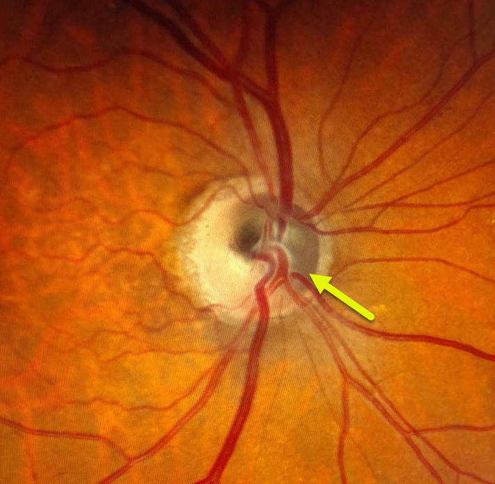
This patient has a melanocytoma, which is a benign melanocytic tumor intrinsic to the optic nerve. There is pigmentation noted within the superonasal portion of the optic disc.
Learning Points:
Melanocytomas can sometimes enlarge and spread into the adjacent retina and choroid, and can very rarely undergo malignant transformation.
Most patients are asymptomatic, but some can develop visual field loss from compressive optic neuropathy.
These lesions should be periodically monitored for any signs of growth.
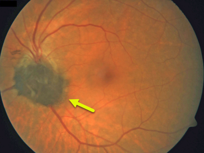
MELANOCYTOMA
Originally posted on @retina.rocks April 15, 2020
This patient has a melanocytoma, which is a benign melanocytic tumor intrinsic to the optic nerve.
They are intensely pigmented with feathery margins, sometimes spreading into the adjacent retina and choroid.
Learning Points:
Although melanocytomas may enlarge over time, malignant transformation is very rare. Spontaneous necrosis may cause a similar appearance to vitreous seeding from a melanoma.
Vision and visual field loss can rarely occur from compressive optic neuropathy.


