

Methamphetamine Toxicity
<< Back to Cases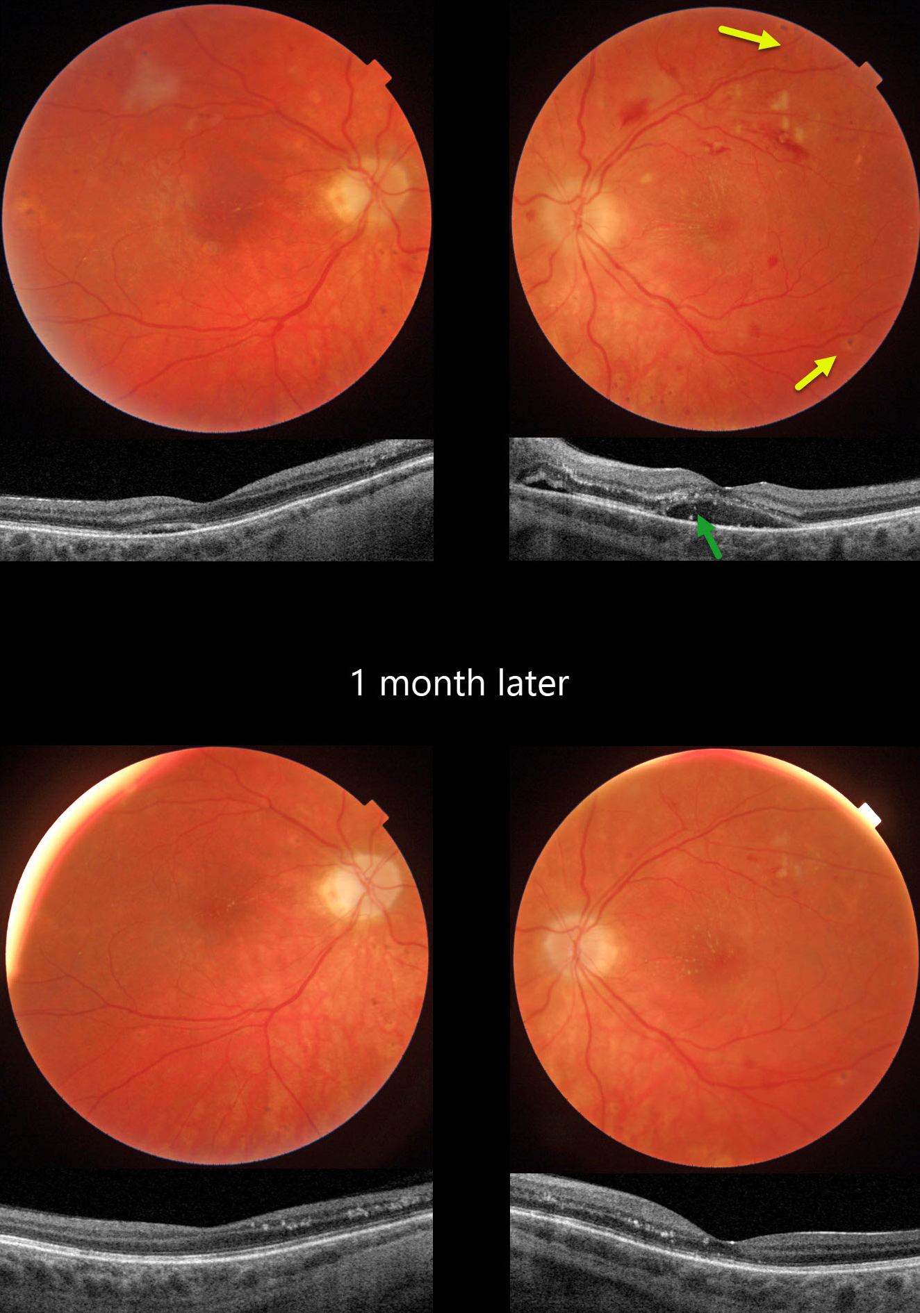
HYPERTENSIVE RETINOPATHY FROM METHAMPHETAMINE
Originally posted on @retina.rocks May 22, 2025
This 21YO female presented with bilateral vision loss. She was admitted to the hospital 2 weeks earlier for malignant hypertension (HTN), likely from methamphetamine abuse. Blood pressure at that time was 292/208. Vision was hand motion OD and 20/80 OS.
Triton color photography of her left eye shows a somewhat pale and swollen nerve, mostly inner retinal hemorrhages, and resolving nerve fiber layer infarcts (cotton-wool spots, CWS). Faint lipid exudates radiate nasally and superiorly from the macular center. Multifocal small hyperpigmented choroidal lesions are noted outside the arcades. Swept-source OCT shows subretinal fluid with hyperreflective lipid in the subretinal space and outer nuclear layer. Similar but much milder findings are noted in her right eye.
When examined 1 month later, vision remained at hand motion OD and improved to 20/40 OS with improved funduscopic and OCT findings.
Learning Points:
Malignant hypertension, defined as blood pressure above 180 systolic and/or 120 diastolic, is a life-threatening medical emergency. Eye doctors are in a unique position to often diagnose this condition. Patients will often present with bilateral optic nerve swelling, flame-shaped retinal hemorrhages, CWS, and, with more chronic disease, lipid precipitates in the nasal macular outer plexiform layer.
Acute hypertensive choroidopathy outside the setting of pre-eclampsia, in our experience, is quite rare. Independent of the disc and neurosensory retinal findings, patients present with localized multifocal serous retinal detachments (de Venecia and Jampol, Arch Ophthalmol 1984;102:68-73). With blood pressure control, these resolve often with minimal funduscopic changes. Focal (Elschnig spots) or linear (Siegrist streaks) choroidal pigmentary changes may result from more severe disease.
METHAMPHETAMINE TOXICITY
Originally posted on @retina.rocks January 28, 2020
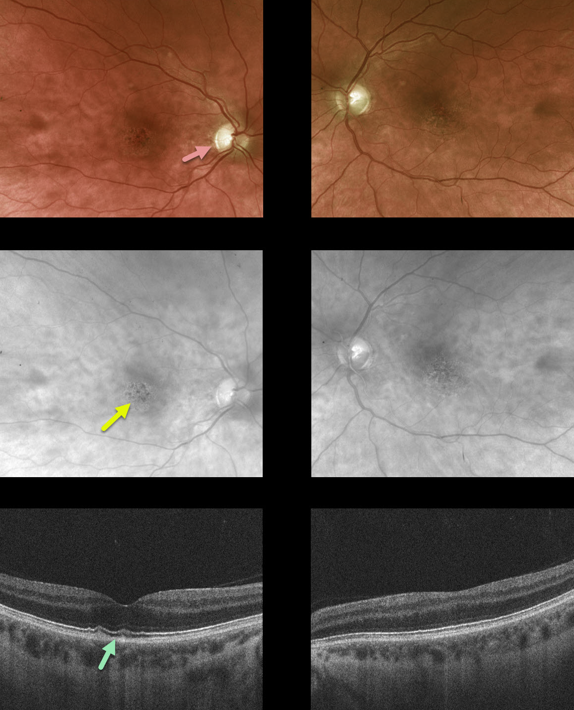
This former methamphetamine and heroin addict presented with 20/50 vision OU, symmetric foveal RPE mottling, mild optic atrophy and some thickening and hyper-reflectivity of the foveal outer segments on OCT.
Learning Points:
Retinal changes that occur due to methamphetamine use include hemorrhages, cotton wool spots and vasculitis. We presume the findings in this patient were a sequela of prior toxicity.
Similar findings can be found in long-term dextroamphetamine/amphetamine (Adderall) users.


