

Morning Glory Syndrome
<< Back to Cases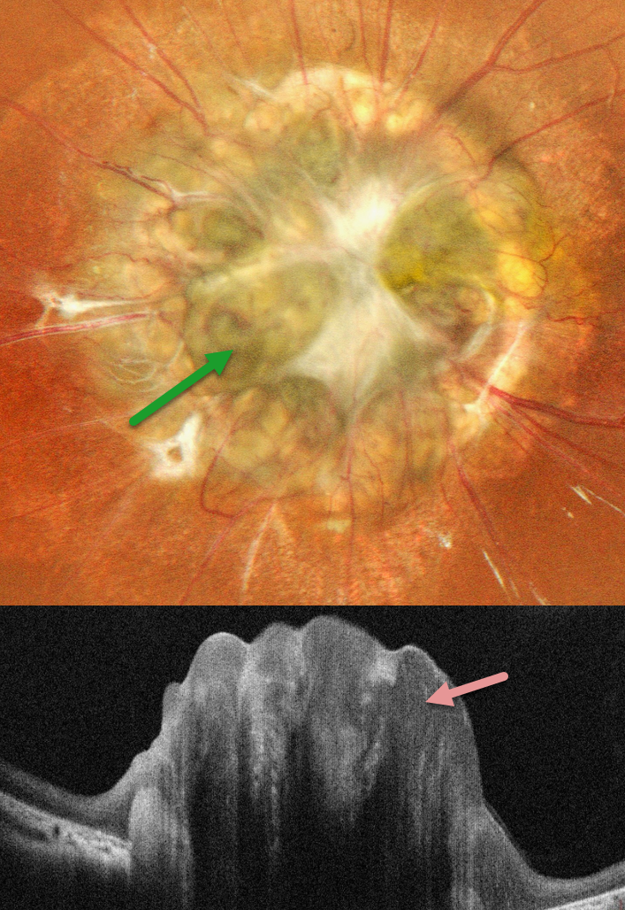
MORNING GLORY DISC WITH EXTENSIVE GLIAL PROLIFERATION OBSCURING DISC EXCAVATION
Shraddha Raj Shrivastava and Manish Nagpal
Originally posted on @retina.rocks September 11, 2026
This healthy 8YO female was born blind in her left eye. Vision was 20/20 in her normal OD and no light perception OS.
Pseudocolor SLO imaging shows a markedly enlarged optic disc with surrounding chorioretinal scarring. Numerous retinal vessels extend outwards from the disc, and extensive fibrotic tissue obscures the disc details. This tissue is markedly elevated and hyperreflective on OCT scanning.
Learning Points:
Morning glory syndrome is a typically unilateral congenital anomalous disc malformation that presents with an enlarged, colobomatous-appearing disc with an increased number of retinal vessels that radiate outwards. The center of the nerve is typically covered by glial tissue. In our patient’s case, this tissue is so extensive that it obscures the disc coloboma.
This abnormality is named for its striking resemblance to the Morning Glory flower. Visual prognosis is usually poor. The condition can be associated with serous macular detachment. Neuroimaging should be considered to rule out transsphenoidal encephalocele and intracranial carotid artery dysgenesis. Fortunately, our patient’s neurologic imaging was negative.
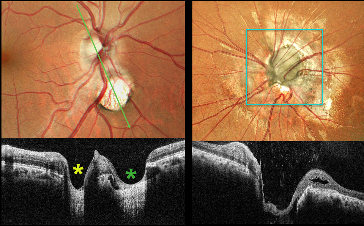
MORNING GLORY DISC AND CHOROIDAL COLOBOMA WITH PSEUDODUPLICATED OPTIC NERVE
Shraddha Raj Shrivastava and Manish Nagpal
Originally posted on @retina.rocks June 30, 2026
This 26YO male presented with a history of stable poor vision and inward deviation of his left eye since childhood. Vision was 20/20 OD and counting fingers in his esotropic, microphthalmic OS.
Color photography of the right posterior pole shows a round coloboma inferonasal to the nerve, giving the appearance of a pseudo-duplicated disc. OCT scanning through the nerve and coloboma reinforces the illusion of a second nerve head. The anomalous left nerve is enlarged and excavated, with numerous vessels radiating outwards from its inferonasal aspect.
Morning glory syndrome is typically unilateral and shares some similarities with optic disc coloboma and juxtapapillary staphyloma. This abnormality is named for its striking resemblance to the Morning Glory flower. Visual prognosis is usually poor. The condition can be associated with serous macular detachment. Neuroimaging should be considered to rule out transsphenoidal encephalocele and intracranial carotid artery dysgenesis, and fortunately, our patient’s imaging was negative. For a great review on the pathogenesis and treatment of maculopathy associated with cavitary optic disc anomalies, see Jain and Johnson, AJO 2014;158:423-435.
The coexistence of morning glory disc anomaly with microphthalmos, and contralateral chorioretinal coloboma in this case, may represent a continuum of optic fissure developmental defects with asymmetric expression, leading to a spectrum of cavitary optic disc anomalies in the same individual.
Pseudoduplication of the optic nerve occurs when a round choroidal coloboma or chorioretinal scar about the size of the optic nerve is associated with overlying radiating blood vessels (Bloom et al, Retinal Cases 2022;16:174-176). In our experience, this fake-out is most common from toxoplasmosis scars located near the optic nerve.
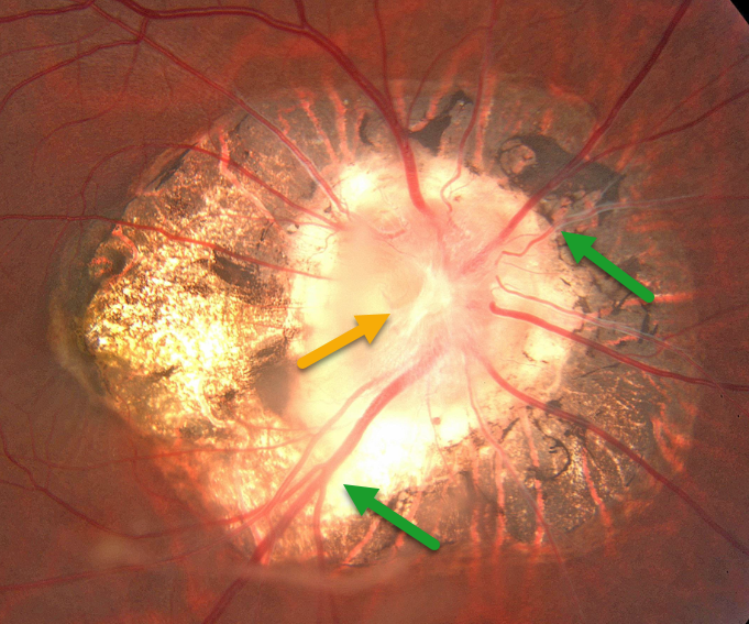
OPTIC NERVE MORNING GLORY
Larissa Cervera-Perez
Originally posted on @retina.rocks July 9, 2024
This 43YO female presented for a routine examination. There was a history of pathological myopia. Vision was 20/70 OD and 20/60 OS.
Optos color imaging shows peripapillary scarring with a morning-glory optic disc, including a large disc, a central glial tissue area, and numerous straight, radiating vessels. Observation was recommended.
Learning Points:
Morning glory syndrome is typically unilateral and shares some similarities with optic disc coloboma and juxtapapillary staphyloma. This abnormality gets its name from its striking resemblance to the Morning Glory flower. Visual prognosis is usually poor. The condition can be associated with serous macular detachment. Neuroimaging should be considered to rule out transsphenoidal encephalocele and intracranial carotid artery dysgenesis.
For a great review on the pathogenesis and treatment of maculopathy associated with cavitary optic disc anomalies, see Jain and Johnson, AJO 2014;158:423-435.
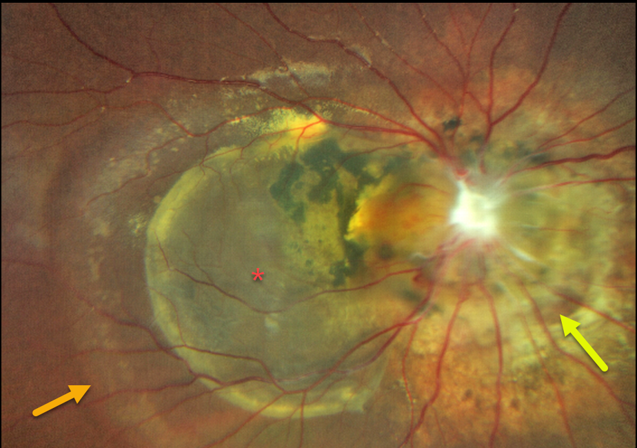
MORNING GLORY + NEUROFIBROMATOSIS TYPE 2
Yuenpang Cheung and Stephanie Choi
Originally posted on @retina.rocks December 12, 2023
This 18YO male, originally from the Dominican Republic, was first seen by an ophthalmologist at 14YO due to strabismus. At that time, he was found to have a significant morning glory anomaly.
Two years later, at age 16, he developed a right-sided facial palsy and, on work-up, was found to have a right optic nerve sheath meningioma. He was then genetically confirmed to have neurofibromatosis 2 (NF-2).
There was an unclear treatment history, and he recently presented to our clinic to establish care. Vision was no light perception in his right eye and 20/25 in his normal left eye.
Optos color RG imaging shows a morning glory disc with peripapillary chorioretinal scarring, an associated chronic retinal detachment, and a large macular cyst.
Learning Points:
Neurofibromatosis type 2 (NF2) can arise spontaneously or be inherited as an autosomal dominant mutation in the NF2 gene located at chromosome 22q12.2.
The hallmark sign of NF2 is bilateral vestibular schwannomas (or acoustic neuromas). Other manifestations of NF2 include cranial meningioma, spinal ependymomas, and peripheral nerve tumors. The most common ocular finding is a posterior subcapsular cataract in young patients.
Less common findings include optic nerve meningiomas, papilledema, strabismus, epiretinal membrane, and combined hamartoma of the retina and RPE.
OPTIC NERVE MORNING GLORY
Originally posted on @retina.rocks July 29, 2022
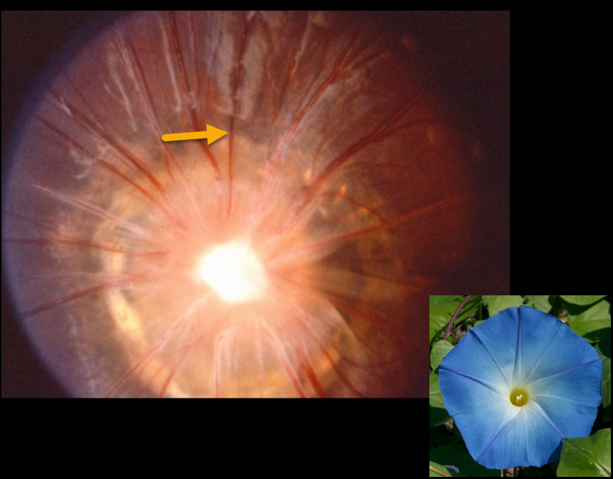
This patient has a classic morning glory optic disc, including a large disc, an area of central glial tissue, and numerous straight radiating vessels.
Learning Points:
Morning glory syndrome is typically unilateral and shares some similarities with optic disc coloboma and juxtapapillary staphyloma. This abnormality gets its name from its striking resemblance to the Morning glory flower. Visual prognosis is usually poor. The condition can be associated with serous macular detachment.
Neuroimaging should be considered to rule out transsphenoidal encephalocele and intracranial carotid artery dysgenesis.
For a great review on the pathogenesis and treatment of maculopathy associated with cavitary optic disc anomalies, see Jain and Johnson, AJO 2014;158:423-435.
OPTIC NERVE MORNING GLORY
Originally posted on @retina.rocks July 15, 2021
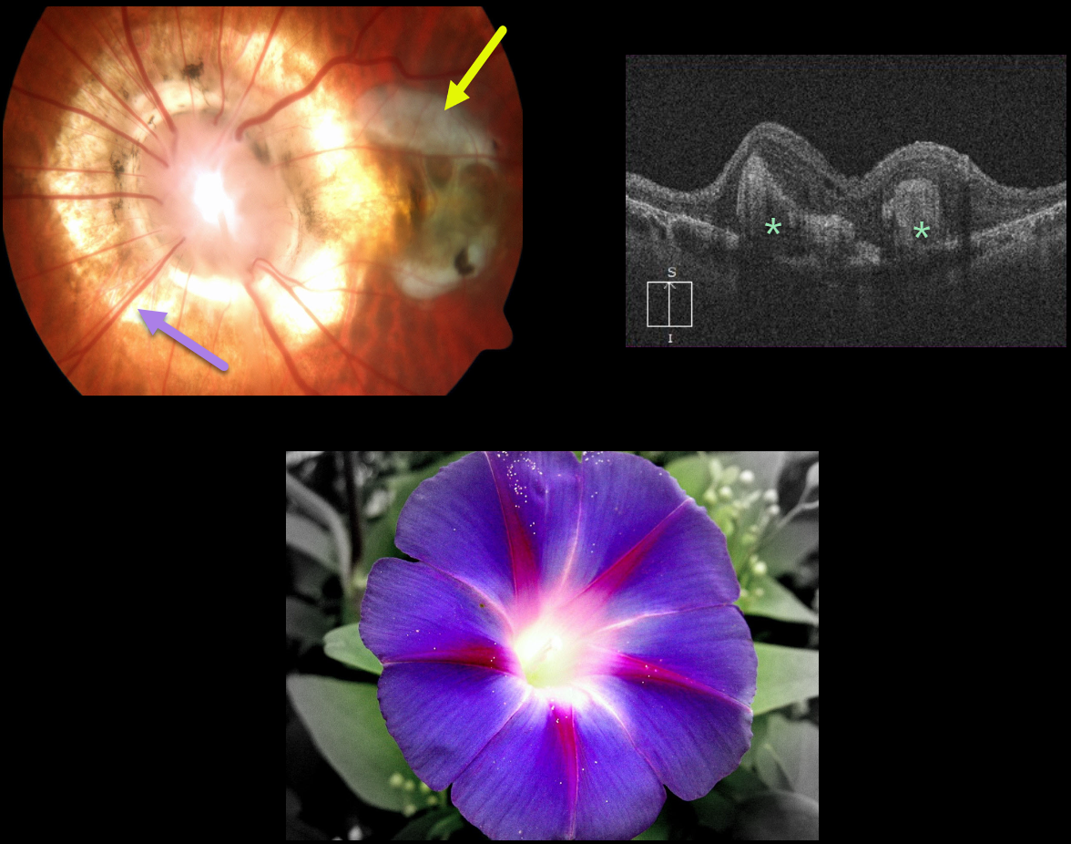
This patient has a unilateral morning glory disc in their left eye with an associated fibrotic disciform scar. OCT scanning shows variable subretinal macular scarring.
Learning Points:
Morning glory syndrome is typically unilateral and shares some similarities with optic disc coloboma and juxtapapillary staphyloma. This abnormality is named for its striking resemblance to the morning glory flower.
Visual prognosis is usually poor. The condition can be associated with serous macular detachment. Neuroimaging should be considered to rule out transsphenoidal encephalocele and intracranial carotid artery dysgenesis.
For a great review on the pathogenesis and treatment of maculopathy associated with cavitary optic disc anomalies, see Jain and Johnson, AJO 2014;158:423-435.
OPTIC NERVE MORNING GLORY
Originally posted on @retina.rocks December 28, 2020
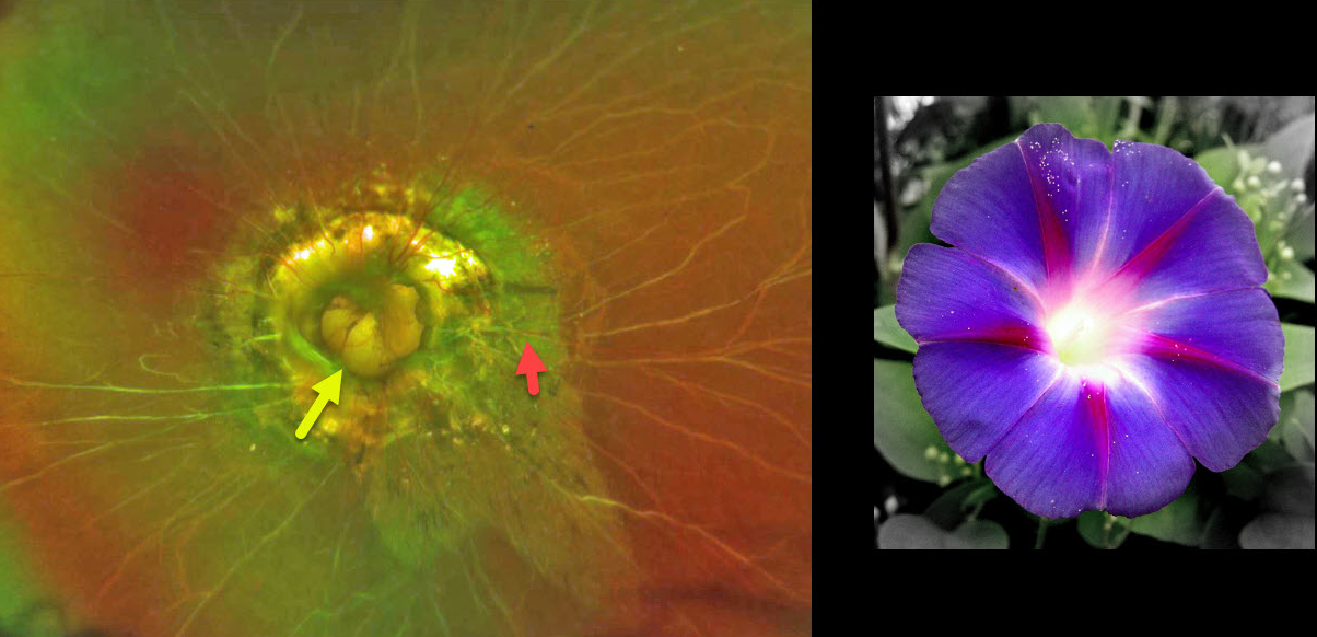
This patient has morning glory syndrome. The left optic nerve appears to have a large optic disc excavation with radial vessels, central glial tissue, and peripapillary atrophy (PPA) and pigmentation.
Visual prognosis is usually poor.
Learning Points:
The morning glory syndrome is typically unilateral and shares some similarities with optic disc coloboma and juxtapapillary staphyloma.
The condition is often associated with serous macular detachment. Neuroimaging should be considered to rule out transsphenoidal encephalocele and intracranial carotid artery dysgenesis.
OPTIC NERVE MORNING GLORY
Originally posted on @retina.rocks June 5, 2020
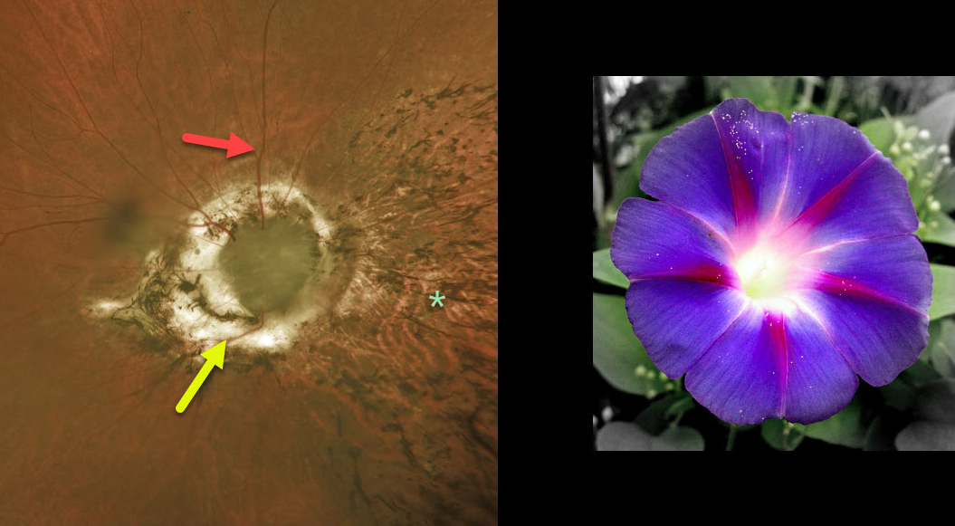
This 66yo male is CF OD and 20/200 OS. He has a history of retinal detachment surgery OD, as well as severe glaucoma.
His right eye has a possible morning glory anomaly (no central vessels with possible overlying glial tissue, radiating numerous retinal vessels) versus severe glaucomatous damage.
The intraretinal pigment migration nasally is from the prior detachment. Although he had prior retinal reattachment surgery (presumed from a retinal break), this may also have been from subretinal fluid that is occasionally seen with these anomalous optic nerves.
Learning Points:
The morning glory syndrome is typically unilateral and shares some similarities with optic disc coloboma and juxtapapillary staphyloma. This syndrome is appropriately named after the Morning Glory flower.
The condition is often associated with serous macular detachment.
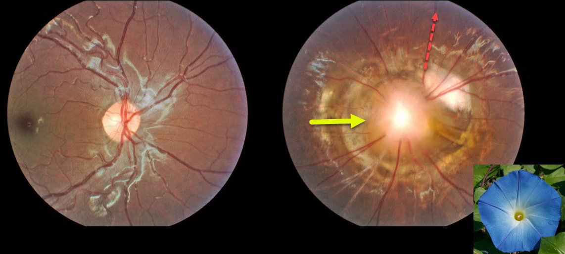
OPTIC NERVE MORNING GLORY
Originally posted on @retina.rocks February 13, 2020
This patient has morning glory syndrome, and the left optic nerve appears to have large optic disc excavation with radial vessels, central glial tissue and peripapillary atrophy and pigmentation.
Visual prognosis is usually poor.
Learning Points:
The morning glory syndrome is typically unilateral and shares some similarities with optic disc coloboma and juxtapapillary staphyloma.
The condition is often associated with serous macular detachment. Neuroimaging should be considered to rule out transsphenoidal encephalocele and intracranial carotid artery dysgenesis.


