

Multifocal Choroiditis
<< Back to Cases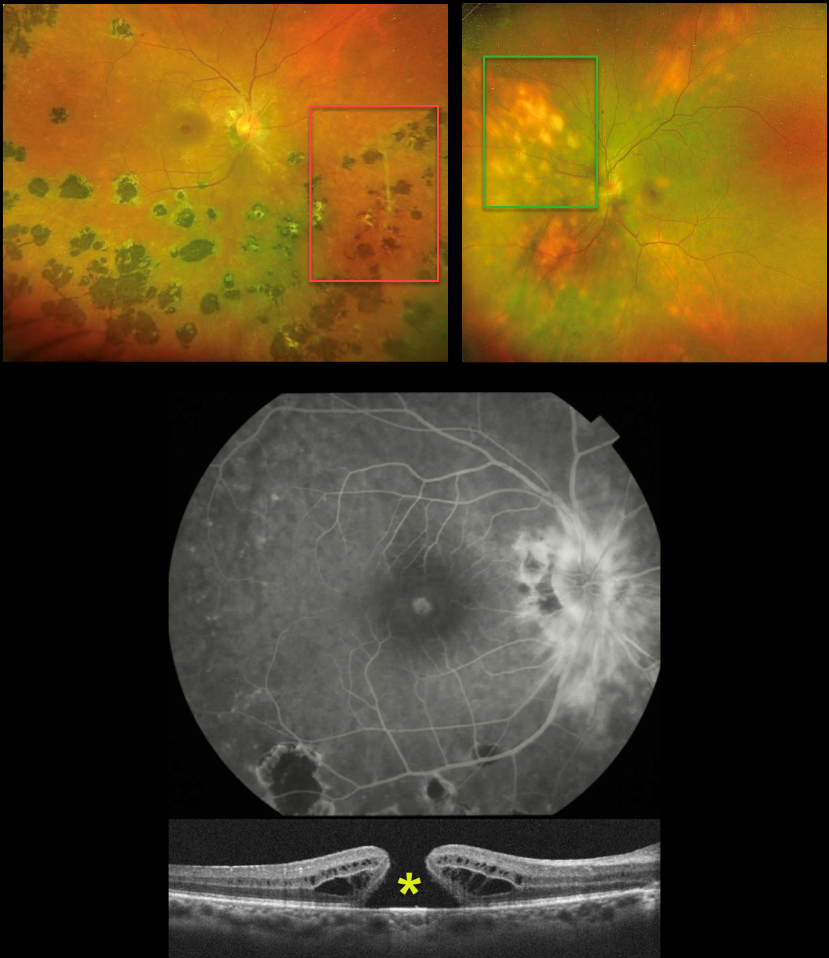
MULTIFOCAL CHOROIDITIS VS BIRDSHOT
Seif Anwar
Originally posted on @retina.rocks June 17, 2026
This healthy 41YO female presented with 6 months of bilateral vision loss. Vision was 20/240 OD and 20/200 OS.
Optos color RG imaging OD shows peripapillary scarring, multifocal large hyperpigmented chorioretinal scars scattered inferiorly and temporally, and some vascular sheathing. A full-thickness macular hole is noted and confirmed on OCT. Fluorescein angiography shows peripapillary leakage.
Her left eye’s Optos shows markedly different findings, with multifocal, creamy-white, depigmented lesions in a pattern radiating away from the disc.
Medical workup was negative, including HLA-A29, QuantiFERON TB Gold, chest X-ray, and serologies for toxoplasmosis and CMV.
Learning Points:
Birdshot chorioretinopathy was appropriately described and named by Drs. Ryan and Maumenee (Am J Ophthlamol 1980;89:31-45) since the lesions look like shotgun birdshot scattered throughout the fundus. Birdshot is typically chronic and bilateral, with about 90-95% of patients being HLA-A29 positive and usually affects women in their 40s to 60s.
Multifocal choroiditis (MFC) is the current preferred term for a group of historically heterogeneous disorders, including multifocal choroiditis, recurrent multifocal choroiditis, multifocal choroiditis and panuveitis, punctate inner choroidopathy, progressive subretinal fibrosis, and pseudo-histoplasmosis (Essex et al, Retina 2013;33:1-4 and Spaide et al, Retina 2013;33:1315-1324). MFC occurs in otherwise healthy adults, often myopic and female. Findings include variably pigmented multifocal scars throughout the fundus, panuveitis, subretinal fibrosis, and macular neovascularization (MNV).
Our patient’s diagnosis is unclear because her right eye shows features suggestive of MFC, while her left eye is fairly pathognomonic for birdshot. In our experience, birdshot lesions never become hyperpigmented, as in our patient’s right eye, although the lesions in her left eye are virtually pathognomic for birdshot.
Following several Remicade treatments, vision remained stable at 20/240 OD and improved to 20/50 OS. We continue to follow her closely.
What’s your diagnosis???
MULTIFOCAL CHOROIDITIS WITH MACULAR NEOVASCULARIZATION
César Adrián Gómez Valdivia
Originally posted on @retina.rocks May 25, 2026
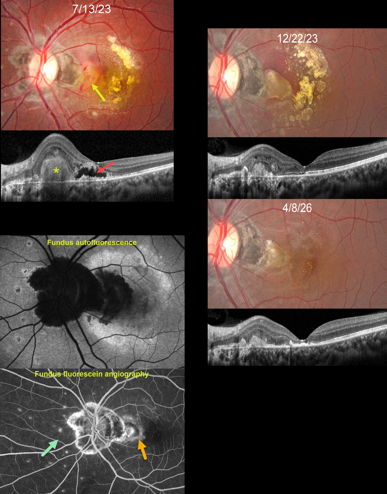
This 21YO female presented with a gradually progressive central scotoma in her left eye for the prior 2 months. Vision was 20/20 in her normal OD and 20/1600 OS.
Fundus photography shows a foveal subretinal fibrotic macular neovascularization (MNV) with surrounding subretinal blood and variably confluent subretinal lipid noted more temporally. Some inner retinal hemorrhages are also noted. Variably pigmented peripapillary scarring is present with multifocal small, depigmented scars nasally. OCT scanning shows a hyperreflective subretinal mound corresponding to the area of subretinal fibrosis. More centrally, there is a fluid-filled pocket of outer retinal hyporeflectivity surrounded by a ragged, variably thickened border of hyperreflective material. Fundus autofluorescence (FAF) shows peripapillary hypo-FAF as does the subretinal fibrosis with surrounding blood. The macula otherwise shows variable hyper-FAF, likely due to unmasking from outer retinal atrophy. The MNV stains angiographically. Multifocal scars stain superiorly, nasally, and inferiorly.
Following 3 monthly injections of Eylea, vision improved to 20/50. The MNV is contracted into an oval-shaped subretinal lesion with surrounding fading subretinal blood and absorbing lipid. She was then lost to follow-up for over 2 years. Thankfully, the MNV showed further contraction, with faint, scattered residual lipid dots. Despite outer retinal atrophy on OCT, vision improved to 20/25.
Learning Points:
Multifocal choroiditis (MFC) is the current preferred term for a group of historically heterogeneous disorders, including multifocal choroiditis, recurrent multifocal choroiditis, multifocal choroiditis and panuveitis, punctate inner choroidopathy, progressive subretinal fibrosis, and pseudo-histoplasmosis (Essex et al, Retina 2013;33:1-4 and Spaide et al, Retina 2013;33:1315-1324).
MFC occurs in otherwise healthy adults, often myopic and female. Findings include variably pigmented multifocal scars throughout the fundus, panuveitis, subretinal fibrosis, and MNV. Unlike ocular histoplasmosis, new scars and uveitis can be seen. MNV can be successfully managed with anti-VEGF therapy.
Our patients’ MNV and peripheral scars most likely represent MFC. We continue to follow her closely
MULTIPLE EVANESCENT WHITE DOT SYNDROME (MEWDS )+ MFC OR POHS
Mattie Adams
Originally posted on @retina.rocks April 16, 2025
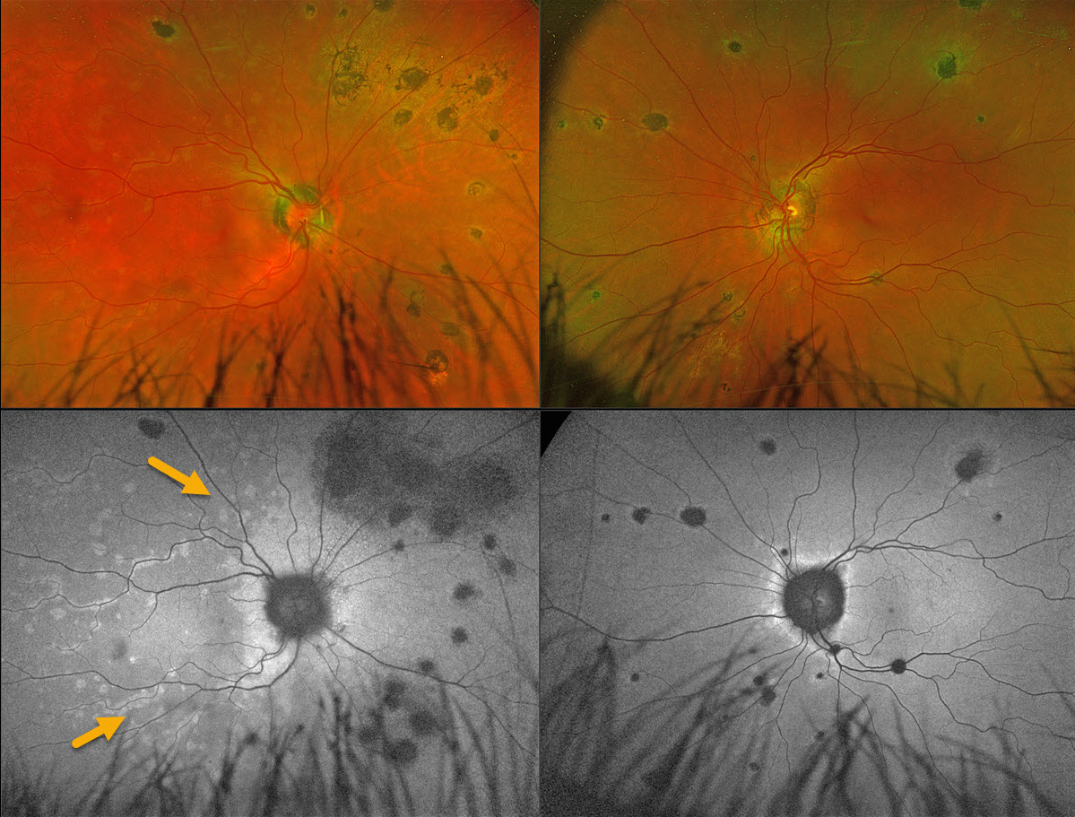
This 56YO female presented with 1 week of flashes and blurred vision in her right eye. Vision was 20/25 OD and 20/20 OS.
Coarse, large pigmented multifocal scars with peripapillary scarring are noted OU. Multiple deep white retinal spots are scattered throughout the right posterior pole. These spots on fundus autofluorescence (FAF) hyper-autofluoresce. Confluent hyper-FAF extends outwards from the optic nerve.
Learning Points:
Multiple evanescent white dot syndrome (MEWDS) is a usually unilateral, idiopathic inflammation of the outer retina that typically occurs in young, healthy myopic females. Acutely, patients will also have an enlarged blind spot, often complaining of temporal visual field loss, due to the peripapillary inflammation. The white dots disappear within a week or two, which is why the lesions are called “evanescent”.
Multifocal choroiditis (MFC) is the current preferred term for a group of historically heterogeneous disorders, including multifocal choroiditis, recurrent multifocal choroiditis, multifocal choroiditis and panuveitis, punctate inner choroidopathy, progressive subretinal fibrosis, and pseudo-histoplasmosis (Essex et al, Retina 2013;33:1-4 and Spaide et al, Retina 2013;33:1315-1324). MFC occurs in otherwise healthy adults, often myopic and female. Findings include variably pigmented multifocal scars throughout the fundus, panuveitis, subretinal fibrosis, and macular neovascularization (MNV). Unlike ocular histoplasmosis, new scars and uveitis can be seen. MNV can be successfully managed with anti-VEGF therapy.
Our patient’s scars represent either preexisting MFC or ocular histoplasmosis, and MEWDS is sometimes found in some of these eyes. Distinct from primary MEWDS, secondary MEWDS may be an epiphenomenon in inflammatory conditions such as MFC that disrupt the choriocapillaris-Bruch membrane-RPE complex (Essilfie et al, Retina 2022;42:227-235).
HODGKIN LYMPHOMA MIMICKING MULTIFOCAL CHOROIDITIS
Originally posted on @retina.rocks December 4, 2024
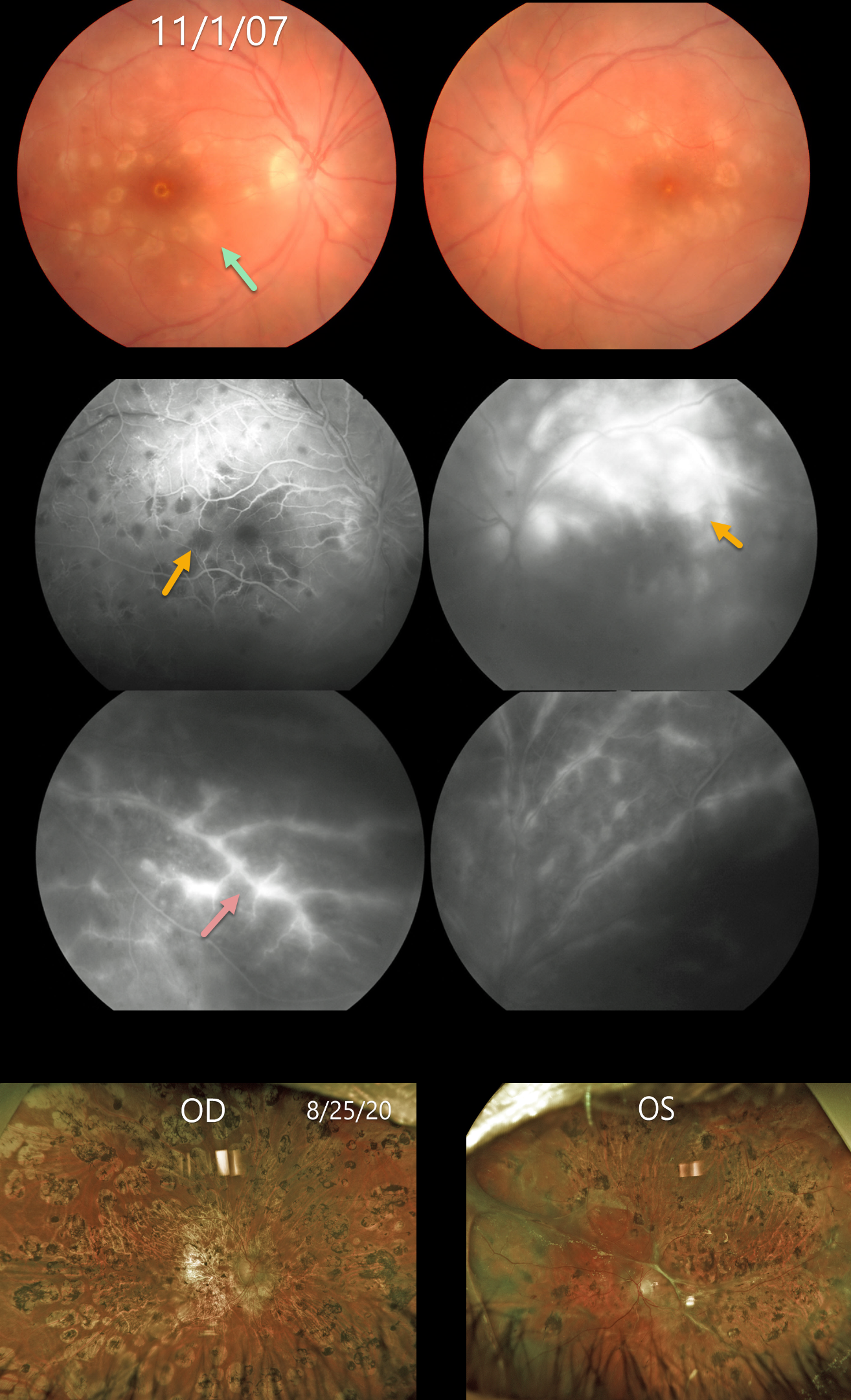
This 18YO female presented on 11/1/07 with acute bilateral panuveitis. Moderate vitreous cells were present along with subretinal multifocal yellow-white choroidal infiltrates. Angiographically, these choroidal lesions blocked early and stained late. A frosted-branch pattern of vascular leakage was noted bilaterally. Following a course of oral prednisone, the acute uveitic findings resolved, leaving behind inactive multifocal scarring. She developed severe recurrent panuveitis a year or two later, leaving her with 20/400 vision bilaterally (images not shown).
Shortly thereafter, she was diagnosed with Hodgkin lymphoma. At almost the exact date of this diagnosis, Baillif et al reported a patient with bilateral angle-closure glaucoma and multifocal choroiditis as the initial presentation for Hodgkin lymphoma (Retinal Cases 2011;5:201-205). The posterior segment findings in this report were virtually identical to those in our patient. The authors felt that the uveitis was a paraneoplastic phenomenon.
She subsequently underwent successful bone marrow transplantation. Her lymphoma and uveitis have both remained in remission since. A rhegmatogenous retinal detachment later developed in her left eye, which was repaired with vitrectomy and silicone oil. Despite successful reattachment, vision subsequently decreased to no light perception. On 8/25/20, vision was 20/400 OD with severe stable bilateral chorioretinal scarring bilaterally.
Learning Points:
Multifocal choroiditis (MFC) is the current preferred term for a group of historically heterogeneous disorders, including multifocal choroiditis, recurrent multifocal choroiditis, multifocal choroiditis and panuveitis, punctate inner choroidopathy, progressive subretinal fibrosis, and pseudo-histoplasmosis (Essex et al, Retina 2013;33:1-4 and Spaide et al, Retina 2013;33:1315-1324). MFC occurs in otherwise healthy adults, often myopic and female. Findings can include variably pigmented multifocal scars throughout the fundus, panuveitis, subretinal fibrosis, and macular neovascularization (MNV). Unlike ocular histoplasmosis, new scars and uveitis can be seen. MNV can be successfully managed with anti-VEGF therapy.
As with any uveitis, the clinician must always include the possibility of a masquerade syndrome as occurred in our patient.
MULTIFOCAL CHOROIDITIS
Originally posted on @retina.rocks August 26, 2022
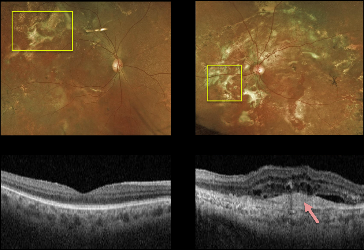
This 58YO male has stable vision of 20/20 OD and light perception OS. Extensive areas of subretinal fibrosis are scattered throughout each fundus, left greater than right.
A hyperreflective subfoveal scar on OCT in his left eye accounts for the poor vision. Fortunately, the right macula is relatively normal, and we are observing him twice yearly.
Learning Points:
Multifocal choroiditis (MFC) is the current preferred term for a group of historically heterogeneous disorders, including multifocal choroiditis, recurrent multifocal choroiditis, multifocal choroiditis and panuveitis, punctate inner choroidopathy, progressive subretinal fibrosis, and pseudo-histoplasmosis (Essex et al, Retina 2013;33:1-4 and Spaide et al, Retina 2013;33:1315-1324).
MFC occurs in otherwise healthy adults, often myopic and female. Findings include variably pigmented multifocal scars throughout the fundus, panuveitis, subretinal fibrosis, and macular neovascularization (MNV). Unlike ocular histoplasmosis, new scars and uveitis can be seen.
MNV can be successfully managed with anti-VEGF therapy.
MULTIFOCAL CHOROIDITIS
Originally posted on @retina.rocks August 6, 2021
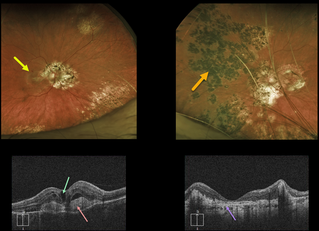
This 57YO female presented with 20/200 vision OD and counting fingers vision OS from bilateral multifocal choroiditis (MFC).
There is a newly symptomatic active macular neovascularization (MNV) in her right eye and an inactive disciform scar in her left eye.
OCT scanning shows intraretinal fluid overlying a hyperreflective MNV in the right macula with variable thickened and atrophic retina in the left macula. The MNV in the right eye is gradually improving with monthly anti-VEGF injections.
Learning Points:
MFC occurs in otherwise healthy adults, often myopic and female. Findings include multifocal variably pigmented scars throughout the posterior pole and periphery.
Unlike ocular histoplasmosis, new scars and uveitis can be seen. MNV can be successfully managed with anti-VEGF therapy.
MULTIFOCAL CHOROIDITIS
Originally posted on @retina.rocks July 16, 2020
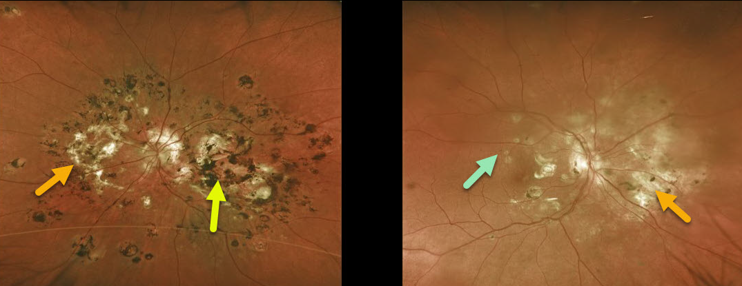
This 59YO female initially had unilateral multifocal choroiditis (MFC) in her left eye. When we started seeing her in the mid-1990s, predating the anti-VEGF era, we battled multiple inflammatory macular neovascularizations (MNV).
Eventually, she developed subfoveal MNV and nasal subretinal fibrosis despite multiple thermal laser treatments, and her vision remains counting fingers OS.
Fourteen years later, she developed asymptomatic hypo and hyperpigmented lesions in her right eye similar to those of her left eye, and she remained 20/20 OD.
Initially treated with intravitreal Avastin for MNV, intravitreal triamcinolone was also added for new macular lesions.
Learning Points:
MFC occurs in otherwise healthy adults, often myopic and female. Findings include multifocal variably pigmented multifocal scars throughout the posterior pole and periphery.
Unlike ocular histoplasmosis, new scars and uveitis can be seen. Punctate inner choroidopathy most likely represents a continuum of MFC rather than a separate disease. MNV can be successfully managed with anti-VEGF therapy.


