

Multiple Evanescent White Dot Syndrome (MEWDS)
<< Back to Cases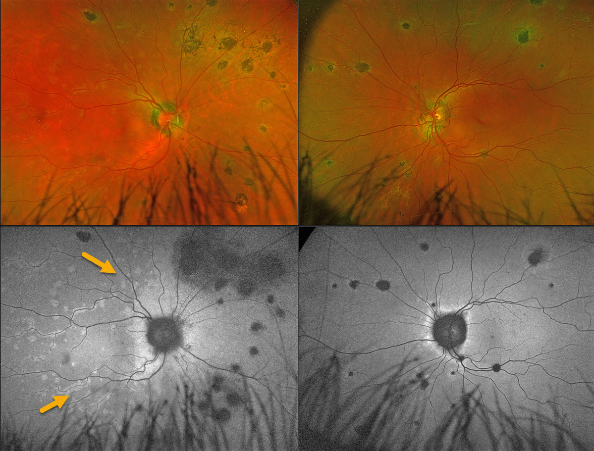
MULTIPLE EVANESCENT WHITE DOT SYNDROME (MEWDS )+ MFC OR POHS
Mattie Adams
Originally posted on @retina.rocks April 16, 2025
This 56YO female presented with 1 week of flashes and blurred vision in her right eye. Vision was 20/25 OD and 20/20 OS.
Coarse, large pigmented multifocal scars with peripapillary scarring are noted OU. Multiple deep white retinal spots are scattered throughout the right posterior pole. These spots on fundus autofluorescence (FAF) hyper-autofluoresce. Confluent hyper-FAF extends outwards from the optic nerve.
Learning Points:
Multiple evanescent white dot syndrome (MEWDS) is a usually unilateral, idiopathic inflammation of the outer retina that typically occurs in young, healthy myopic females. Acutely, patients will also have an enlarged blind spot, often complaining of temporal visual field loss, due to the peripapillary inflammation. The white dots disappear within a week or two, which is why the lesions are called “evanescent”.
Multifocal choroiditis (MFC) is the current preferred term for a group of historically heterogeneous disorders, including multifocal choroiditis, recurrent multifocal choroiditis, multifocal choroiditis and panuveitis, punctate inner choroidopathy, progressive subretinal fibrosis, and pseudo-histoplasmosis (Essex et al, Retina 2013;33:1-4 and Spaide et al, Retina 2013;33:1315-1324). MFC occurs in otherwise healthy adults, often myopic and female. Findings include variably pigmented multifocal scars throughout the fundus, panuveitis, subretinal fibrosis, and macular neovascularization (MNV). Unlike ocular histoplasmosis, new scars and uveitis can be seen. MNV can be successfully managed with anti-VEGF therapy.
Our patient’s scars represent either preexisting MFC or ocular histoplasmosis, and MEWDS is sometimes found in some of these eyes. Distinct from primary MEWDS, secondary MEWDS may be an epiphenomenon in inflammatory conditions such as MFC that disrupt the choriocapillaris-Bruch membrane-RPE complex (Essilfie et al, Retina 2022;42:227-235).
MULTIPLE EVANESCENT WHITE DOT SYNDROME (MEWDS)
Yuenpang Cheung
Originally posted on @retina.rocks February 26, 2025
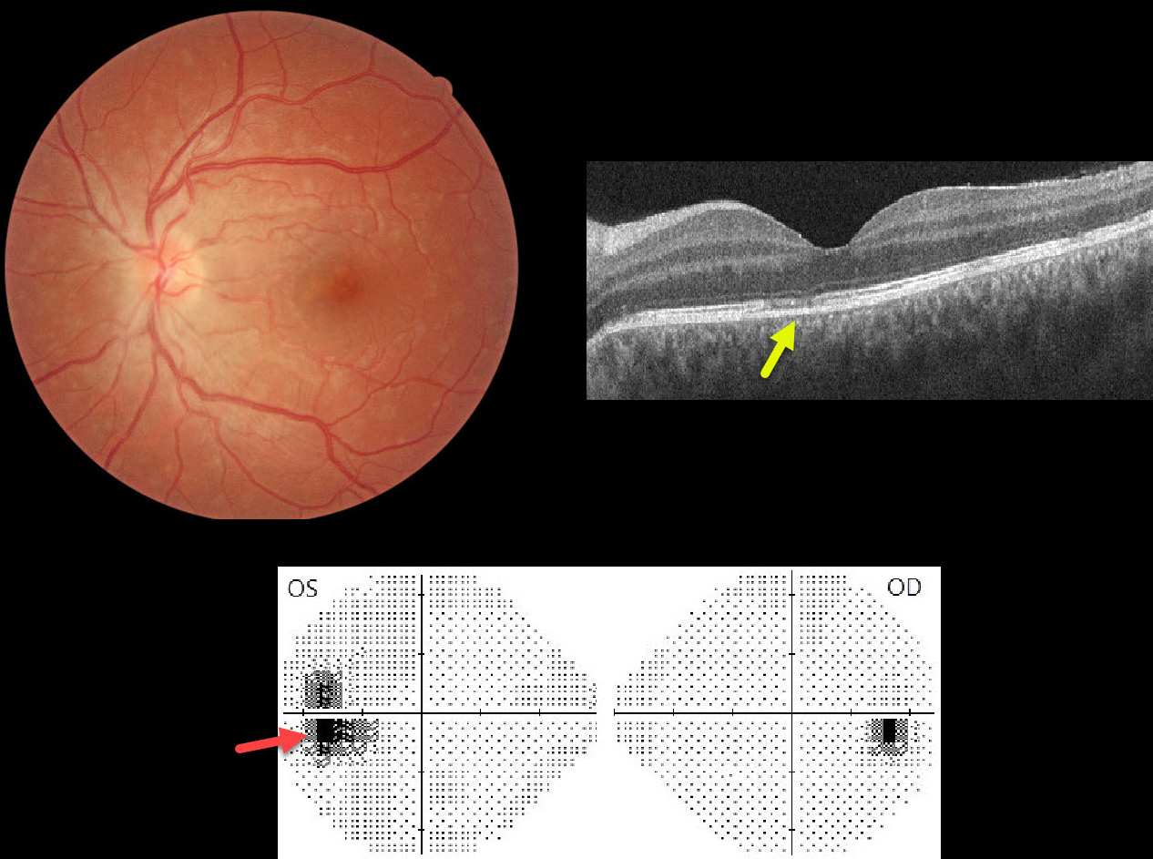
This healthy 27YO female presented with 2 weeks of photopsias OS. Vision was 20/20 in her normal OD and 20/25 OS.
Color photography shows multiple faint subretinal white dots scattered throughout the posterior pole. The nerve is slightly edematous, and the fovea has a granular appearance. OCT scanning shows scattered focal areas of ellipsoid zone and outer retinal band loss. Visual field testing shows an enlarged blind spot OS.
Learning Points:
Multiple evanescent white dot syndrome (MEWDS) is a usually unilateral, idiopathic inflammation of the outer retina that typically occurs in young, healthy myopic females. The granular fovea (Jampol spots, named in honor of Dr. Lee Jampol, who first described this syndrome in 1984) is a classic pathognomonic finding. Acutely, patients will also have an enlarged blind spot, often complaining of temporal visual field loss, due to the peripapillary inflammation.
The white dots disappear within a week or two, which is why the lesions are called “evanescent”. However, the blind spot may persist for many months, well after the fundus appearance has normalized. This stage of MEWDS was originally called the Acute idiopathic blind spot enlargement (AIBSE) syndrome before Gass recognized that the two disorders were the same disease but with distinct findings at different points in time.
ACUTE IDIOPATHIC BLIND SPOT ENLARGEMENT SYNDROME
Originally posted on @retina.rocks February 20, 2023
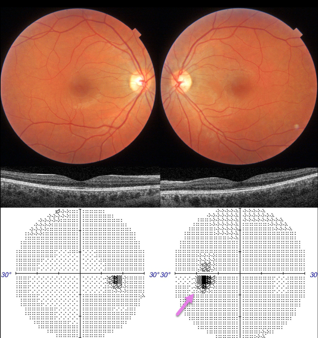
This 36YO female presented with 1 month of a paracentral scotoma in her left eye. There was no prior ocular or past medical history. Vision was 20/20 OU.
Ocular examinations were completely normal OU. Color imaging and OCT were normal. Visual fields were normal OD with a slightly enlarged blind spot OS.
Learning Points:
Multiple evanescent white dot syndrome (MEWDS) presents with usually unilateral idiopathic inflammation of the outer retina that typically occurs in young, healthy myopic females.
The granular fovea (Jampol spots, named after Dr. Lee Jampol, who first described this syndrome in 1984) is a classic pathognomonic finding that was not seen in this patient.
Acute cases will also have an enlarged blind spot, often with temporal visual field loss, due to peripapillary inflammation. The white dots disappear within a week or two, which is why the lesions are called “evanescent”. However, the blind spot may persist for many months, well after the fundus appearance has normalized.
This stage of MEWDS was originally called the Acute idiopathic blind spot enlargement (AIBSE) syndrome before Gass recognized that the two disorders were the same disease but with distinct findings at different points in time.
Observation for our patient was recommended. We expect her symptomatic enlarged blind spot will gradually resolve over the coming months.
MULTIPLE EVANESCENT WHITE DOT SYNDROME (MEWDS)
Originally posted on @retina.rocks January 13, 2023
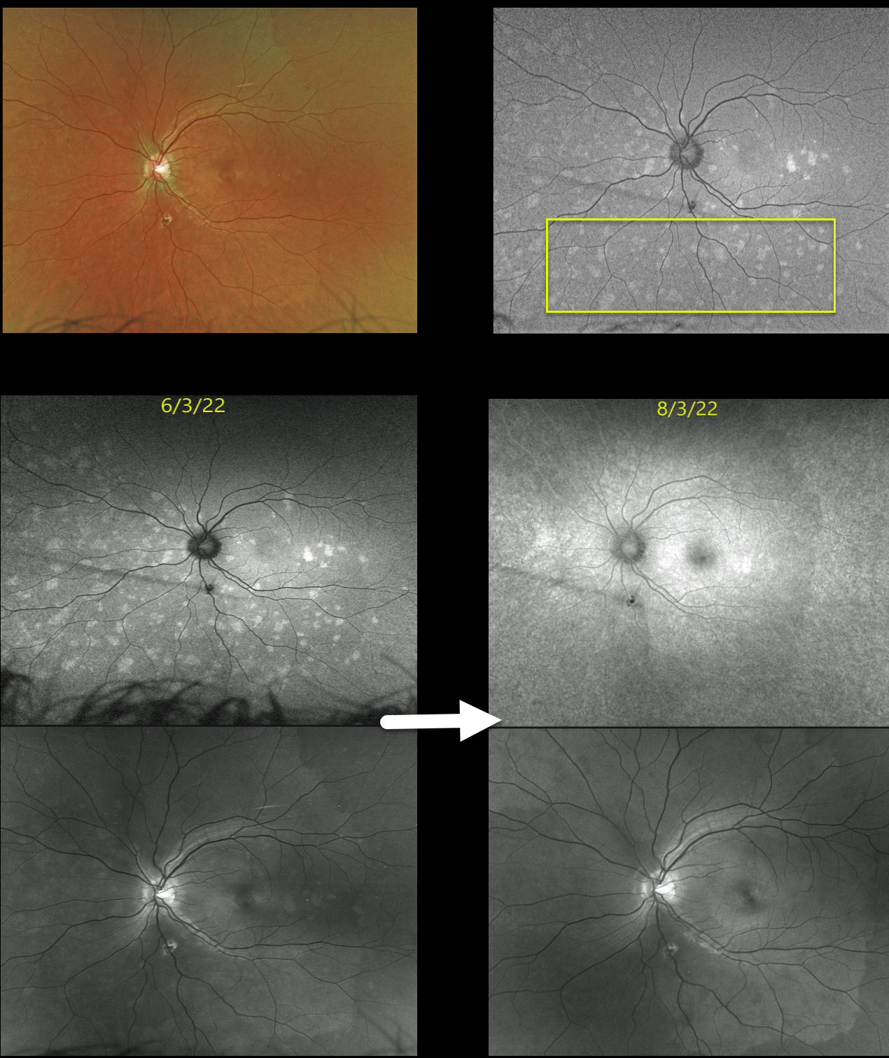
This 22YO female presented with blurred vision in her left eye for 2 weeks. Vision was 20/20 in her normal right eye and 20/70 in her left eye.
Optos color imaging of her left eye shows multiple faint, deep retinal white dots scattered throughout, which are hyper-autofluorescent. Macular OCT scanning was virtually normal (image not shown).
Two months later, the fundoscopic changes resolved, although fundus autofluorescence and Optos green channel imaging show more subtle residual findings.
Learning Points:
Multiple evanescent white dot syndrome (MEWDS) presents with usually unilateral idiopathic inflammation of the outer retina that typically occurs in young, healthy myopic females.
The granular fovea (Jampol spots, named after Dr. Lee Jampol, who first described this syndrome in 1984) is a classic pathognomonic finding that was not seen in this patient. Acute cases will also have an enlarged blind spot, often with temporal visual field loss, due to peripapillary inflammation.
The white dots disappear within a week or two, which is why the lesions are called “evanescent”. However, the blind spot may persist for many months, well after the fundus appearance has normalized.
MULTIPLE EVANESCENT WHITE DOT SYNDROME (MEWDS)
Originally posted on @retina.rocks April 13, 2022
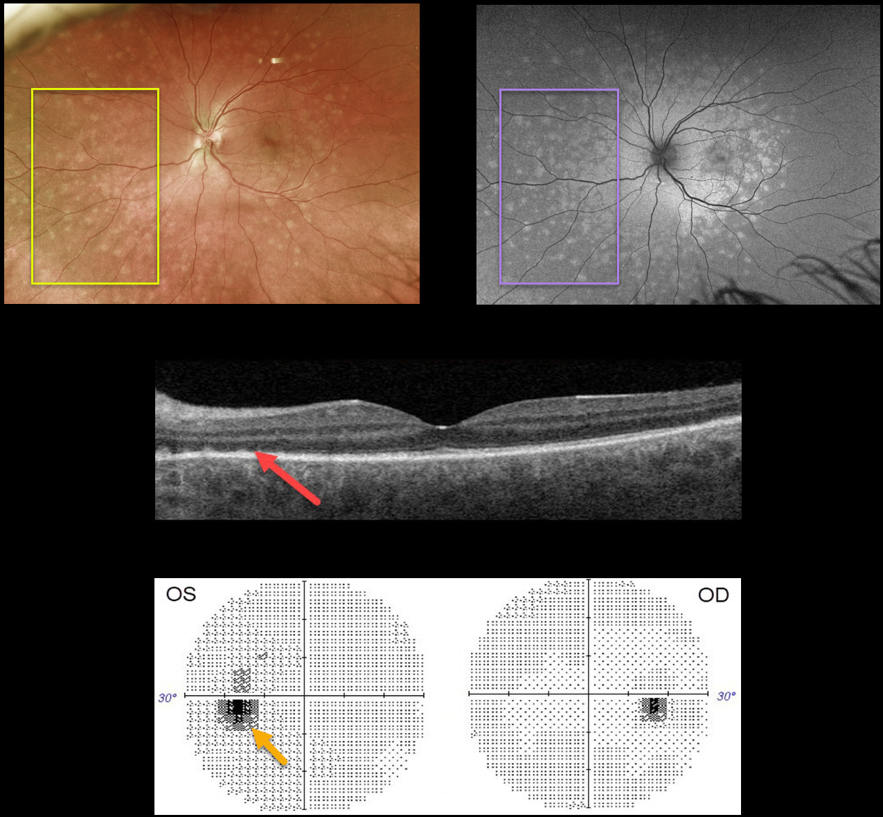
This 36YO male presented with flashes and a temporal scotoma in his left eye. Vision was 20/20 OD and 20/25 OS.
Optos color imaging shows multiple deep retinal white dots scattered throughout the posterior pole, which are hyper-autofluorescent.
Swept-source OCT shows nasal loss of the outer retinal bands with some hyperreflective spots. Visual fields show an enlarged left blind spot, consistent with the peripapillary hyperautofluorescence.
Learning Points:
Multiple evanescent white dot syndrome (MEWDS) presents with usually unilateral idiopathic inflammation of the outer retina that typically occurs in young, healthy myopic females.
The granular fovea (Jampol spots, named in honor of Dr. Lee Jampol, who first described this syndrome in 1984), noted in our patient, is a classic pathognomonic finding.
Acutely, patients will also have an enlarged blind spot, often complaining of temporal visual field loss, due to the peripapillary inflammation. The white dots disappear within a week or two, which is why the lesions are called “evanescent”.
However, the blind spot may persist for many months, well after the fundus appearance has normalized. This stage of MEWDS was originally called the Acute idiopathic blind spot enlargement (AIBSE) syndrome before Gass recognized that the two disorders were the same disease but with distinct findings at different points in time.
MULTIPLE EVANESCENT WHITE DOT SYNDROME (MEWDS)
Originally posted on @retina.rocks December 28, 2021
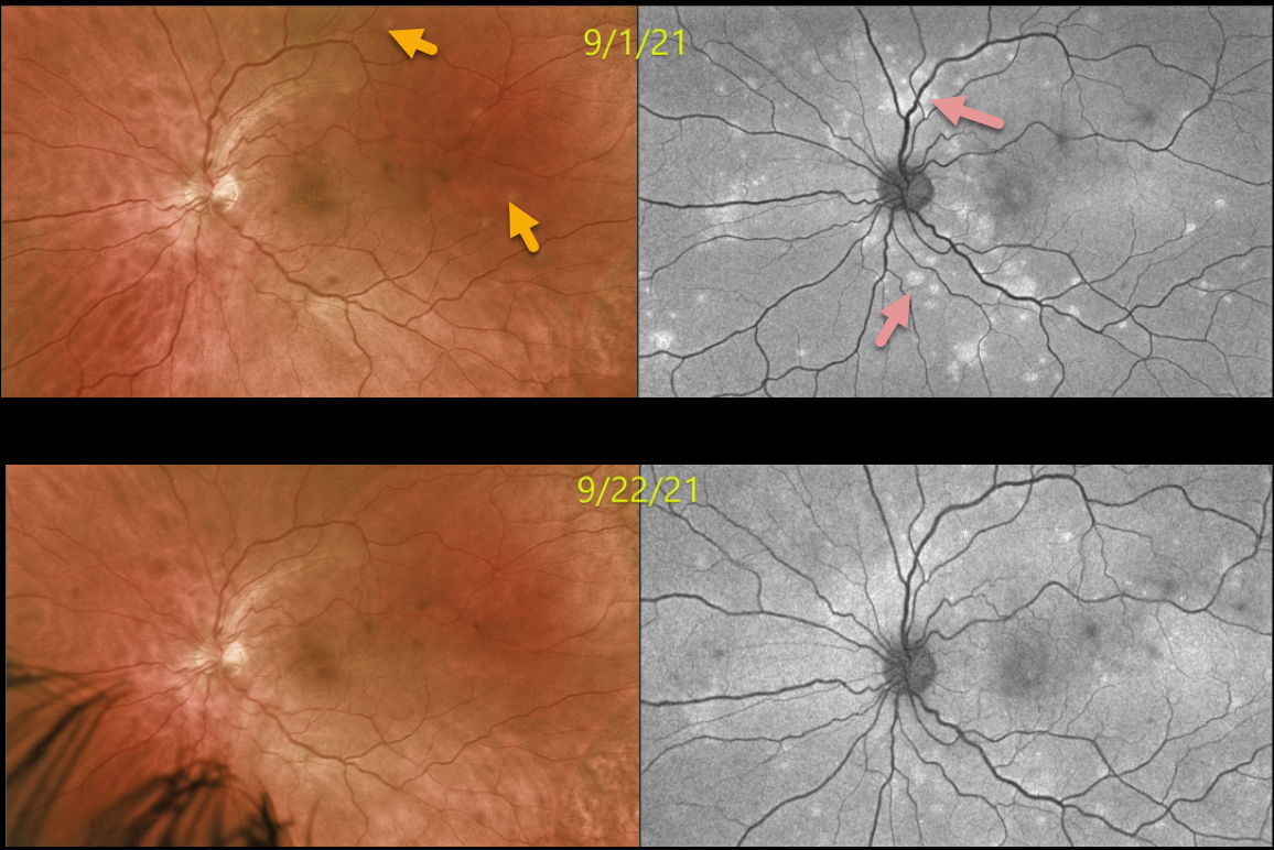
This 35YO male presented with 1 week of bilateral vision loss. Vision was 20/30 OD and 20/60 OS. Optos color imaging shows multiple deep retinal white dots scattered throughout the posterior pole (left eye only). Fundus autofluorescence (FAF) more dramatically shows these hyper-FAF lesions.
Three weeks later, vision improved to 20/30 OD and 20/25 OS as the lesions started to regress.
Learning Points:
Multiple evanescent white dot syndrome (MEWDS) presents with usually unilateral idiopathic inflammation of the outer retina that typically occurs in young, healthy myopic females.
The granular fovea (Jampol spots, named after Dr. Lee Jampol, who first described this syndrome in 1984) is a classic pathognomonic finding.
Acute cases will also have an enlarged blind spot, often with temporal visual field loss, due to peripapillary inflammation.
The white dots disappear within a week or two, which is why the lesions are called “evanescent”. However, the blind spot may persist for many months, well after the fundus appearance has normalized.
This stage of MEWDS was originally called the Acute Idiopathic Blind Spot Enlargement (AIBSE) syndrome before Gass recognized that the 2 disorders were the same disease but with distinct findings at different points in time.
MULTIPLE EVANESCENT WHITE DOT SYNDROME (MEWDS)
Originally posted on @retina.rocks September 17, 2021
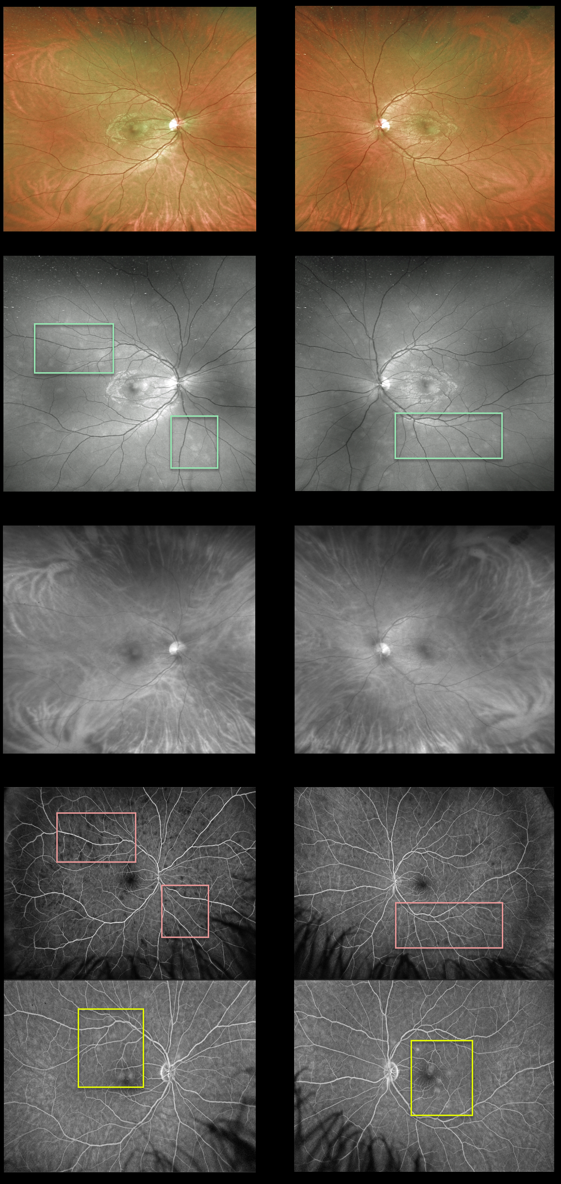
This 30YO male presented with a one-day history of seeing “dots” in his vision. Vision was 20/25 OD and 20/20 OS.
There are multiple deep retinal white dots scattered throughout the posterior pole of each eye. The Optos green channel best shows these white dots compared to the color images. The spots disappear with the Optos red channel, confirming their retinal localization.
Fluorescein angiography shows variable early hypofluorescence with late staining of the lesions.
Learning Points:
Multiple evanescent white dot syndrome (MEWDS) presents with usually unilateral idiopathic inflammation of the outer retina that typically occurs in young, healthy myopic females.
The granular fovea (Jampol spots, named in honor of Dr. Lee Jampol, who first described this syndrome in 1984), not well imaged in our case, is a classic pathognomonic finding.
Acute cases will also have an enlarged blind spot, often with temporal visual field loss, due to peripapillary inflammation.
The white dots disappear within a week or two, which is why the lesions are called “evanescent”. However, the blind spot may persist for many months, well after the fundus appearance has normalized.
This stage of MEWDS was originally called the Acute idiopathic blind spot enlargement (AIBSE) syndrome before Gass recognized that the 2 disorders were the same disease but with distinct findings at different points in time.
MULTIPLE EVANESCENT WHITE DOT SYNDROME (MEWDS)
Originally posted on @retina.rocks February 11, 2021
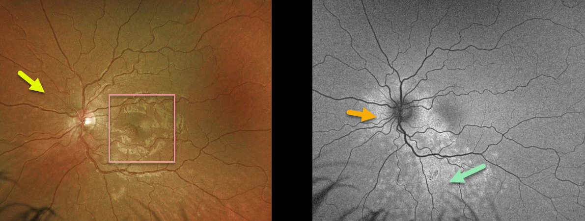
This 25YO female presented with 20/25 vision OU and a four-day history of temporal field loss in her left eye. Examination of the right eye was normal.
There was a granular appearance to the left fovea with some deep retinal white dots, most visible just nasal to the nerve. Fundus autofluorescence (FAF) more dramatically showed a ring of hyper-FAF encircling the nerve with multiple hyper-FAF spots extending inferiorly.
Learning Points:
Multiple evanescent white dot syndrome (MEWDS) presents with unilateral idiopathic inflammation of the outer retina, typically occurring in young, healthy myopic females.
The granular fovea (Jampol spots, named in honor of Dr. Lee Jampol, who first described this syndrome in 1984) noted in our case is a classic pathognomonic finding.
Acute cases will also have an enlarged blind spot, often with temporal visual field loss, due to peripapillary inflammation.
The white dots disappear within a week or two, which is why the lesions are called “evanescent”. However, the blind spot may persist for many months, well after the fundus appearance has normalized.
This stage of MEWDS was originally called the acute idiopathic blind spot enlargement (AIBSE) syndrome before Dr. Gass recognized that the 2 disorders were the same disease but with distinct findings at different points in time.
MULTIPLE EVANESCENT WHITE DOT SYNDROME (MEWDS)
Originally posted on @retina.rocks November 11, 2020
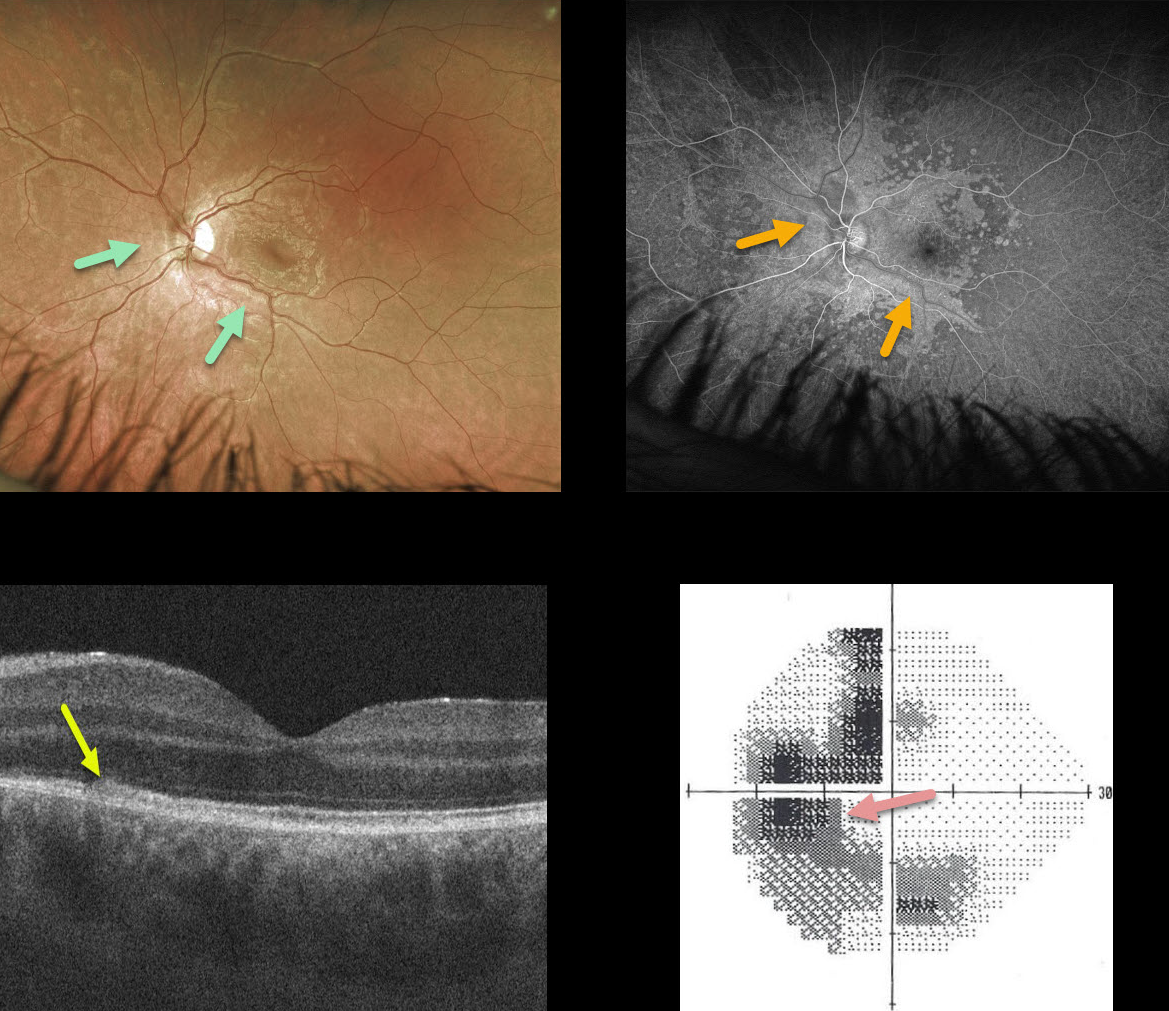
This 23YO patient presented with a few days of a temporal blurred spot in his left eye. Vision was 20/20 OD and 20/25 OS.
The left fundus has confluent outer retinal white spots centered around the optic nerve and extending into the macula. These lesions also had an unusual perivascular distribution more peripherally.
Fluorescein angiography shows hyperfluorescence staining in these lesions. Although this appearance suggested a possible vasculitis, angiography revealed absolutely no vascular changes. We are not sure why the outer retinal spots appeared in this vascular distribution.
OCT shows some hyperreflectivity and loss of detail of the ellipsoid zone and outer retina, and visual field testing shows an enlarged blind spot with temporal and inferior loss.
Learning Points:
We initially considered the possibility of syphilis given the pseudo-vasculitis and placoid confluence of the outer retinal lesions. However, the patient is in a long-term monogamous relationship and declined initial RPR/FTA testing.
Visual symptoms improved without any treatment. When the patient returned 6 weeks later, the white spots had disappeared clinically and on fundus autofluorescence, and his scotoma and symptoms had improved.
MULTIPLE EVANESCENT WHITE DOT SYNDROME (MEWDS)
Originally posted on @retina.rocks September 25, 2020
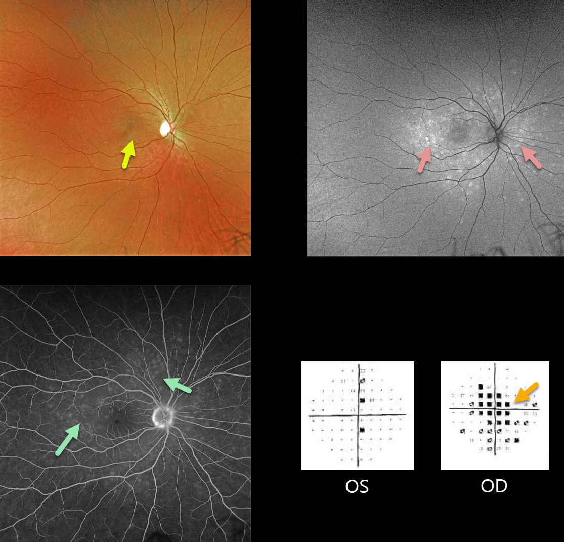
Our patient is a 31YO female with acute vision loss of 20/60 in her right eye with classic MEWDS findings.
Granular foveal changes (Jampol spots) and scattered white spots are seen in the outer retina. The spots appear hyper-autofluorescent on fundus autofluorescence imaging.
Fluorescein angiography shows multifocal wreath-like hyperfluorescent staining lesions.
Acute cases will also have an enlarged blind spot, often with temporal visual field loss.
Learning Points:
The MEWDS white dots usually disappear within a week or two, which is why the lesions are called evanescent. However, the blind spot may persist for many months, well after the fundus appearance has normalized.
This stage of MEWDS was originally called the Acute idiopathic blind spot enlargement (AIBSE) syndrome before Gass recognized that the 2 disorders were the same disease but with distinct findings at different points in time.
MULTIPLE EVANESCENT WHITE DOT SYNDROME (MEWDS)
Originally posted on @retina.rocks January 15, 2020
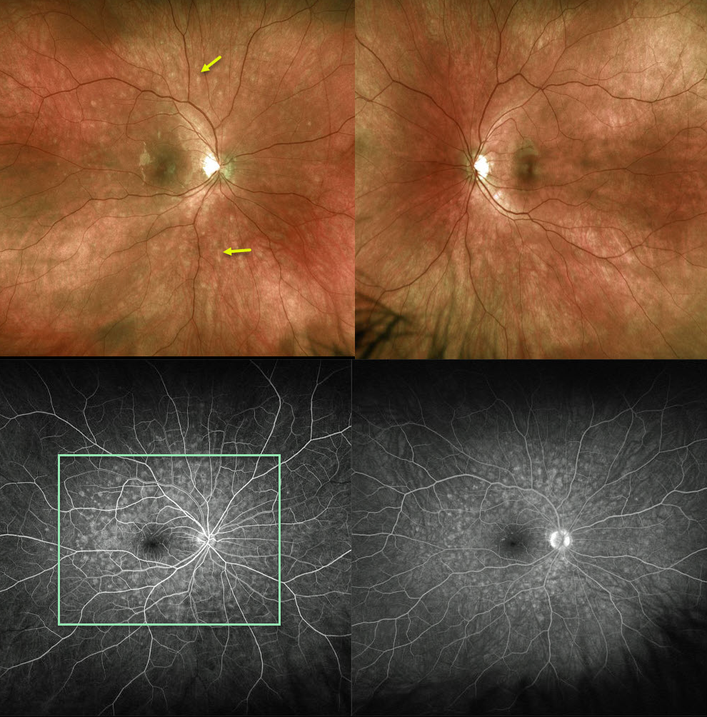
The Optos fundus photo of this 20YO female shows 100-200 micron outer retinal white spots scattered around the posterior pole, characteristic of multiple evanescent white dot syndrome (MEWDS).
Fluorescein angiography shows a wreath-like pattern of hyperfluorescence.
Learning Points:
MEWDS is an idiopathic, usually unilateral inflammation of the outer retina.
Most common in young women, patients often have a preceding viral syndrome and complain of blurred vision, photopsias, and temporal visual field loss.
Initially presenting with scatttered outer retinal white spots, the spots resolve within a few weeks making the diagnosis elusive. However, the enlarged blind spot often remains for months.
The condition is self-limited and usually resolves within several months without treatment.


