

Myelinated NFL
<< Back to Cases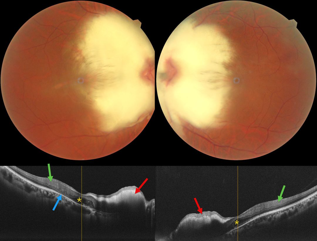
MYELINATED NERVE FIBER LAYER
Aarzoo Juneja and Lalit Singhal
Originally posted on @retina.rocks March 10, 2026
This healthy 36YO female presented with high myopia of -11.50 -1.75 x 50 OD and -11.00 -1.00 x 170 OS. Vision was 20/80 OD and 20/60 OS. Color photography shows a bilaterally symmetric myelinated nerve fiber layer (NFL) covering each nerve and the nasal macula. The discs may also be hypoplastic, although the margins are difficult to discern due to the myelination.
On OCT, the myelination appears as a markedly hyperreflective, thickened inner retina with posterior shadowing. The foveal outer nuclear layers (ONL) appear thickened OU. There is hyperreflective material in the right nasal foveal ONL with disorganization of the underlying outer retinal bands. The inner retinal bands are disorganized more temporally OU, with outer retinal atrophy OD.
Learning Points:
Myelinated NFL occurs when retinal nerve fibers develop a myelin sheath, which usually stops posterior to the lamina cribrosa. The myelination may represent an oligodendrocytic choristoma (Rao et al, Retina 2019;39:1125-1132).
Myelinated NFL is a relatively uncommon, unilateral or bilateral, usually isolated finding present at birth. The myelinated NFL is often connected to the optic nerve. Most patients are asymptomatic, although some may have a corresponding relative scotoma. The myelination can progress in about 10% of cases and can also resolve following retinal ischemic events. Our patient’s findings were consistent with Straatsma syndrome, which consists of the triad of bilateral myopia, amblyopia, and myelinated NFL.
Myelinated NFL is reported to anatomically affect just the NFL (Rao et al, Retina 2019;39:1125-1132). Our patient’s OCT findings showing both inner and outer retinal changes outside the area of myelination has not been previously reported.
MYELINATED NERVE FIBER LAYER
Originally posted on @retina.rocks January 7, 2026
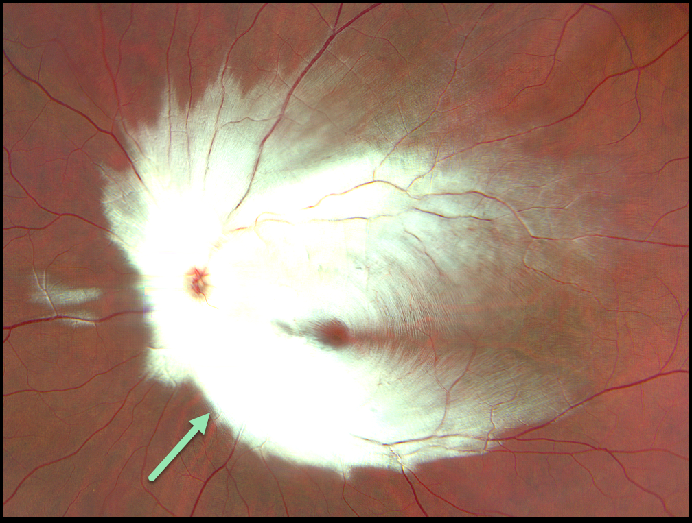
This healthy 17YO male presented with a history of decreased vision in the left eye since birth. Family and developmental history were unremarkable. Vision was 20/20 in his normal right eye and counting fingers at 10 feet in his left eye.
Color photography shows the myelinated nerve fiber layer (NFL) covering nearly the entire posterior pole and peripapillary region.
Learning Points:
Myelinated NFL occurs when retinal nerve fibers develop a myelin sheath, which usually stops posterior to the lamina cribrosa. The myelination may represent an oligodendrocytic choristoma (Rao et al, Retina 2019;39:1125-1132).
Myelinated NFL is a relatively uncommon, unilateral or bilateral, usually isolated finding present at birth. The myelinated NFL is often connected to the optic nerve. Most patients are asymptomatic, although some may have a corresponding relative scotoma. The myelination can progress in about 10% of cases and can also resolve following retinal ischemic events.
BEST DISEASE WITH MYELINATED NERVE FIBER LAYER
The European Society (EVRS) and Vaibhav Sethi
Originally posted on @retina.rocks July 25, 2025
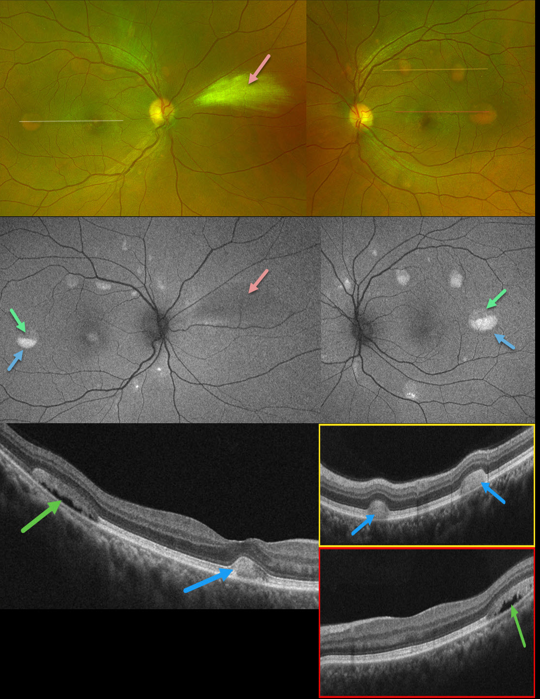
This 39YO male presented with asymptomatic retinal lesions. Family history was negative. Vision was 20/20 OU.
Optos color RG imaging shows bilateral partially scrambled vitelliform lesions in each posterior pole. On OCT, the superior, scrambled portion of these lesions is hyporeflective, and the inferior, yellow portion is hyperreflective. On fundus autofluorescence (FAF), the superior portions are hypo-FAF, and the inferior vitelliform material is hyper-FAF. The incidental area of the nasal myelinated nerve fiber layer OD is hypo-FAF from blocking the underlying RPE.
Learning Points:
Vitelliform lesions are characterized by the accumulation of yellow subretinal material thought to be caused by impaired metabolism of photoreceptors or retinal pigment epithelium. They can be associated with a wide variety of retinal disorders, including Best vitelliform macular dystrophy (BVD, our patient), adult-onset foveomacular dystrophy, acute exudative polymorphous vitelliform maculopathy, as well as paraneoplastic, toxic, tractional, and degenerative etiologies (Iovino et al, Surv Ophthalmology 2023;68:361-379).
BVD is usually transmitted as an autosomal-dominant disorder due to a mutation in the BEST1 gene, which encodes bestrophin-1. Bestrophin-1, a calcium-activated chloride channel, is primarily found in the basolateral plasma membrane of the RPE. Unfortunately, our patient denied genetic testing due to cost.
BRVO + MYELINATED NERVE FIBER LAYER
Evgenii Chernov
Originally posted on @retina.rocks September 30, 2024
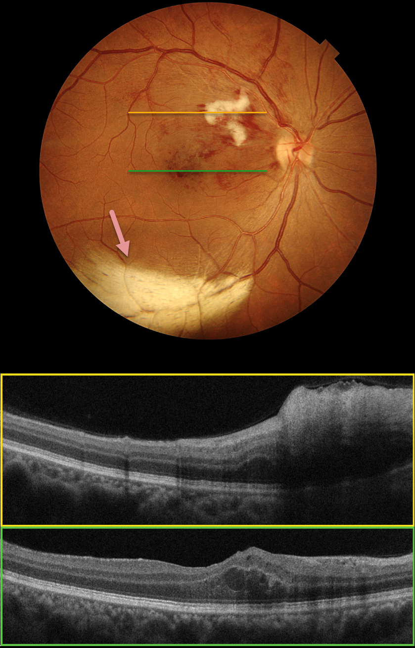
Color photography of this 48YO female’s right eye shows a major superotemporal branch retinal vein occlusion (BRVO) with foveal blood. A prominent nerve fiber layer infarct (cotton-wool spot, CWS) is present in the superonasal macula. OCT scanning shows diffuse hyperreflective inner retinal thickening through the CWS and some central foveal fluid in the outer nuclear layer. An unrelated patch of myelinated nerve fiber layer (NFL) is noted along the inferotemporal arcade. The macular edema resolved following 3 monthly intravitreal Lucentis injections (not shown).
Our patient’s eye shows two different pathologies that each involve the NFL. Myelinated NFL occurs when retinal nerve fibers develop a myelin sheath, which usually stops posterior to the lamina cribrosa. The white myelin beautifully displays the normally transparent NFL. Myelination may represent an oligodendrocytic choristoma (see Rao et al Retina 2019;39:1125-1132). Nerve fiber layer infarcts cause a more fluffy, white ischemic opacification of the NFL and are a relatively nonspecific finding in numerous disorders, including diabetes, retinal venous occlusion, hypertension, and HIV/AIDS.
CHOROIDAL COLOBOMA + MYELINATED NERVE FIBER LAYER
Originally posted on @retina.rocks July 31, 2024
This healthy 8YO girl was referred for an asymptomatic retinal finding in her right eye. Vision was 20/30 in her right eye and 20/30 in her left eye. Anterior segments were normal.
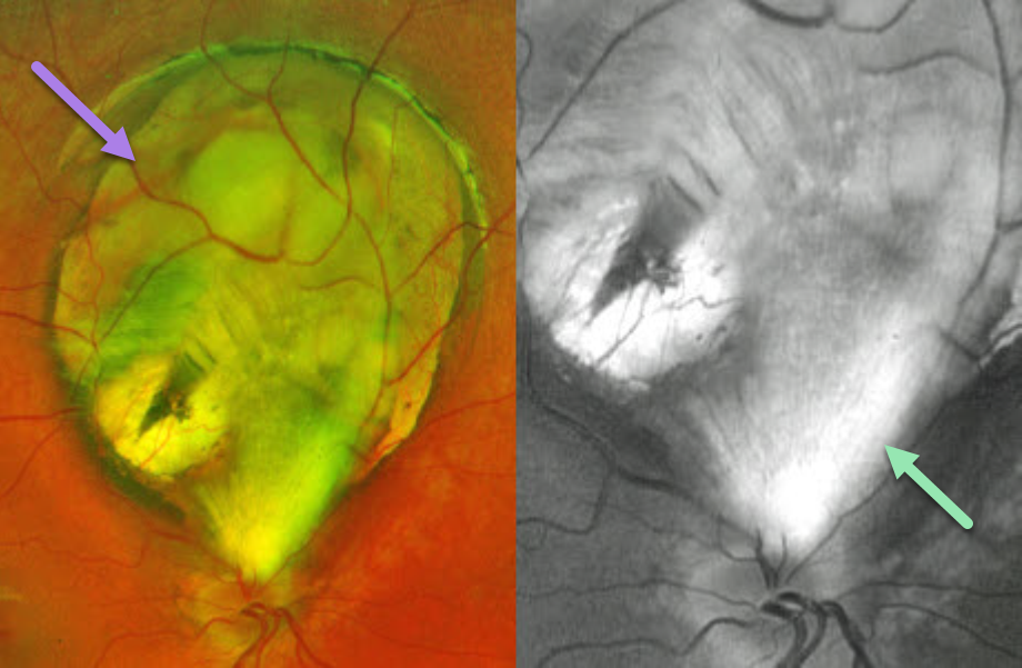
Optos color RG imaging shows a superior choroidal coloboma that almost extends to the disc margin. An incidental patch of myelinated nerve fiber layer (NFL), best imaged in the green channel, extends from the superior nerve into the inferior aspect of the coloboma.
Learning Points:
Choroidal colobomas are caused by failure of the optic vesicle and choroidal fissure to close during fetal development. Choroidal coloboma can present as solitary or multiple lesions. Visual prognosis depends on the location and involvement of the optic nerve and macula. We will follow our patient yearly due to her increased risk of retinal detachment.
Myelinated NFL is a relatively uncommon and usually isolated finding present at birth. Most patients are asymptomatic, although some may have a corresponding relative scotoma. Myelination can progress in about 10% of cases and can also resolve following retinal ischemic events. Myelinated NFL occurs when retinal nerve fibers develop a myelin sheath, which usually stops posterior to the lamina cribrosa. The myelination may represent an oligodendrocytic choristoma (see Rao et al Retina 2019;39:1125-1132).
MYELINATED NERVE FIBER LAYER + WWP
Originally posted on @retina.rocks May 18, 2023
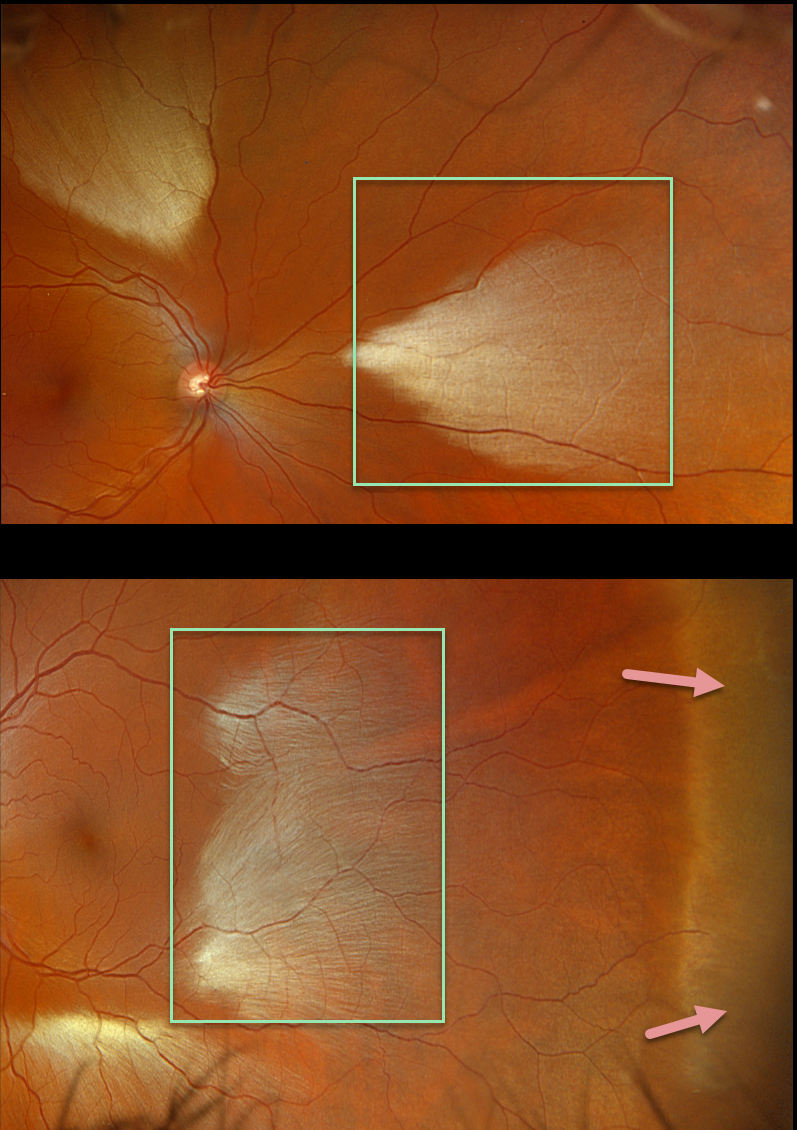
This healthy 35YO male was referred for asymptomatic fundoscopic changes. Vision was 20/20 OU.
Opto imaging shows large multifocal patches of myelinated nerve fiber layer (NFL) in both eyes. Temporal white without pressure (WWP) is also noted in his left eye.
Learning Points:
Myelinated NFL is a relatively uncommon, unilateral or bilateral, usually isolated finding present at birth. The myelinated NFL is often connected to the optic nerve, but not necessarily as in this case. Most patients are asymptomatic, although some may have a corresponding relative scotoma. The myelination can progress in about 10% of cases and can also resolve following retinal ischemic events.
Myelinated NFL occurs when retinal nerve fibers develop a myelin sheath, which usually stops posterior to the lamina cribrosa. The myelination may represent an oligodendrocytic choristoma (see Rao et al Retina 2019;39:1125-1132).
WWP is a relatively common benign peripheral finding, most often seen in darkly pigmented individuals. Typically, there are bilateral peripheral geographic areas of whitish retinal discoloration. WWP gets its name because the area appears white without indentation, unlike scleral depression (white with pressure). It can sometimes be confused with retinal breaks or detachment.
The exact cause of this phenomenon is unknown. Historically, it was felt to be due to a benign vitreoretinal interface change, but more recent OCT findings show increased reflectivity in the outer retina (see Diaz et al, Retina 2014;34:1020-1021).
MYELINATED NERVE FIBER LAYER
Originally posted on @retina.rocks February 8, 2023
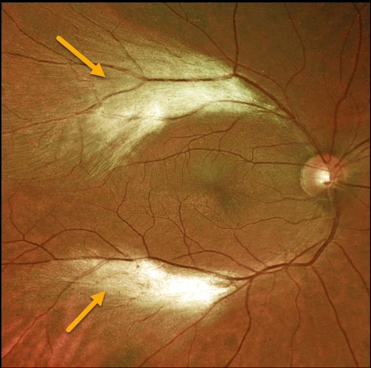
This 47YO female presented with two distinct patches of myelinated nerve fiber layer (NFL) along the distal temporal arcades in the right eye. Her ocular examinations were otherwise normal.
Learning Points:
Myelinated NFL is a relatively uncommon and usually isolated finding present at birth. Most patients are asymptomatic, although some may have a corresponding relative scotoma. Myelination can progress in about 10% of cases and can also resolve following retinal ischemic events.
Myelinated NFL occurs when retinal nerve fibers develop a myelin sheath, which usually stops posterior to the lamina cribrosa. The myelination may represent an oligodendrocytic choristoma (see Rao et al Retina 2019;39:1125-1132).
CRVO + CHRPE + MYELINATED NERVE FIBER LAYER
Will Gibson
Originally posted on @retina.rocks October 27, 2022
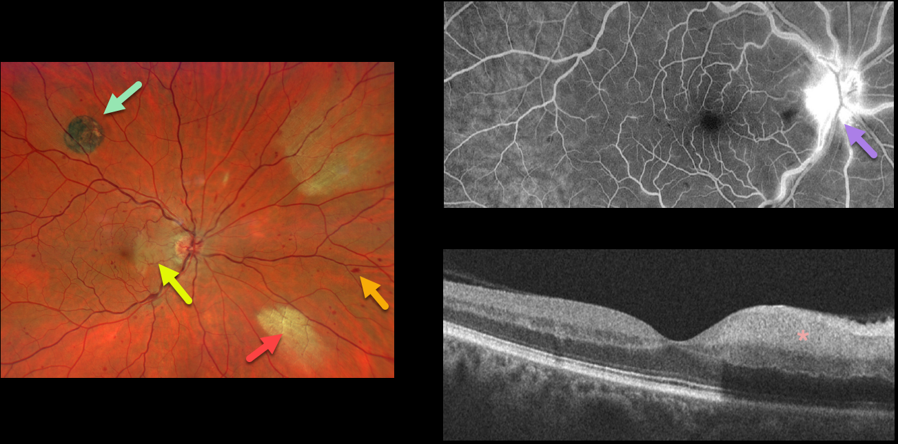
This 52YO male presented with acute visual symptoms in his right eye. Vision was 20/20.
Optos imaging shows a non-ischemic central retinal vein occlusion (CRVO) with mild scattered retinal hemorrhages in all quadrants. An acute nasal macular branch retinal artery occlusion (BRAO) is noted, along with a small round patch of congenital hypertrophy of the RPE (CHRPE) superotemporally, and two patches of myelinated nerve fiber layer (NFL) nasally.
Fluorescein angiography shows a well-perfused central retinal vein occlusion (CRVO) with late optic nerve leakage. OCT scanning shows hyperreflectivity of the inner nasal retina.
MYELINATED NERVE FIBER LAYER
Originally posted on @retina.rocks September 15, 2021
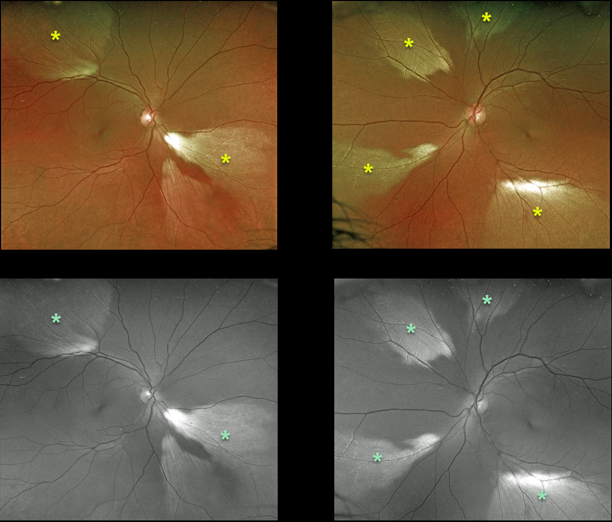
This 50YO female presented with multiple bilateral patches of myelinated nerve fiber layer (NFL). The findings are beautifully imaged using both the Optos color RG and green channels.
Myelinated NFL is a relatively uncommon, unilateral or bilateral, usually isolated finding present at birth. The myelinated NFL is often connected to the optic nerve, but not necessarily as in this case. Most patients are asymptomatic, although some may have a corresponding relative scotoma. Myelination can progress in about 10% of cases and can also resolve following retinal ischemic events. Myelinated NFL occurs when retinal nerve fibers develop a myelin sheath, which usually stops posterior to the lamina cribrosa. The myelination may represent an oligodendrocytic choristoma (see Rao et al, Retina 2019;39:1125-1132).
MYELINATED NERVE FIBER LAYER
Kaitlin Spikes
Originally posted on @retina.rocks May 7, 2021
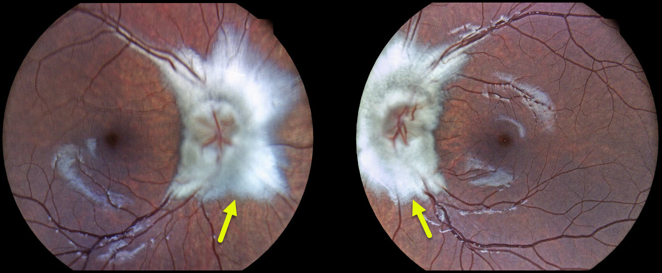
This 9YO girl presented with bilateral optic nerve myelination. The patient had no visual complaints and had 20/20 vision in both eyes.
Learning Points:
Myelinated NFL is a relatively uncommon, unilateral or bilateral, usually isolated finding present at birth. Most patients are asymptomatic, although some may have a corresponding relative scotoma.
Myelination can progress in about 10% of cases and can also resolve following retinal ischemic events.
Myelinated NFL occurs when retinal nerve fibers develop a myelin sheath, which usually stops posterior to the lamina cribrosa. The myelination may represent an oligodendrocytic choristoma.
MYELINATED NERVE FIBER LAYER
Originally posted on @retina.rocks February 19, 2021
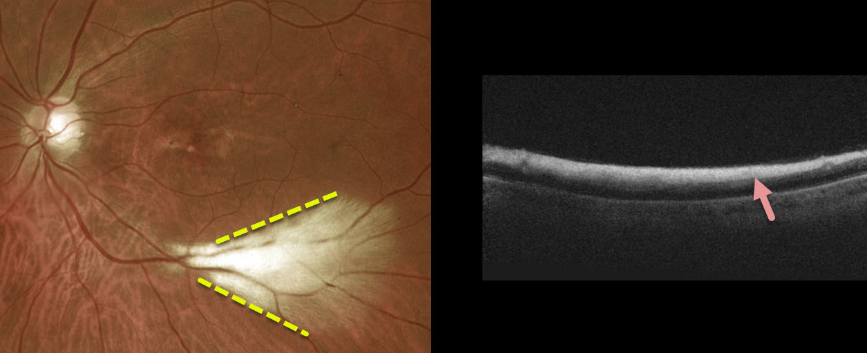
This patient had a large area of myelinated nerve fiber layer (NFL) in the distal inferotemporal left macula. OCT scanning through this area showed marked inner retinal hyperreflectivity.
Learning Points:
Myelinated NFL is a relatively uncommon and usually isolated finding present at birth. Most patients are asymptomatic, although some may have a corresponding relative scotoma.
Myelination can progress in about 10% of cases and can also resolve following retinal ischemic events.
Myelinated NFL occurs when retinal nerve fibers develop a myelin sheath, which usually stops posterior to the lamina cribrosa. The myelination may represent an oligodendrocytic choristoma (Rao et al, Retina 2019;39:1125-1132).
MYELINATED NERVE FIBER LAYER
Originally posted on @retina.rocks October 6, 2020
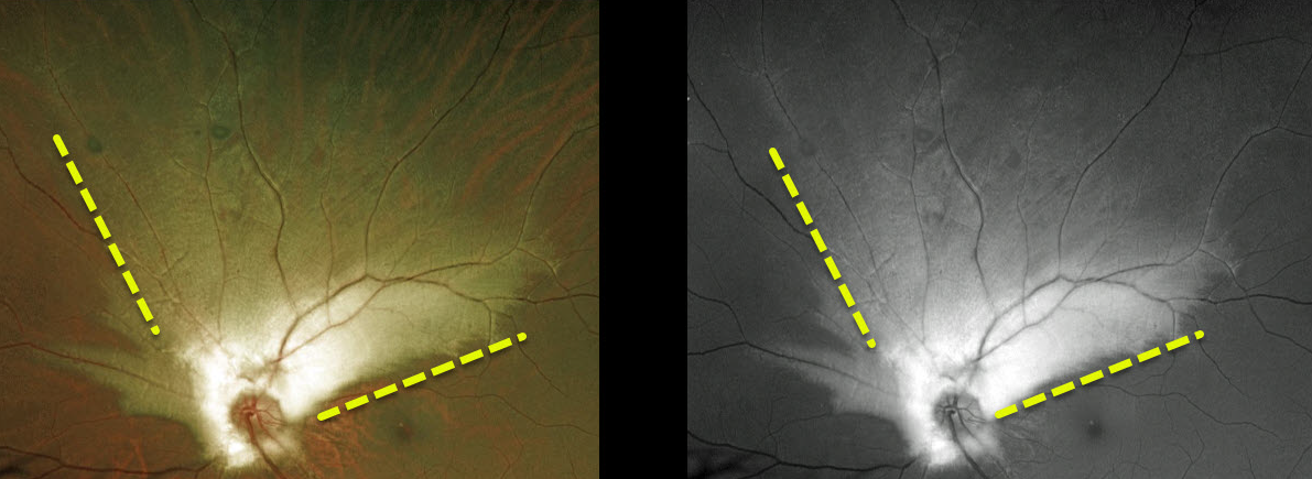
This 20 YO male has a large area of myelinated nerve fiber layer (NFL) extending superiorly from the left optic nerve.
This relatively uncommon and usually isolated finding is present at birth. Most patients are asymptomatic, although some may have a corresponding relative scotoma.
Myelination can progress in about 10% of cases and can also resolve following retinal ischemic events.
Learning Points:
Myelinated NFL occurs when retinal nerve fibers develop a myelin sheath, which usually stops posterior to the lamina cribrosa. The myelination may represent an oligodendrocytic choristoma.
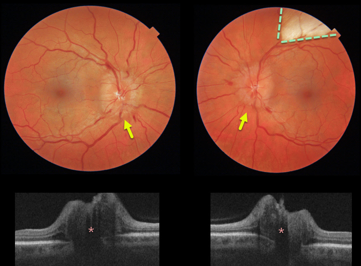
PAPILLEDEMA
Originally posted on @retina.rocks June 26, 2020
This healthy 40yo female presented with chronic headaches. She was on no systemic medications, and her blood pressure was normal. Her nerves were swollen bilaterally, and a myelinated nerve fiber layer was also noted in her left eye.
The retinal nerve fiber layer on OCT was severely thickened bilaterally.
Learning Points:
The diagnosis of idiopathic intracranial hypertension (pseudotumor cerebri) was made after an MRI scan was normal and a lumbar puncture revealed a markedly elevated opening pressure.
She is currently being successfully managed with oral acetazolamide.


