

Diabetic Retinopathy Non-Proliferative (NPDR)
<< Back to Cases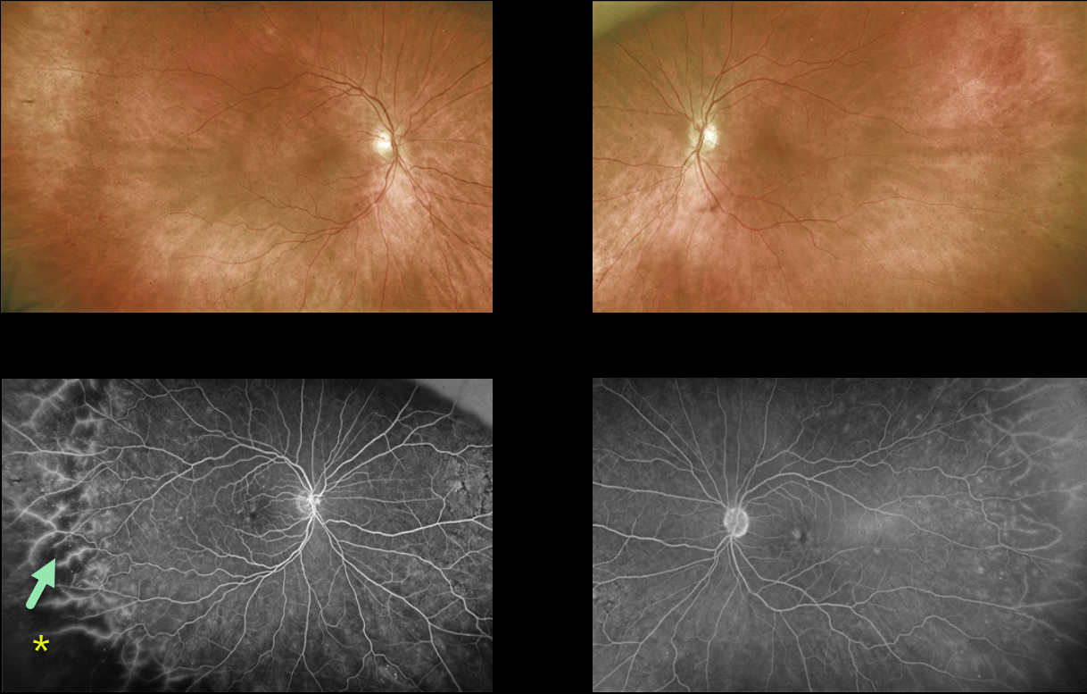
NON-PROLIFERATIVE DIABETIC RETINOPATHY (NPDR)
Originally posted on @retina.rocks November 20, 2023
This 69YO female with a history of type 2 diabetes was referred for asymptomatic retinal changes. Vision was 20/25 OU.
Optos color RG imaging shows virtually normal posterior poles with temporal peripheral retinal hemorrhages.
Fluorescein angiography shows temporal ischemia with diffuse vascular staining.
Learning Points:
The DRCR Retina Network’s Protocol AA found that ultrawidefield (UWF) imaging, compared to the standard ETDRS 7-field images, increases the frequency of diabetic retinopathy identification by nearly two fold (JAMA Ophthalmology 2019;137:65-73).
UWF fluorescein angiography further increases the sensitivity for assessing retinal ischemia, with about 70% of nonperfusion located outside the posterior pole (Retina 2022;42:1302-1310).
Finally, eyes with angiographically predominantly peripheral lesions, as in our patient, are at a higher 4-year risk of worsening disease, independent of the baseline diabetic retinopathy severity score (JAMA Ophthalmology 2022;140:946-954).
UWF fundus imaging has transformed the field of retina. In our practice, we are constantly humbled by the superiority of UWF imaging in finding peripheral pathology compared to our clinical examination. And increasingly, this technology is becoming the gold standard for identifying pathology and guiding our treatment decisions.
NON-PROLIFERATIVE DIABETIC RETINOPATHY (NPDR)
Originally posted on @retina.rocks December 5, 2022
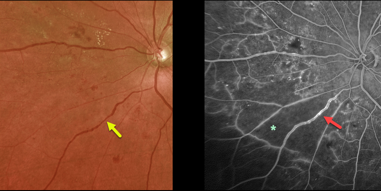
This 64YO diabetic female presented with asymptomatic severe nonproliferative diabetic retinopathy (NPDR) in her left eye. Vision was 20/40.
Optos color imaging shows venous beading inferonasally. Fluorescein angiography shows severe retinal ischemia with venous staining.
Our patient’s ischemia was localized just to one quadrant with good perfusion elsewhere, so we are following her closely.
Learning Points:
Severe NPDR is defined as severe (at least 20) retinal hemorrhages in all quadrants, beading in at least 2 quadrants, or intraretinal microvascular abnormalities in 1 quadrant. These eyes are at significant risk for progressing to proliferative disease, so we will often treat with panretinal photocoagulation before neovascularization develops.
In our experience, eyes with significant venous beading or venous loops more often than not already have proliferative disease.
DIABETIC MACULAR ISCHEMIA
Originally posted on @retina.rocks November 23, 2022
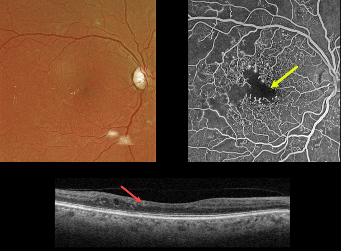
This 67YO male has a history of type 2 diabetes. Vision is 20/40.
Optos color imaging is fairly unremarkable, with some mild temporal foveal lipid. However, fundus fluorescein angiography shows foveal ischemia with an enlarged, irregular foveal avascular zone along with more peripheral superotemporal retinal ischemia.
Swept-source OCT shows a thinned, relatively featureless inner retina, also known as disorganization of inner retinal layers (DRIL).
Observation was recommended. The ischemic retina and DRIL will not improve with anti-VEGF therapy.
Learning Points:
DRIL is seen in various retinal vascular disorders, including diabetic macular edema and retinal vein occlusions. It is generally associated with worse vision and increased anti-VEGF treatment burden (see Babiuch et al, JAMA Ophthalmol 2019;137:38-46).
NON-PROLIFERATIVE DIABETIC RETINOPATHY (NPDR)
Originally posted on @retina.rocks March 17, 2020
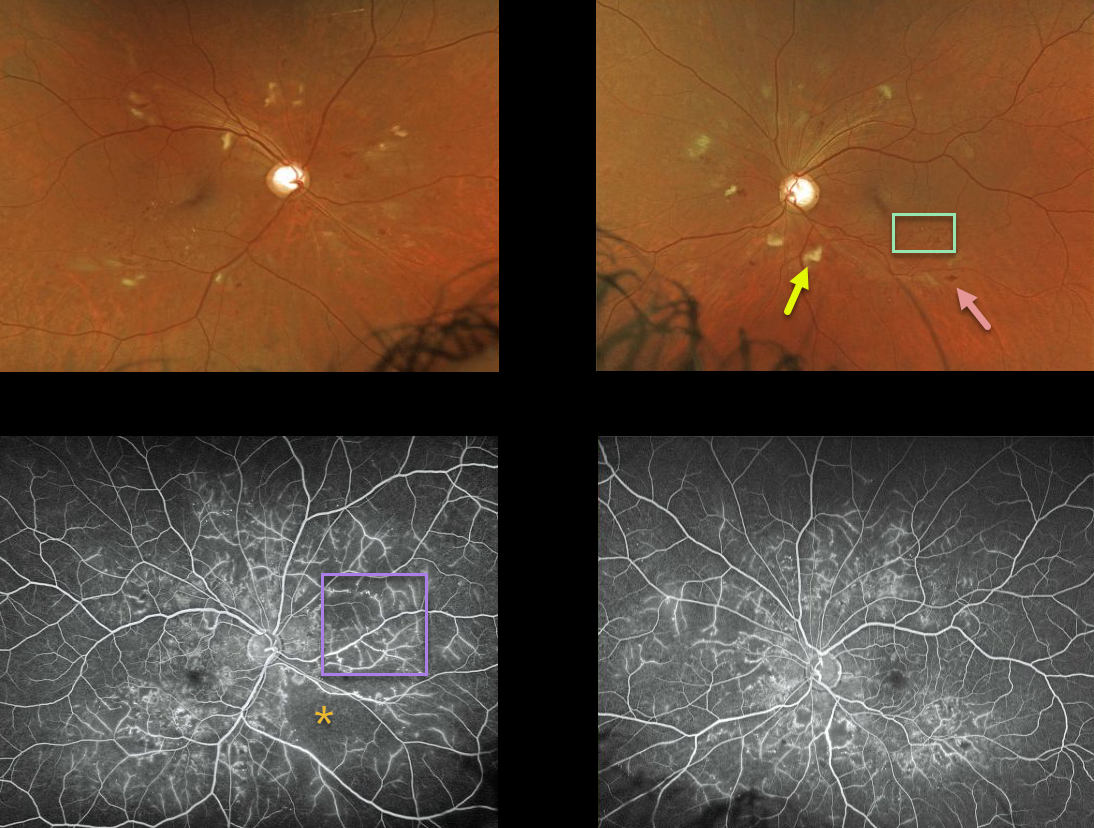
The fundus photos demonstrate severe NDPR with cotton wool spots, microaneurysms, and intraretinal hemorrhages.
Our patient’s ultra wide-field fundus fluorescein angiography (UW-FFA) shows significant areas of non-perfusion with vascular staining but no neovascularization.
Learning Points:
In some cases of moderate or severe retinopathy, a baseline fluorescein angiography is performed. UW-FFA can be useful to detect clinically silent proliferative disease and monitor these patients for conversion from NPDR to PDR.


