

Ocular Histoplasmosis (OHS)
<< Back to Cases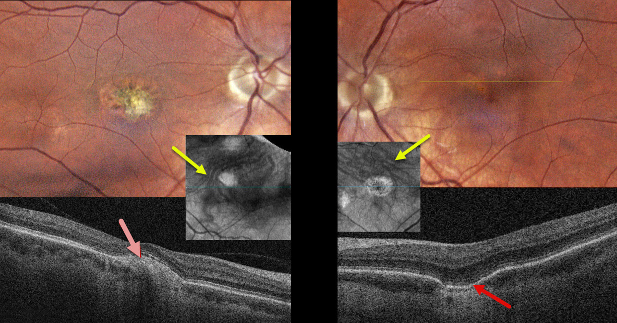
PACHYCHOROID SPECTRUM PREVIOUSLY MISDIAGNOSED AS OCULAR HISTOPLASMOSIS
Steven Bloom
Originally posted on @retina.rocks April 22, 2026
This healthy 39YO female gave a history 18 years earlier of prior anti-VEGF therapy OD for ocular histoplasmosis. She was visually asymptomatic, and vision was 20/40 OD and 20/30 OS.
Color photography of her left eye shows a patchy area of pigment loss in the superonasal fovea. OCT scanning through this area shows a choroidal excavation. The right macula has an overall mostly depigmented area of scarring, which appears as a mound of subretinal hyperreflectivity on OCT. Choroidal en face OCT shows dilated pachyvessels OU that cross the horizontal meridian (vortex anastomoses). There were no areas of peripheral chorioretinal scarring in either eye. Observation was recommended.
Learning Points:
Pachychoroid, meaning “thick” choroid, is secondary to large, dilated outer choroidal vessels. Increased oncotic pressure from these dilated vessels can cause RPE dysfunction, serous detachment, and macular neovascularization (MNV). Pachychoroid disease spectrum is a growing list of findings, including idiopathic central serous chorioretinopathy (ICSC), pachychoroid neovascularization, polypoidal choroidal vasculopathy, pachychoroid pigment epitheliopathy, pachydrusen, peripapillary pachychoroid syndrome, choroidal excavation, and pachyvitelliform maculopathy.
Our guess is that our patient had active ICSC when she was treated for her ‘ocular histoplasmosis.’ The lesion in the right macula is atypical for ICSC. But the other findings, including dilated choroidal vessels with vortex anastomoses and a choroidal excavation in her left eye, argue for pachychoroid as her unifying diagnosis.
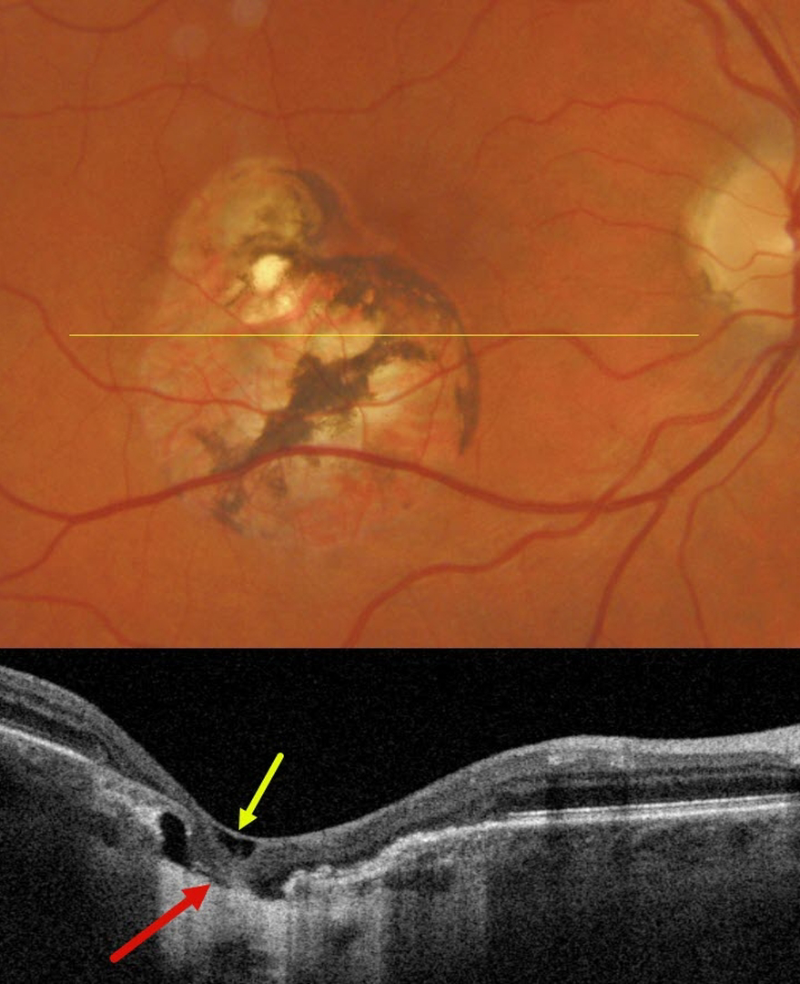
OCULAR HISTOPLASMOSIS LASER SCAR
Originally posted on @retina.rocks January 8, 2025
This 52YO male has been followed by us for many years for ocular histoplasmosis (OHS). Decades earlier, he received thermal laser from an outside practice for macular neovascularization (MNV) in his right eye. When recently seen for his yearly examination, vision was 20/20 OD, and he remained completely visually asymptomatic.
Triton color imaging shows a variably pigmented inferotemporal macular laser scar just sparing the foveal center. Swept-source OCT through this scar shows internal limiting membrane draping, variable outer- and mid-retinal atrophy, variable RPE loss, and more centrally a complete foveal cavitation with draping of the featureless neurosensory retina onto bare sclera.
Learning Points:
Thermal laser photocoagulation was the only treatment for MNV prior to the introduction of photodynamic therapy (PDT) in 2000 and anti-VEGF therapy in 2005. Unlike age-related MNV, which had an extremely high failure rate due to persistent/recurrent lesions, treating OHS-related lesions was often a one-and-done. The Macular Photocoagulation Study found that 74% of extrafoveal OHS MNV were without recurrences at year 5 (Arch Ophthalmology 1991;109:1109-1114). It is always amazing to us how few patients, including this patient, ever complained of a laser-induced scotoma, even when treatment extended close to the macular center.
We currently rarely perform thermal laser for extrafoveal MNV and prefer either PRN anti-VEGF monotherapy or combined anti-VEGF with PDT.
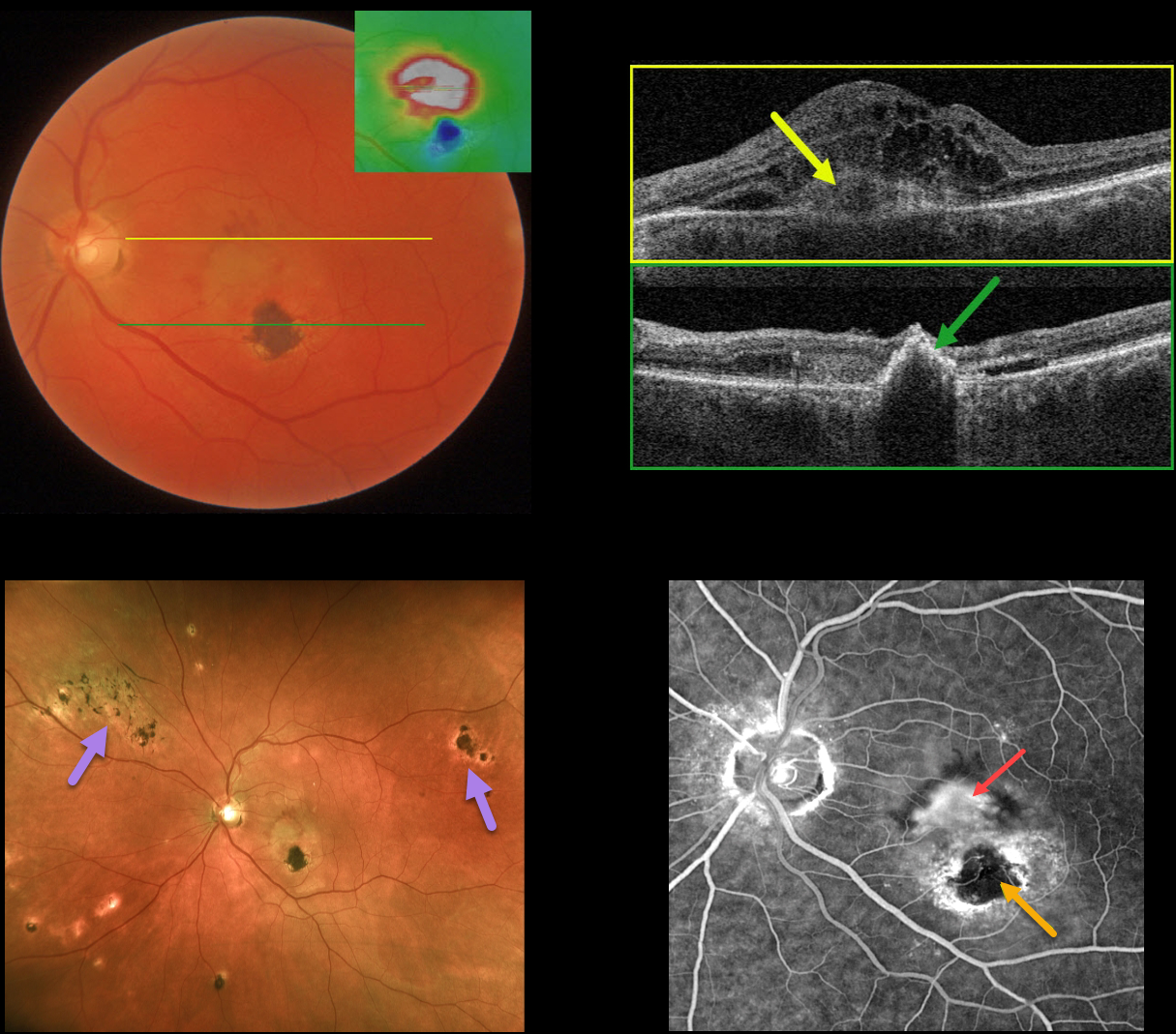
OCULAR HISTOPLASMOSIS
Originally posted on @retina.rocks January 3, 2023
This 62YO male presented with acute vision loss of 20/400 in his left eye. He gave a history of thermal laser in this eye about 25 years earlier for ocular histoplasmosis.
Color photography shows opaque macular fluid with some dots of superior subretinal blood.
OCT map shows central and superior retinal thickening. OCT B-scan through the macular center shows a type 2 macular neovascularization (MNV, located above the RPE) with marked overlying macular edema and trace nasal subretinal fluid. A B-scan through the inferior macular laser scar shows a full-thickness, markedly hyperreflective lesion.
Fluorescein angiography shows a leaking subfoveal MNV with a central hypofluorescent laser scar with surrounding staining.
Optos ultrawidefield imaging provides a broader perspective on macular, peripapillary, and peripheral chorioretinal scarring.
An intravitreal Avastin injection was given, and this will be followed by half-fluence photodynamic therapy to try to minimize the treatment burden.
Learning Points:
Ocular histoplasmosis is caused by the Histoplasmosis capsulatum fungus, which is commonly found in soil contaminated by bird or bat droppings.
It is seen worldwide, but in the United States, it is most often seen in the Ohio-Mississippi River valley. Most people in endemic areas inhale the organism’s spores early in life, with only a few percent developing asymptomatic chorioretinal scars.
A small percentage of these will develop MNV later in life. Treatment is similar to wet AMD, with anti-VEGF therapy usually being the first-line therapy. Unlike AMD, these patients often do not require ongoing injections.
Photodynamic therapy can help minimize treatment burden, and thermal laser therapy remains a good option for more peripheral macular lesions.
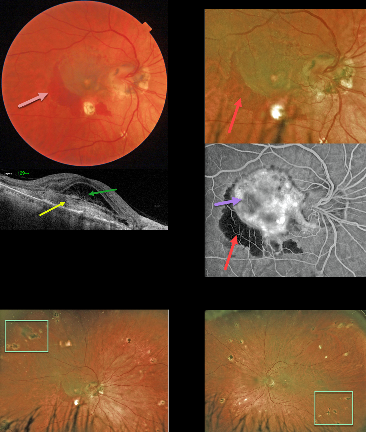
OCULAR HISTOPLASMOSIS
Originally posted on @retina.rocks December 16, 2022
This 52 YO female presented to the clinic with vision of counting fingers OD and 20/25 OS. She reported that her vision had decreased 3 weeks earlier.
Color imaging shows a somewhat pigmented macular neovascularization (MNV) with associated subretinal blood.
Swept-source OCT shows a hyperreflective, laminated type 2 (located above the RPE) MNV. Overlying fibrinous-appearing subretinal fluid vs a bacillary layer detachment is seen.
Optos fluorescein angiography, magnified to show the macular details, show a leaking, hyperfluorescent MNV with adjacent blockage from the subretinal hemorrhage.
Ultrawidefield Optos imaging shows the extent of the bilateral peripheral multifocal chorioretinal scarring. Anti-VEGF therapy was started.
Learning Points:
Ocular histoplasmosis is caused by the Histoplasmosis capsulatum fungus, which is commonly found in soil contaminated by bird or bat droppings.
It is seen worldwide, but in the United States is most often seen in the Ohio-Mississippi River valley. Most people in endemic areas inhale the organism’s spores early in life, with only a few percent developing asymptomatic chorioretinal scars.
A small percentage of these will develop MNV later in life. Treatment is similar to wet AMD, with anti-VEGF therapy usually being the first-line therapy. Unlike AMD, these patients often do not require ongoing injections. Photodynamic therapy can help minimize the treatment burden, and thermal laser is still a good option for more peripheral macular lesions.
OCULAR HISTOPLASMOSIS
Originally posted on @retina.rocks September 28, 2022
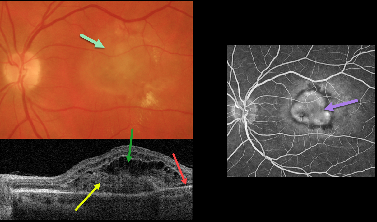
This 54YO female presented with recent vision loss in her left eye. Vision was 20/400 from an ocular histoplasmosis (OHS) related macular neovascularization (MNV).
Optos color imaging shows opaque macular fluid with a few dots of peripheral blood. Multifocal peripheral OHS scars were noted (not shown).
Swept-source OCT shows a type 2 MNV (located above the RPE), overlying outer retinal fluid and trace temporal subretinal fluid.
Fluorescein angiography shows a well-defined subfoveal lesion. Anti-VEGF therapy was recommended.
Learning Points:
Ocular histoplasmosis is caused by the Histoplasmosis capsulatum fungus which is commonly found in soil contaminated by bird or bat droppings.
It is seen worldwide, but in the United States is most often seen in the Ohio-Mississippi River valley. Most people in endemic areas inhale the organism’s spores early in life with only a few percent developing asymptomatic chorioretinal scars.
A small percentage of these will develop MNV later in life. Treatment is similar to wet AMD, with anti-VEGF therapy usually being the first line therapy. Unlike AMD, these patients often do not require ongoing injections.
Photodynamic therapy can help minimize the treatment burden, and thermal laser is still a good option for more peripheral macular lesions.
OCULAR HISTOPLASMOSIS
Originally posted on @retina.rocks May 13, 2022
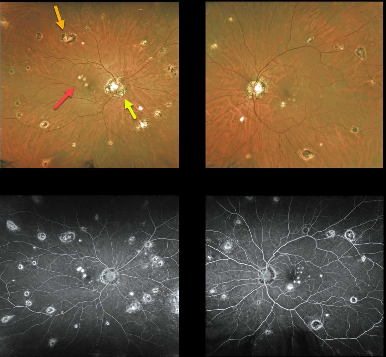
This 45YO female has classic bilateral findings for atrophic ocular histoplasmosis (OHS), including the triad of peripapillary scarring, macular lesions, and scattered peripheral scars. Vision is 20/25 OD and 20/30 OS. All scars stain on fluorescein angiography.
We like to add a fourth criterion, a quiet vitreous, to this classic triad. The presence of vitreous inflammation or new lesions is not seen in OH and would suggest an alternative condition, such as multifocal choroiditis.
Learning Points:
Atrophic OHS does not require treatment, but patients with macular or peripapillary scarring need to be monitored due to the risk for macular neovascularization.
Treatment is similar to wet AMD, with anti-VEGF therapy usually being the first-line therapy. Unlike AMD, these patients often do not require ongoing injections.
Photodynamic therapy can help minimize treatment burden, and thermal laser therapy remains a good option for more peripheral macular lesions.
OCULAR HISTOPLASMOSIS
Originally posted on @retina.rocks March 2, 2022
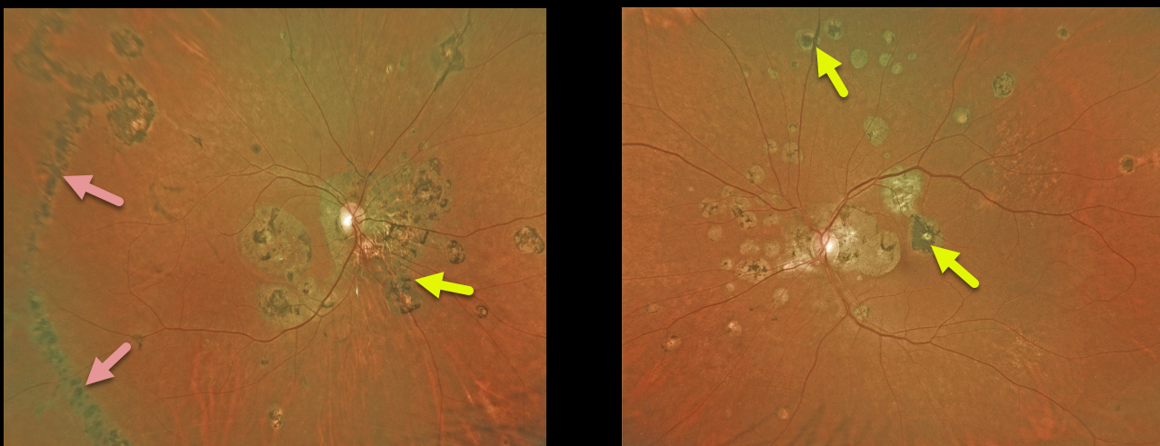
This 66YO female with a history of ocular histoplasmosis has extensive bilateral macular, peripapillary, and peripheral scarring. The right eye shows a large hyperpigmented temporal streak lesion. Vision is stable at counting vision OD and 20/40 OS.
Learning Points:
The incidence of streak lesions in OHS, also known as Schlaegel lines, is estimated at 5% (Arch Ophthalmol 1981;99:246-248), and in our experience, they are more common in areas of more extensive peripheral scarring. They appear as linear aggregations of midperipheral OHS scars that run parallel to the ora serrata.
They somewhat resemble lattice degeneration in location and appearance, but do not increase the patient’s risk of ocular complications.
OCULAR HISTOPLASMOSIS
Originally posted on @retina.rocks August 16, 2021
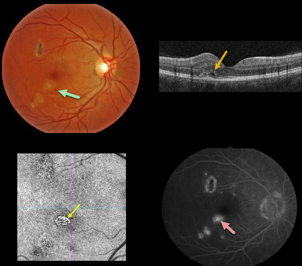
This 30YO male presented with 20/25 vision in his right eye from an ocular histoplasmosis-related macular neovascularization (MNV). Some fluid and blood were associated with a macular histoplasmosis scar in the inferotemporal fovea.
A type 2 MNV, located above the RPE, was noted on OCT. The MNV was best imaged on OCT angiography, with macular leakage noted on fluorescein angiography.
Anti-VEGF therapy was recommended.
Learning Points:
Ocular histoplasmosis is caused by the fungus Histoplasma capsulatum, which is commonly found in soil contaminated by bird or bat droppings. It is found worldwide, but in the United States, it is most common in the Ohio-Mississippi River valleys.
The vast majority of people in endemic areas inhale the organism’s spores early in life, and only a few percent will develop asymptomatic chorioretinal scars.
A small percentage of these will develop MNV later in life. Treatment is similar to wet AMD, with anti-VEGF therapy usually being the first-line therapy. Unlike AMD, these patients often do not require ongoing injections.
Photodynamic therapy can help minimize treatment burden, and thermal laser therapy remains a good option for more peripheral macular lesions.
OCULAR HISTOPLASMOSIS
Originally posted on @retina.rocks April 21, 2021
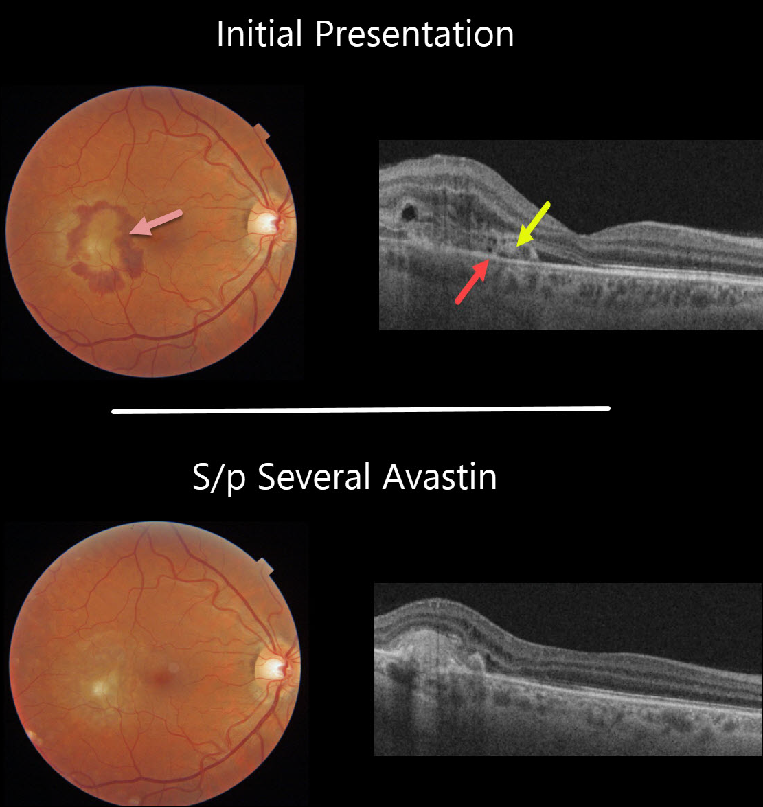
This 44YO male presented with a symptomatic macular neovascularization (MNV) from ocular histoplasmosis. Vision was 20/40.
Subretinal blood surrounds the MNV in the temporal macula. On OCT, the MNV is located above the RPE, indicating a type 2 lesion. Some retinal thickening is noted above the MNV.
The blood and fluid resolved completely following several additional monthly Avastin injections.
Learning Points:
There are three types of MNV: type 1 MNV is below the RPE, type 2 is between the RPE and outer segments, and type 3 is intraretinal.
OCULAR HISTOPLASMOSIS
Originally posted on @retina.rocks March 10, 2021
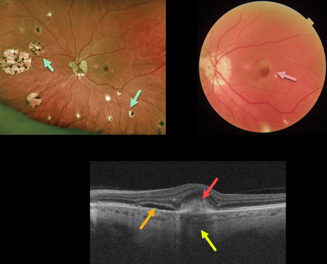
This 65YO presented with 20/400 vision in the left eye. Optos imaging shows classic ocular histoplasmosis (OHS) peripapillary, macular, and peripheral chorioretinal scarring. A more close-up macular image from the Triton swept-source OCT shows a greenish macular neovascularization (MNV) with associated subretinal blood.
The OCT B-scan shows subretinal fluid and hyperreflective subretinal blood, which causes choroidal shadowing.
Learning Points:
Ocular histoplasmosis is caused by the Histoplasmosis capsulatum fungus, which is commonly found in soil contaminated by bird or bat droppings. It is found worldwide, but in the United States, it is most common in the Ohio-Mississippi River valleys.
The vast majority of people in endemic areas inhale the organism’s spores early in life, and only a few percent will develop asymptomatic chorioretinal scars. A small percentage of these will develop MNV later in life.
Originally described by Woods and Wahlen in 1959, the findings include the classic triad of peripapillary atrophy, peripheral multifocal punched-out scars, and a macular scar or neovascularization.
We like to add a fourth criterion: a quiet vitreous, since numerous inflammatory disorders can simulate ocular histoplasmosis.
OCULAR HISTOPLASMOSIS
Originally posted on @retina.rocks October 29, 2020
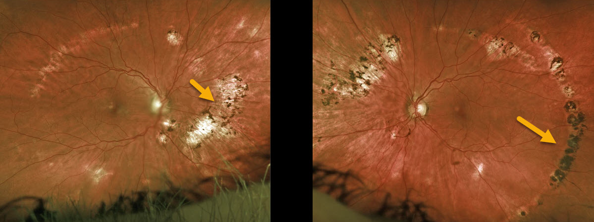
This is a beautiful example of bilateral streak lesions in a patient with the ocular histoplasmosis syndrome (OHS).
Learning Points:
The incidence of streak lesions (Schlaegel lines) in OHS is estimated at about 5% (Arch Ophthalmol 1981;99:246-248), and in our experience, they are more common in areas of more extensive peripheral scarring.
They appear as linear aggregations of midperipheral OHS scars that run parallel to the ora serrata.
They somewhat resemble lattice degeneration in location and appearance, but do not increase the patient’s risk of ocular complications.
OCULAR HISTOPLASMOSIS
Originally posted on @retina.rocks May 29, 2020
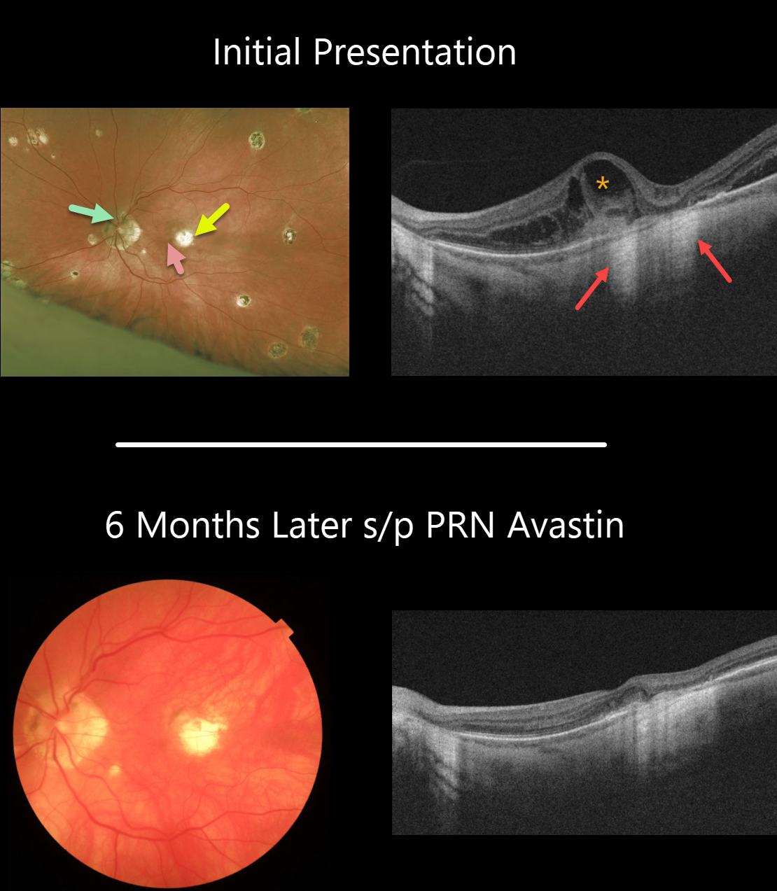
Our patient has 20/20 vision OD and 20/400 OS. He has classic peripapillary atrophy, macular and peripheral scarring.
He came in with symptomatic vision loss in his left eye with a subtle nasal foveal subretinal hemorrhage from an acute macular neovascularization (MNV) adjacent to a pre-existing chorioretinal scar.
The OCT line scan surprisingly shows significant intraretinal fluid overlying the hemorrhage and temporal to the clinical scar. The loss of the RPE at the chorioretinal scar allows OCT to show increased hyperreflectance of the underlying choroid and sclera. This is the opposite of shadowing.
Six months later, vision in the left eye is 20/200, and the blood and fluid remain completely resolved with PRN Avastin.
Learning Points:
Ocular histoplasmosis is caused by the Histoplasmosis capsulatum fungus, which is commonly found in soil contaminated by bird or bat droppings. It is found worldwide, but in the United States, it is most common in the Ohio-Mississippi River valleys.
The vast majority of people in endemic areas inhale the organism’s spores early in life, and only a few percent will develop asymptomatic chorioretinal scars. A small percentage of these will develop MNV later in life.
Originally described by Woods and Wahlen in 1959, the findings include the classic triad of peripapillary atrophy, peripheral multifocal punched-out scars, and a macular scar or neovascularization.
We like to add a fourth criterion: a quiet vitreous, since numerous inflammatory disorders can simulate ocular histoplasmosis.
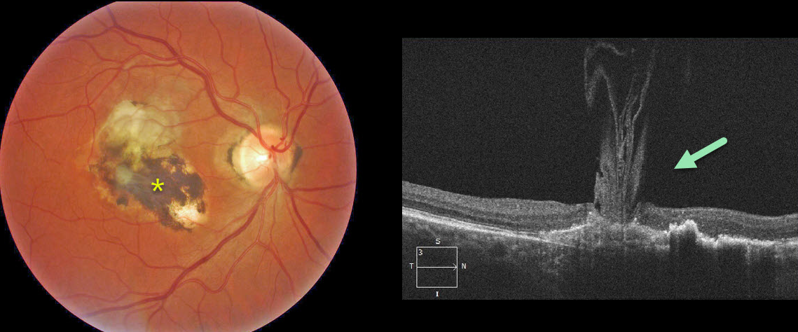
VOLCANIC MACULOPATHY
Originally posted on @retina.rocks January 20, 2020
The OCT scan shows unusual vitreous strands adhering to an inactive histoplasmosis scar resembling an erupting volcano.


