

Ocular Ischemia
<< Back to Cases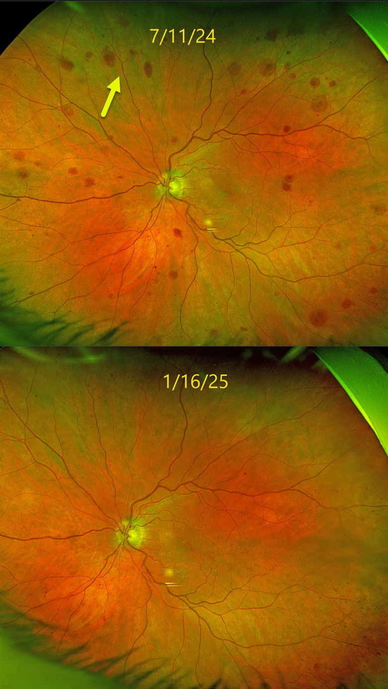
OCULAR ISCHEMIA
Originally posted on @retina.rocks March 3, 2025
This 83YO male was referred for asymptomatic changes in his left fundus. Vision was 20/30 in his healthy OD and 20/100 OS.
Optos color RG imaging shows scattered deep, variably round retinal hemorrhages extending from the peripheral macula into the midperiphery. Carotid Doppler testing revealed a critical stenosis of the left internal carotid artery, and he was emergently referred to his cardiologist. The findings dramatically improved within months of carotid surgery.
Learning Points:
The retinal hemorrhages in the ocular ischemic syndrome (OIS) are unique. Retinal vascular disorders like diabetic retinopathy, retinal vein occlusions, and hypertensive retinopathy usually have flame- and dot-shaped inner retinal hemorrhages concentrated around the posterior pole.
Our patient’s findings are classic for OIS, with hemorrhages in the deeper peripheral retina. The retinal periphery is literally the end of the line for the retinal arterial circulation. In an eye receiving less blood supply due to carotid occlusive disease, the more proximal retina has first dibs on the available oxygen. There are penetrating capillaries that dive radially from the nerve fiber and ganglion cell layers into the deeper retina. The deep vascular complex that supplies the inner and outer plexiform layers is thus furthest downstream, and these endothelial cells are likely damaged by OIS. Blood cells leaking into this space accumulate, giving the large outer blot hemorrhages.
RETINAL HEMORRHAGES
Originally posted on @retina.rocks February 12, 2024
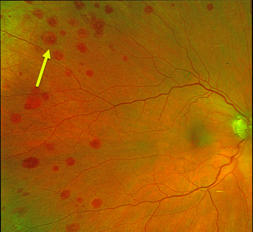
This 79YO female was referred for asymptomatic retinal findings. She has a history of diet-controlled type 2 diabetes.
Optos color RG imaging shows scattered, round, deep retinal hemorrhages, some with white centers. Most of the midperipheral hemorrhages are much more extensive in her right eye.
Bloodwork, including CBC, serum viscosity, and serum protein electrophoresis, was normal. We suspect ocular ischemia as the cause of her findings, but despite numerous attempts, the patient continues to refuse carotid testing.
Learning Points:
The term “Roth spot” was named after Dr. Moritz Roth, who, in 1872, noted white-centered hemorrhages in patients with bacterial endocarditis. He felt that these white spots were septic emboli that originated from an infected cardiac valve. However, similar lesions can be seen in many other disorders, including leukemia, hyperviscosity, anemia, diabetes, and hypertension (Duane et al Ophthalmology 1980;87:66-69).
The white material may represent a variety of causes, including fibrin. So these lesions are best called white-centered hemorrhages, unless one is specifically referring to one of these lesions in a patient with bacterial endocarditis.
OCULAR ISCHEMIC SYNDROME
Originally posted on @retina.rocks April 10, 2023
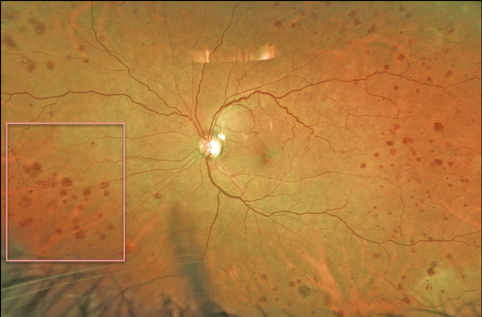
This 70YO female was referred for asymptomatic unilateral retinal hemorrhages in her left eye. Vision was 20/30. Optos imaging shows encircling midperipheral deep outer retinal hemorrhages.
Since our patient had a known history of complete left internal carotid artery blockage, no further testing was ordered. We will follow her several times yearly due to the risk of ocular neovascularization.
Learning Points:
The retinal hemorrhages in the ocular ischemic syndrome (OIS) are unique. Retinal vascular disorders like diabetic retinopathy, retinal vein occlusions, and hypertensive retinopathy usually have flame- and dot-shaped inner retinal hemorrhages concentrated around the posterior pole.
Our patient’s findings are classic for OIS, with hemorrhages in the deeper peripheral retina. The retinal periphery is literally the end of the line for the retinal arterial circulation.
In an eye receiving less blood supply due to carotid occlusive disease, the more proximal retina has first dibs on the available oxygen. There are penetrating capillaries that dive radially from the nerve fiber and ganglion cell layers into the deeper retina. The deep vascular complex that supplies the inner and outer plexiform layers is thus furthest downstream, and these endothelial cells are likely damaged in OIS. Blood cells leaking into this space accumulate, causing the large outer blot to hemorrhage.
OCULAR ISCHEMIA
Originally posted on @retina.rocks July 18, 2022
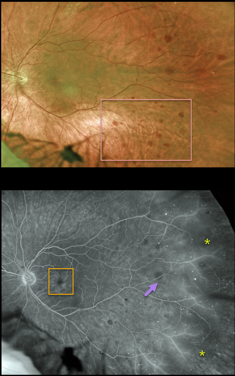
This 69YO male presented with a few months of blurred vision in his left eye. Vision was 20/40 in his normal right eye and 20/100 in his left eye.
Scattered deep retinal hemorrhages are noted temporally. Fluorescein angiography shows temporal midperipheral microaneurysms, more distal ischemia with vascular leakage, and moderate cystoid macular leakage.
Since our patient had a history of a complete left internal carotid artery blockage, no further testing was ordered. He is currently monitored regularly by a vascular surgeon.
Learning Points:
The retinal hemorrhages in the ocular ischemic syndrome (OIS) are unique. Retinal vascular disorders like diabetic retinopathy, retinal vein occlusions, and hypertensive retinopathy usually have flame- and dot-shaped inner retinal hemorrhages concentrated around the posterior pole.
Our patient’s findings are classic for OIS, with hemorrhages in the deeper peripheral retina. The retinal periphery is literally the end of the line for the retinal arterial circulation. In an eye receiving less blood supply due to carotid occlusive disease, the more proximal retina has first dibs on the available oxygen.
There are penetrating capillaries that dive radially from the nerve fiber and ganglion cell layers into the deeper retina. The deep vascular complex that supplies the inner and outer plexiform layers is thus furthest downstream, and these endothelial cells are likely damaged in OIS. Blood cells leaking into this space accumulate, causing the large outer blot to hemorrhage.
These patients need to have carotid Doppler and ultrasound and referred for medical or surgical intervention if they have a critical stenosis. They may also develop anterior or posterior segment neovascularization requiring photocoagulation and/or anti-VEGF injections.
OCULAR ISCHEMIA
Originally posted on @retina.rocks August 17, 2021
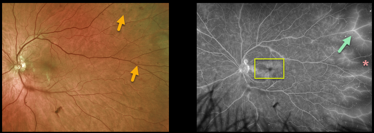
This 62YO male presented with a few months of non-specific blurred vision in his left eye. Vision was 20/30 in his normal right eye and 20/40 in his left eye.
Scattered deep retinal hemorrhages were noted temporally OS. Optos fluorescein angiography showed temporal ischemia with diffuse vascular staining, along with cystoid macular edema.
On further questioning, he described a several-minute episode of transient vision loss in his left eye a few days earlier. An emergent carotid Doppler was performed, which revealed a critical stenosis of his left internal carotid artery. He was immediately referred to a vascular surgeon.
Learning Points:
The retinal hemorrhages in ocular ischemic syndrome (OIS) are unique. Retinal vascular disorders like diabetic retinopathy, retinal vein occlusions, and hypertensive retinopathy usually have flame- and dot-shaped inner retinal hemorrhages concentrated in the posterior pole.
Our patient’s findings are classic for OIS, with hemorrhages in the deeper peripheral retina. The retinal periphery is literally the end of the line for the retinal arterial circulation. In an eye receiving less blood supply due to carotid occlusive disease, the more proximal retina has first dibs on the available oxygen. There are penetrating capillaries that dive radially from the nerve fiber and ganglion cell layers into the deeper retina. The deep vascular complex that supplies the inner and outer plexiform layers is thus the furthest downstream, and these endothelial cells are likely damaged in OIS. Blood cells leaking into this space accumulate, causing the large outer blot to hemorrhage.
These patients need to have a carotid Doppler and be referred for medical or surgical intervention if they have a critical stenosis. They may also develop anterior or posterior segment neovascularization requiring photocoagulation and/or anti-VEGF injections. Iris neovascularization can develop, leading to neovascular glaucoma. Hypotony can also occur.
OCULAR ISCHEMIA
Originally posted on @retina.rocks April 7, 2021
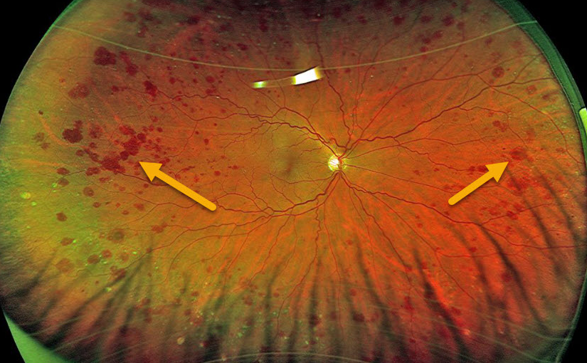
This patient presented with these asymptomatic, unilateral, peripheral, large, outer retinal hemorrhages.
The patient was later diagnosed with a 100% internal carotid artery occlusion. Carotid surgery was not recommended, but we are monitoring the patient due to the risk for anterior or posterior segment neovascularization.
Learning Points:
The retinal hemorrhages in ocular ischemic syndrome (OIS) are somewhat unique. Retinal vascular disorders like diabetic retinopathy, retinal vein occlusions, and hypertensive retinopathy usually have flame- and dot-shaped inner retinal hemorrhages concentrated around the posterior pole.
Our patient’s findings are classic for OIS, with hemorrhages in the deeper peripheral retina. The retinal periphery is literally the end of the line for the retinal arterial circulation. In an eye receiving less blood supply due to carotid occlusive disease, the more proximal retina has first dibs on the available oxygen. There are penetrating capillaries that dive radially from the nerve fiber and ganglion cell layers into the deeper retina. The deep vascular complex that supplies the inner and outer plexiform layers is thus furthest downstream, and these endothelial cells are likely damaged in OIS. Blood cells leaking into this space accumulate, causing the large outer blot to hemorrhage.
These patients need to have a carotid Doppler and ultrasound and be referred for medical or surgical intervention if they have a critical stenosis. They may also develop anterior or posterior segment neovascularization requiring photocoagulation and/or anti-VEGF injections. Iris neovascularization can develop, leading to neovascular glaucoma. Hypotony can also occur.
OCULAR ISCHEMIA WITH OZURDEX IMPLANTS
Originally posted on @retina.rocks February 4, 2021
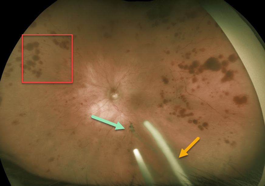
This 86YO patient was receiving Ozurdex (dexamethasone 0.7%) injections every 8 weeks in her left eye for macular edema from an inferior hemiretinal vein occlusion.
She subsequently developed ocular ischemic syndrome (OIS) like changes with scattered peripheral large outer retinal hemorrhages. Inferiorly, more recent Ozurdex implants are noted, along with another ghost-like remnant of an older implant. Although the Ozurdex implants are supposed to completely degrade, these remnants may persist beyond 1 year (see Kim et al Retina 2020;40;2226-2231).
Learning Points:
The retinal hemorrhages in ocular ischemic syndrome (OIS) are unique. Retinal vascular disorders like diabetic retinopathy, retinal vein occlusions, and hypertensive retinopathy usually have flame- and dot-shaped inner retinal hemorrhages concentrated around the posterior pole.
Our patient’s findings are classic for OIS, with hemorrhages in the deeper peripheral retina. The retinal periphery is literally the end of the line for the retinal arterial circulation. In an eye receiving less blood supply due to carotid occlusive disease, the more proximal retina has first dibs on the available oxygen. There are penetrating capillaries that dive radially from the nerve fiber and ganglion cell layers into the deeper retina. The deep vascular complex that supplies the inner and outer plexiform layers is thus furthest downstream, and these endothelial cells are likely damaged in OIS. Blood cells leaking into this space accumulate, causing the large outer blot to hemorrhage.
These patients need to have a carotid Doppler and ultrasound and be referred for medical or surgical intervention if they have a critical stenosis. They may also develop anterior or posterior segment neovascularization requiring photocoagulation and/or anti-VEGF injections. Iris neovascularization can develop, leading to neovascular glaucoma. Hypotony can also occur.
OCULAR ISCHEMIA
Originally posted on @retina.rocks September 8, 2020
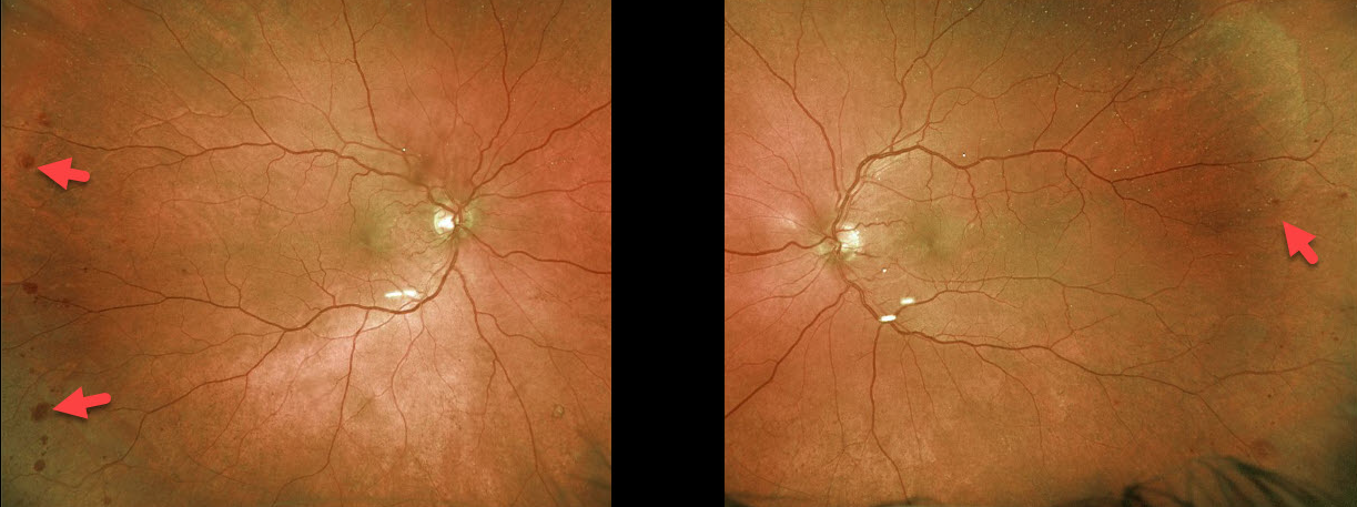
This 71YO patient has bilateral, mostly temporal, peripheral deep blot retinal hemorrhages. He had a known history of bilateral carotid stenosis.
Our patient’s findings are classic for ocular ischemic syndrome (OIS), with hemorrhages in the deeper retina of the temporal periphery.
These patients need to have a carotid Doppler and ultrasound and be referred for medical or surgical intervention for critical stenosis. They may also develop anterior or posterior segment neovascularization requiring photocoagulation and/or anti-VEGF injections. Neovascular glaucoma or hypotony can also occur.
Learning Points:
The retinal hemorrhages in OIS are somewhat unique. Retinal vascular disorders such as diabetic retinopathy, central retinal vein occlusion, and hypertensive retinopathy typically present with flame- and dot-shaped retinal hemorrhages concentrated around the posterior pole, located in the inner retina.
The retinal periphery is literally the end of the line for the retinal arterial circulation. In an eye receiving less blood supply due to carotid occlusive disease, the more proximal retina has first dibs on the available oxygen. There are penetrating capillaries that dive radially from the nerve fiber and ganglion cell layers into the deeper retina. The deep vascular complex that supplies the inner and outer plexiform layers is thus furthest downstream, and these endothelial cells are likely damaged in OIS. Blood cells leaking into this space accumulate, causing the large outer blot to hemorrhage.


