

Optic Nerve Coloboma
<< Back to Cases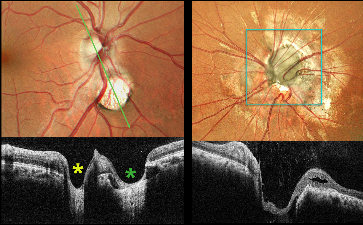
MORNING GLORY DISC AND CHOROIDAL COLOBOMA WITH PSEUDODUPLICATED OPTIC NERVE
Shraddha Raj Shrivastava and Manish Nagpal
Originally posted on @retina.rocks June 30, 2026
This 26YO male presented with a history of stable poor vision and inward deviation of his left eye since childhood. Vision was 20/20 OD and counting fingers in his esotropic, microphthalmic OS.
Color photography of the right posterior pole shows a round coloboma inferonasal to the nerve, giving the appearance of a pseudo-duplicated disc. OCT scanning through the nerve and coloboma reinforces the illusion of a second nerve head. The anomalous left nerve is enlarged and excavated, with numerous vessels radiating outwards from its inferonasal aspect.
Morning glory syndrome is typically unilateral and shares some similarities with optic disc coloboma and juxtapapillary staphyloma. This abnormality is named for its striking resemblance to the Morning Glory flower. Visual prognosis is usually poor. The condition can be associated with serous macular detachment. Neuroimaging should be considered to rule out transsphenoidal encephalocele and intracranial carotid artery dysgenesis, and fortunately, our patient’s imaging was negative. For a great review on the pathogenesis and treatment of maculopathy associated with cavitary optic disc anomalies, see Jain and Johnson, AJO 2014;158:423-435.
The coexistence of morning glory disc anomaly with microphthalmos, and contralateral chorioretinal coloboma in this case, may represent a continuum of optic fissure developmental defects with asymmetric expression, leading to a spectrum of cavitary optic disc anomalies in the same individual.
Pseudoduplication of the optic nerve occurs when a round choroidal coloboma or chorioretinal scar about the size of the optic nerve is associated with overlying radiating blood vessels (Bloom et al, Retinal Cases 2022;16:174-176). In our experience, this fake-out is most common from toxoplasmosis scars located near the optic nerve.
OPTIC NERVE PIT AND COLOBOMA
Kanwaljeet Harjot Madan
Originally posted on @retina.rocks June 13, 2024
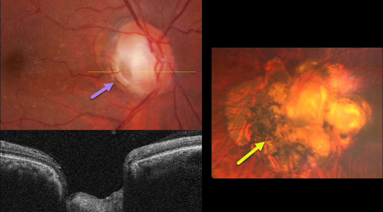
This healthy 63YO male was referred for cataract surgery in his right eye. He had a lifelong history of poor vision in his left eye. Vision was 20/60 OD and counting vision OS.
Color fundus imaging and OCT of the right eye show a grey inferotemporal optic nerve pit. Marked peripapillary scarring surrounds the left nerve, which is anomalous with an inferotemporal disc coloboma.
Learning Points:
An optic nerve pit and coloboma are cavitated abnormalities that can be a unilateral or bilateral condition and are caused by incomplete closure of the embryonic fissure. While most lesions are asymptomatic, they can cause vision loss by forcing fluid into the inner and outer retina, and finally into the subretinal space (optic pit maculopathy). Our patient currently shows no evidence of this.
Cataract surgery was performed on his right eye, and vision improved to 20/20. Observation for the disc abnormalities was recommended.
CHOROIDAL COLOBOMA WITH OPTIC NERVE PSEUDODUPLICATION
Originally posted on @retina.rocks March 25, 2024
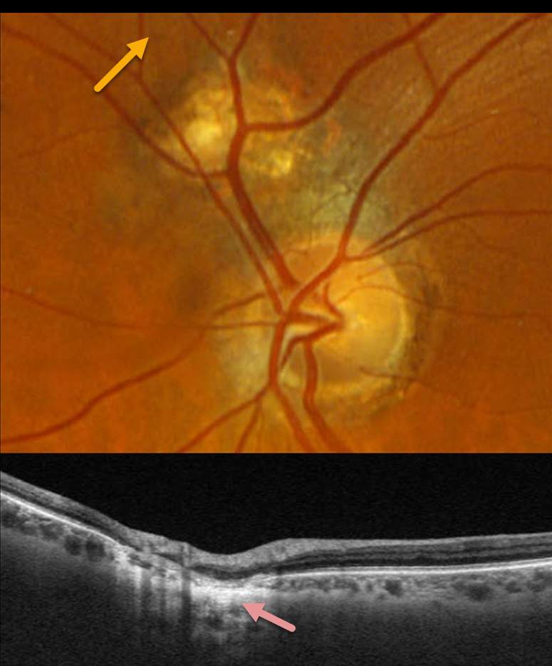
This 66YO male was referred for an asymptomatic lesion in his left fundus. Vision was 20/25 in his normal right eye and 20/25 in his left eye.
Optos color RGB imaging of the left eye shows a normal optic nerve. Just superonasal to the nerve is a round, colobomatous scar.
Triton swept-source OCT through this lesion shows disorganized neurosensory thinning with an underlying choroidal coloboma.
Learning Points:
Pseudoduplication of the optic nerve occurs when a round choroidal coloboma or chorioretinal scar about the size of the optic nerve is associated with overlying radiating blood vessels (Bloom et al, Retinal Cases 2022;16:174-176). In our experience, it is most common for toxoplasmosis scars to be located near the optic nerve.
OPTIC NERVE COLOBOMA AND PIT
Originally posted on @retina.rocks December 2, 2022
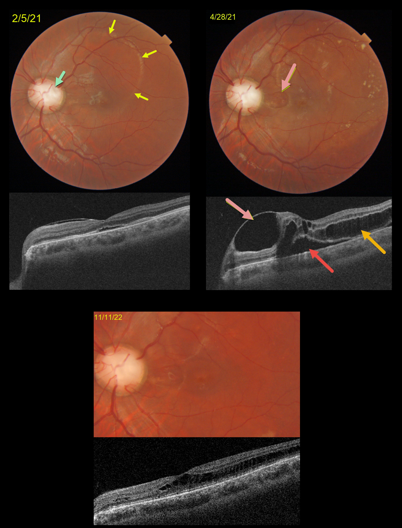
This is a follow-up to our 2/5/21 and 4/28/21 posts of a patient with bilateral optic nerve coloboma with optic pit maculopathy in his left eye.
On 2/5/21, vision was 20/70 with an optic pit and macular fluid. Despite the mostly peripheral fluid, the macular center was normal except for a trace amount of subretinal fluid (SRF).
Two months later, the peripheral fluid was better, but the central OCT findings were markedly different, with a nasal inner cyst, temporal outer schisis, and increased SRF. Although vision had decreased to 20/200, he subjectively felt his vision was better and elected continued observation.
When examined 1.5 years later on 11/11/22, the OCT findings had spontaneously improved. Temporal outer retinal schisis extends through the macular center, which is now free of SRF. Vision was 20/100, and he remained visually asymptomatic.
Learning Points:
An optic nerve coloboma can be a unilateral or bilateral condition caused by incomplete closure of the embryonic fissure.
While most optic pits are asymptomatic, they can cause vision loss due to fluid being forced into the inner retina, outer retina, and finally the subretinal space (optic pit maculopathy). The source of the fluid continues to be a point of ongoing debate, coming from either the vitreous or subarachnoid space.
A host of treatments for optic pit maculopathy have been suggested over the years. However, a recent meta-analysis suggested that a plain old vanilla vitrectomy without gas tamponade is as successful as any other procedure (Zheng et al Ophthalmology Retina 2020;4:289-299).
Our case illustrates how this fluid can spontaneously wax and wane, and that sometimes simple observation is the best option for some patients.
OPTIC NERVE PIT
Originally posted on @retina.rocks October 20, 2022
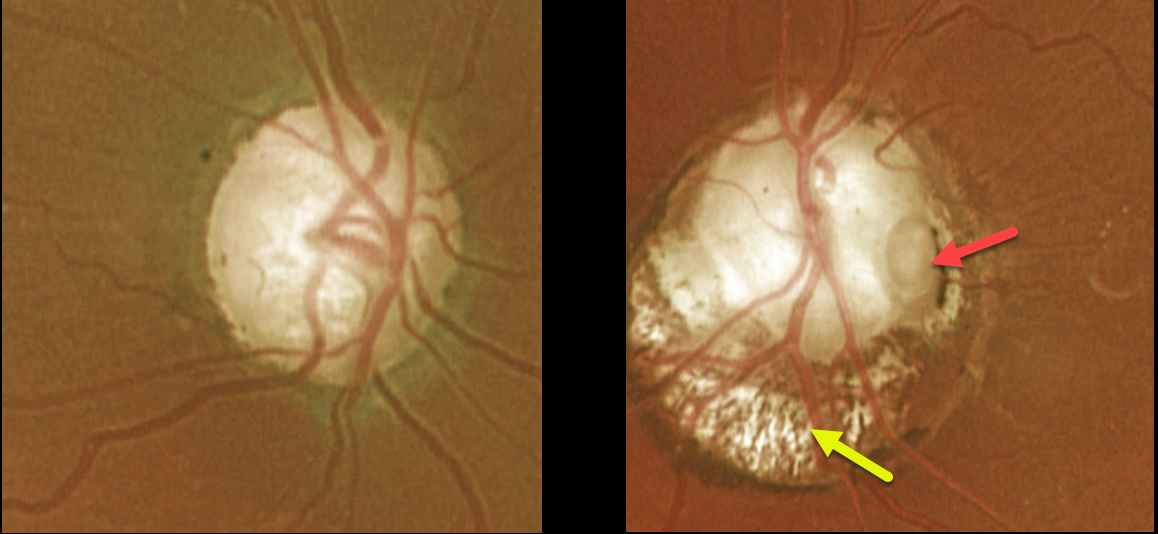
This 66YO female presented with an asymptomatic left optic nerve coloboma with a large temporal pit. The mildly colobomatous portion of the nerve is enlarged and slightly excavated inferiorly. Vision was 20/25 OD and 20/200 OS. Observation was recommended.
Learning Points:
Optic pits are most commonly located within or along the inferior-temporal disc margin and usually appear as a gray depression. They are thought to be caused by incomplete closure of the optic fissure during gestation.
While most optic pits are asymptomatic (as in this case), they can cause vision loss by forcing fluid into the inner and outer retina and, ultimately, the subretinal space (optic pit maculopathy).
The source of the fluid remains a subject of ongoing debate, with origins in either the vitreous or the subarachnoid space.
OPTIC NERVE COLOBOMA
Originally posted on @retina.rocks May 9, 2022
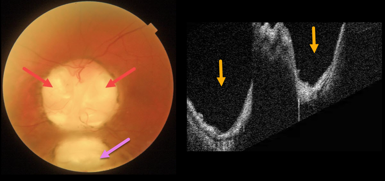
This 66YO male has mild lifelong decreased vision in his left eye. Vision was 20/70.
Triton imaging of the optic nerve coloboma shows optic nerve pits temporally and nasally, as well as a more inferior choroidal coloboma. Swept-source OCT dramatically reveals the pits.
We are following him yearly.
Learning Points:
Both optic nerve and choroidal colobomas can be unilateral or bilateral and are caused by incomplete closure of the embryonic fissure.
Patients with choroidal colobomas are at increased lifetime risk of rhegmatogenous retinal detachment due to breaks in the overlying thinned retina.
Optic nerve pits are also associated with serous macular detachment and macular neovascularization.
For a great review of cavitary optic nerve abnormalities, see Jain and Johnson, AJO 2014;158:423-435.
OPTIC NERVE COLOBOMA
Originally posted on @retina.rocks April 4, 2022
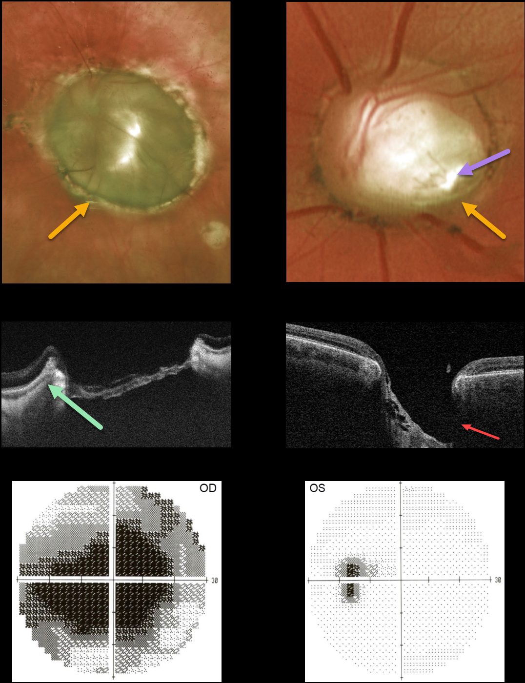
This 45YO female presented with bilateral optic nerve colobomas. Vision was hand motion OD and 20/25 OS.
Triton swept-source OCT shows a relatively featureless retina spanning the coloboma. The much smaller left nerve coloboma also contains an inferotemporal optic disc pit. The pit is also quite evident on OCT. The left nerve has a morning glory appearance with multiple vessels radiating outward from the peripheral disc margin, although there is no central glial tissue.
Visual fields show a large central defect OD with an enlarged blind spot OS.
Learning Points:
Optic nerve cavitary abnormalities can be unilateral or bilateral and result from incomplete closure of the embryonic fissure.
Our patient shows a spectrum of these abnormalities, including a huge optic disc coloboma OD, a much smaller disc coloboma OS with morning glory-like features, and an optic pit.
For a great review of cavitary optic nerve abnormalities, see Jain and Johnson, AJO 2014;158:423-435.
OPTIC NERVE COLOBOMA
Originally posted on @retina.rocks August 25, 2021
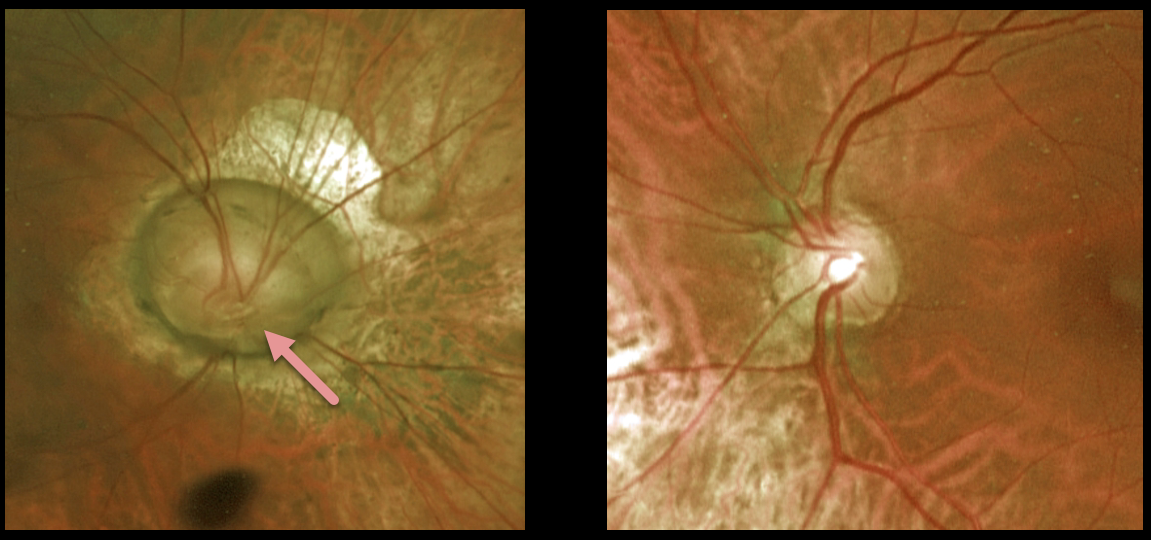
This 55YO female presented with a unilateral optic nerve coloboma in her right eye. Vision was a remarkable 20/50 in her right eye and 20/30 in her normal left eye.
Learning Points:
Optic nerve colobomas can be a unilateral or bilateral condition, and, similar to other cavitary optic disc abnormalities, are caused by incomplete closure of the embryonic fissure. They can have an associated optic nerve pit, which was not seen in this patient. For a great review of cavitary optic nerve abnormalities, see Jain and Johnson, AJO 2014;158:423-435.
OPTIC NERVE COLOBOMA
Originally posted on @retina.rocks April 28, 2021
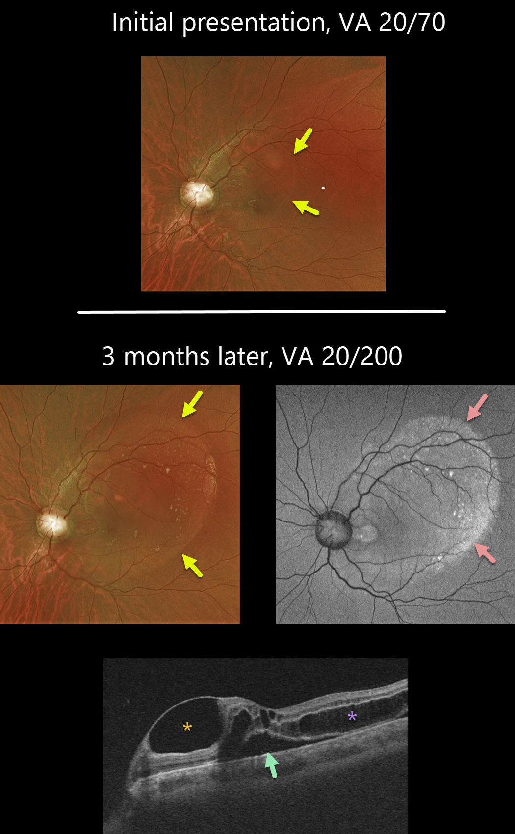
This 19YO male has bilateral optic nerve coloboma with optic pit maculopathy in his left eye.
On initial presentation, vision was 20/70, which decreased to 20/200 at 3-month follow-up. The extent of the macular schisis/fluid dramatically enlarged, accounting for the drop in vision.
Optos fundus autofluorescence (FAF) showed variable hyper-FAF along the temporal edge of the fluid.
Triton OCT imaging showed an unusual inner retinal cyst, as well as subretinal fluid and outer macular schisis.
Remarkably, the patient felt his vision had improved subjectively and did not wish to consider the recommended vitrectomy procedure.
Learning Points:
While most optic pits are asymptomatic, they can cause vision loss by causing fluid to accumulate in the inner and outer retina and the subretinal space (optic pit maculopathy). The source of the fluid remains a point of ongoing debate, with potential contributions from the vitreous and/or subarachnoid space.
OPTIC NERVE COLOBOMA
Originally posted on @retina.rocks February 5, 2021
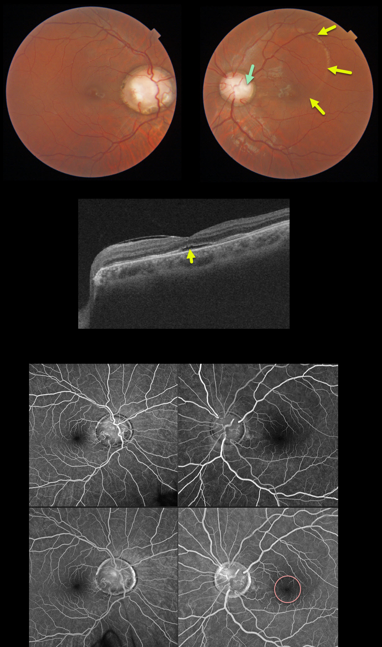
Our patient had bilateral optic nerve colobomas with an optic pit in the left eye. Vision was 20/40 OD and 20/70 OS.
The serous fluid from the optic pit is in the superior macula with mild fluid in the subfoveal space, best noted on the macular OCT. Fluorescein angiography shows no leakage, indicating that this fluid is not exudative.
Our patient opted for no treatment due to minimal symptoms and fairly good vision.
Learning Points:
An optic nerve coloboma can be unilateral or bilateral and is caused by incomplete closure of the embryonic fissure.
While most optic pits are asymptomatic, they can cause vision loss by forcing fluid into the inner and outer retina and, ultimately, the subretinal space (optic pit maculopathy). The source of the fluid remains a point of ongoing debate, with the fluid originating either from the vitreous or the subarachnoid space.
A host of treatments for optic pit maculopathy have been suggested over the years. However, a recent meta-analysis suggested that a plain old vanilla vitrectomy without gas tamponade is as successful as any other procedure (Zheng et al Ophthalmology Retina 2020;4;289-299).


