

Optic Nerve Conus
<< Back to Cases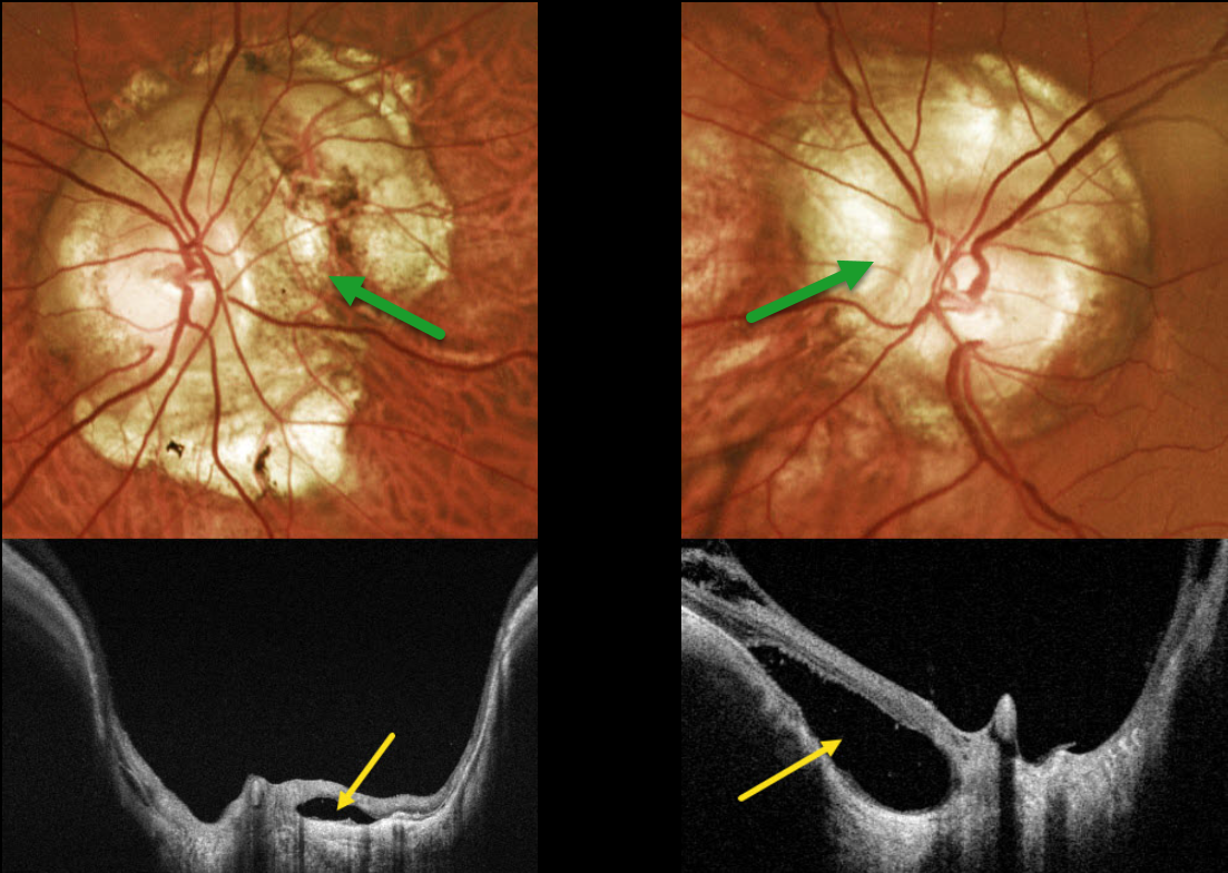
OPTIC NERVE CONUS + PERIPAPILLARY RETINAL DETACHMENT
Originally posted on @retina.rocks March 13, 2024
This 42YO male with high myopia was referred for asymptomatic optic nerve changes. Vision was 20/30 OD and 20/25 OS.
Optos color RG imaging shows bilateral peripapillary atrophy. Clinically, this scarring is markedly excavated from the surrounding retinal plane. Triton swept-source OCT confirms peripapillary staphyloma. Localized retinal detachments are noted bilaterally, particularly within the nasal aspect of atrophy around the left nerve. Within these detachments, there is variable loss of RPE, absence of chorioid, and variable retinal disorganization.
Learning Points:
An optic nerve conus, most commonly found in high myopes, is a staphylomatous-like depression of the optic nerve and surrounding tissue.
OPTIC NERVE CONUS
Will Gibson
Originally posted on @retina.rocks November 7, 2022
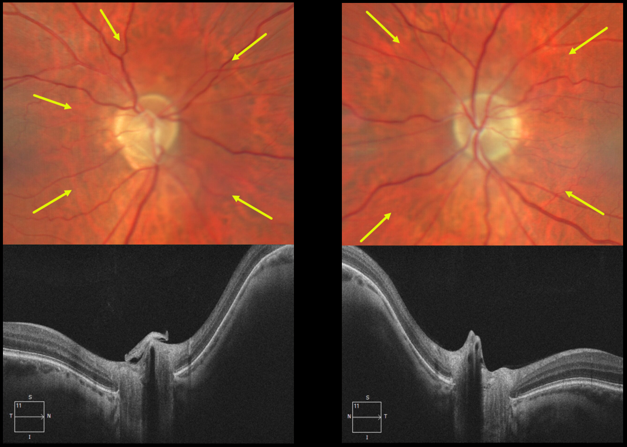
This 54YO male patient presented with asymptomatic bilateral optic nerve conus. Optos color imaging of each eye shows each optic nerve and peripapillary tissue to be sunken from the plane of the surrounding fundus. These findings are captured on OCT.
Learning Points:
An optic nerve conus is a staphylomatous-like depression of the optic nerve and surrounding tissue. This finding is usually seen in high myopia, although our patient had mild bilateral hyperopia.


