

Optic Nerve Drusen
<< Back to Cases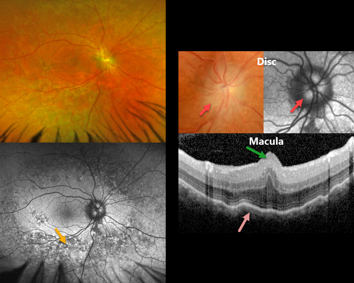
MFRP RETINITIS PIGMENTOSA
João Pedro Marques
Originally posted on @retina.rocks June 9, 2026
This 49YO male has a history of MFRP-related retinitis pigmentosa (RP). We have followed him for 13 years with stable findings of bilateral nanophthalmos (+18D hyperopia, axial lengths 14.82mm OD and 15.14 OS), optic disc drusen (ODD), and choroidal folds. He has compound heterozygosity for 2 pathogenic variants in the MFRP gene.
Optos color RG imaging in his OD shows pigmentary changes throughout the midperiphery along with an ill-defined disc margin. Fundus autofluorescence (FAF) shows variable hyper- and hypo-FAF in the midperiphery. Disc drusen are noted on a more magnified view, which hyper-FAF. Macular OCT shows a central fold, choroidal thickening, and chorioretinal folds. Identical findings were present in his OD (not shown).
Learning Points:
The MFRP (membrane frizzled-related protein) is expressed in the RPE and ciliary epithelium. MFRP functions as a molecular hub on the RPE apical membrane, coordinating protein trafficking and lipid homeostasis. Loss of MFRP leads to DHA accumulation in the RPE, downregulation of visual cycle genes and phototransduction genes, and progressive photoreceptor degeneration.
Biallelic MFRP mutations cause a well-characterized autosomal recessive retinitis pigmentosa (RP) syndrome with nanophthalmos and ODD (Li et al, BJO 2024;108;1679-1687). These eyes are at high risk of angle-closure glaucoma, usually requiring prophylactic laser peripheral iridotomy.
OPTIC NERVE DRUSEN
Ayushi Gupta
Originally posted on @retina.rocks November 25, 2025
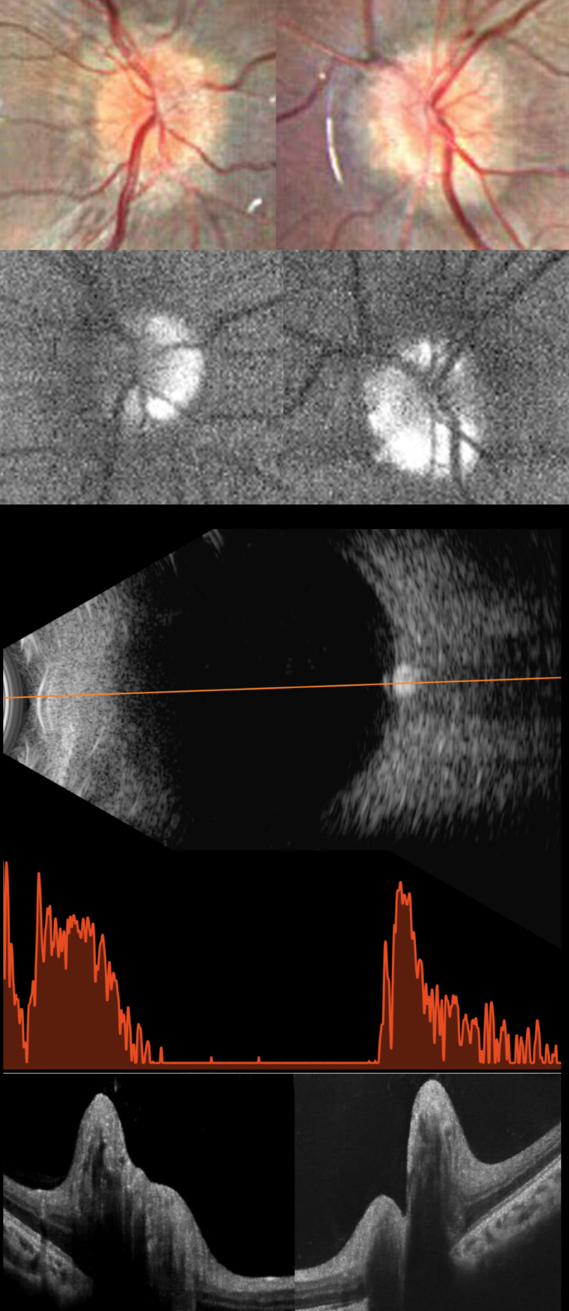
This healthy 11YO girl was diagnosed elsewhere 2 months earlier with papilledema and precocious puberty, which was being treated with oral acetazolamide. Workup included a normal brain MRI and lumbar puncture with normal opening pressure and CSF analysis. Vision in our office was 20/20 OU.
Fundus photography shows pseudopapilledema with extensive optic disc drusen (ODD). On fundus autofluorescence (FAF), the drusen are hyper-FAF. On B-scan ultrasonography, the discs are markedly hyperreflective, indicating calcification. OCT scanning shows marked, irregular disc thickening due to drusen.
Learning Points:
Optic disc drusen (ODD) are refractile calcific concretions that form anterior to the lamina cribrosa. They are thought to represent extruded, extracellular, calcified mitochondria and may be inherited as an autosomal dominant trait.
Porphyrins, organic molecules composed of four interconnected pyrrole subunits, are present within mitochondria. When these compounds break down, they become autofluorescent, which is likely why drusen autofluoresce. Porphyrins are also part of the heme molecule in hemoglobin, which explains why devitalized blood autofluoresces (Bloom and Spaide, Retinal Cases 2022;16:401-402).
ODD can be confused with true disc swelling, as occurred in our patient, which led to an erroneous diagnosis of papilledema. In our experience, the pathognomonic B-scan ultrasonographic disc calcification is rarely seen in patients this young, but is extremely helpful in establishing the diagnosis when present. We find FAF to be a more reliable test when the ODD are not obvious clinically. After speaking with her neurologist, the acetazolamide was discontinued, and observation was recommended.
OPTIC NERVE DRUSEN
Originally posted on @retina.rocks July 24, 2024
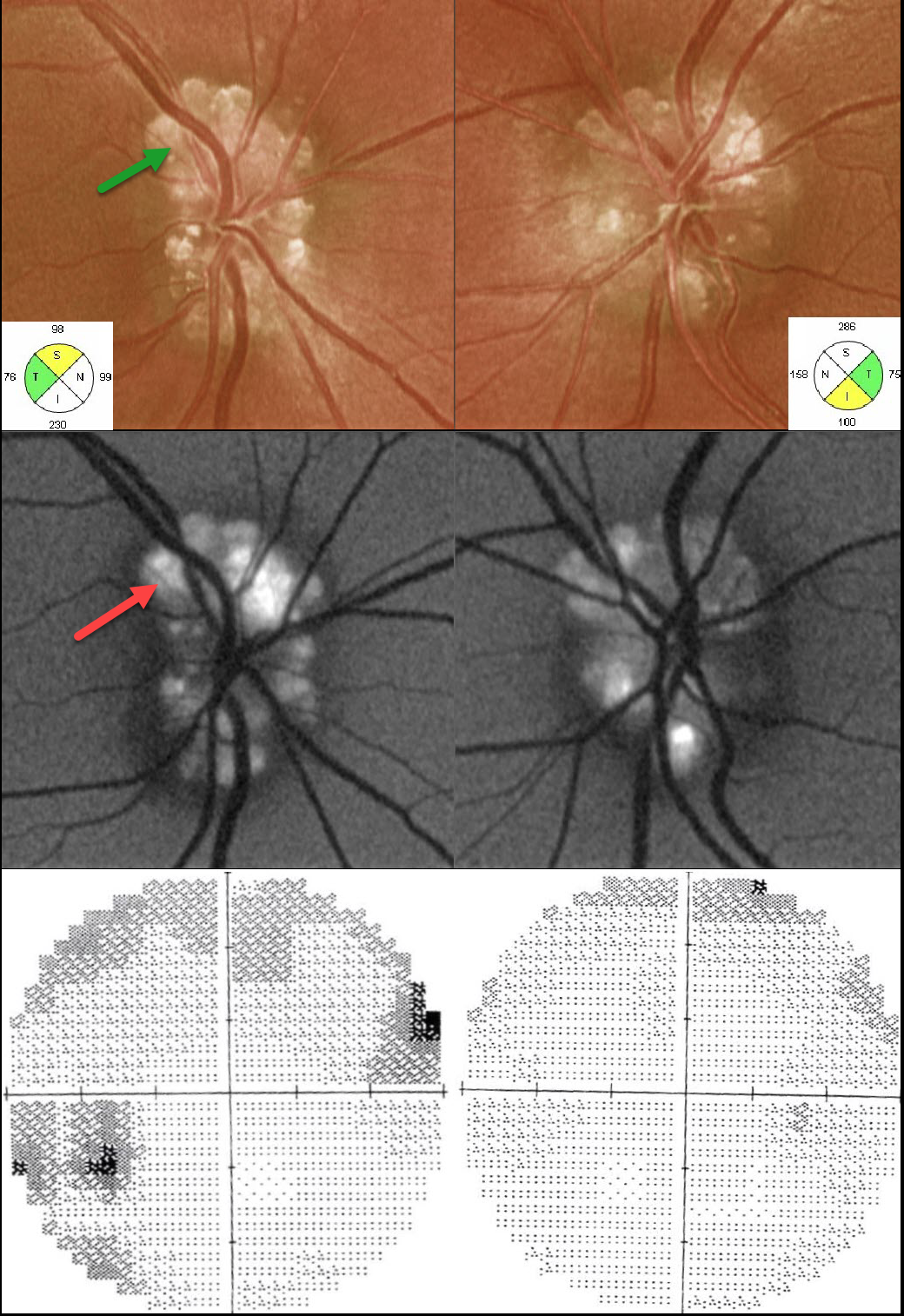
This 29YO male presented with bilateral optic disc drusen (ODD). Vision was 20/40 OD and 20/30 OS. The drusen are markedly hyper-autofluorescent. The retinal nerve fiber layer is variably thickened and thinned. Humphrey visual fields show scattered defects OD with a full field OS. Observation was recommended.
Learning Points:
Optic disc drusen (ODD) are refractile calcific concretions that form anterior to the lamina cribrosa. They are thought to be extruded, extracellular, calcified mitochondria and may be inherited as an autosomal dominant trait.
Porphyrins, organic molecules composed of four interconnected pyrrole subunits, are present within mitochondria. When these compounds break down, they become autofluorescent, which is likely why drusen autofluoresce. Porphyrins are also part of the heme molecule in hemoglobin, accounting for why devitalized blood also autofluoresces (Bloom and Spaide, Retinal Cases 2022;16:401-402).
OPTIC NERVE DRUSEN + RETINITIS PIGMENTOSA
Originally posted on @retina.rocks April 17, 2023
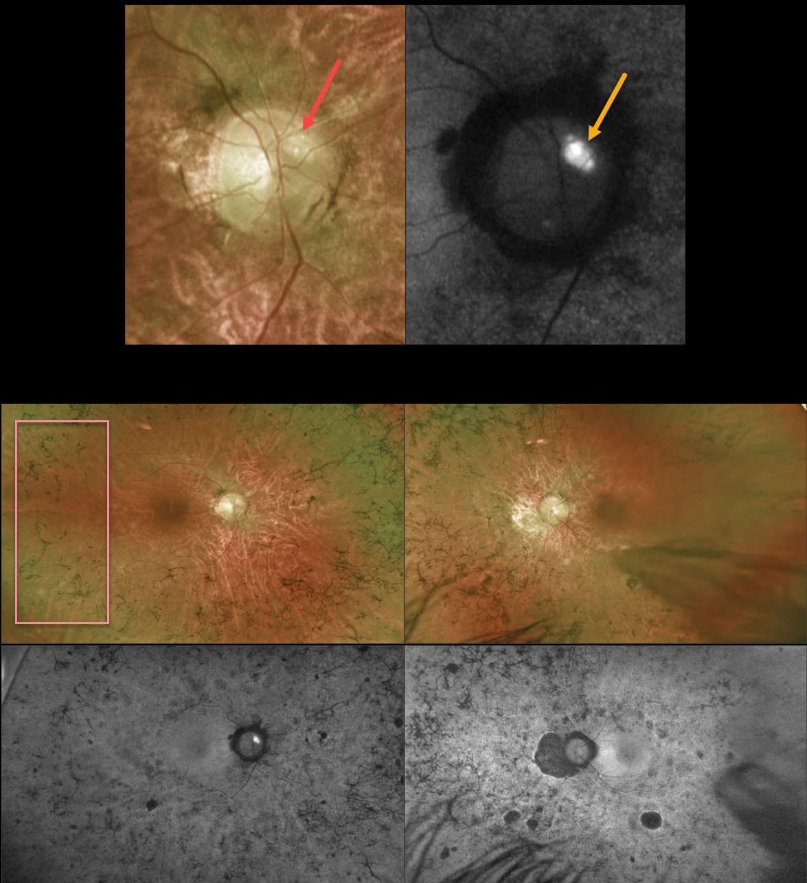
This 43YO male with a known history of retinitis pigmentosa (RP) was referred for further evaluation. He noted a gradual worsening of vision bilaterally. Vision was 20/80 OD and 20/200 OS.
Optos color imaging of the right optic nerve shows several large optic disc drusen (ODD) that markedly hyperautofluoresce. Ultrawide-field imaging shows diffuse, classic bilateral RP scarring.
Learning Points:
ODD are refractile calcific concretions that form anterior to the lamina cribrosa. They are thought to be extruded, extracellular, calcified mitochondria and may be inherited as an autosomal dominant trait. They are present in about 3% of RP patients (Russell et al, Eye 2022;36:2213).
Most patients are asymptomatic, but some can develop progressive visual field defects or central vision loss. There is no treatment for the drusen themselves, although symptomatic macular neovascularization can be treated with thermal laser, photodynamic therapy, or anti-VEGF injections.
OPTIC NERVE DRUSEN
Barbara Parolini and Veronika Matello
Originally posted on @retina.rocks March 17, 2023
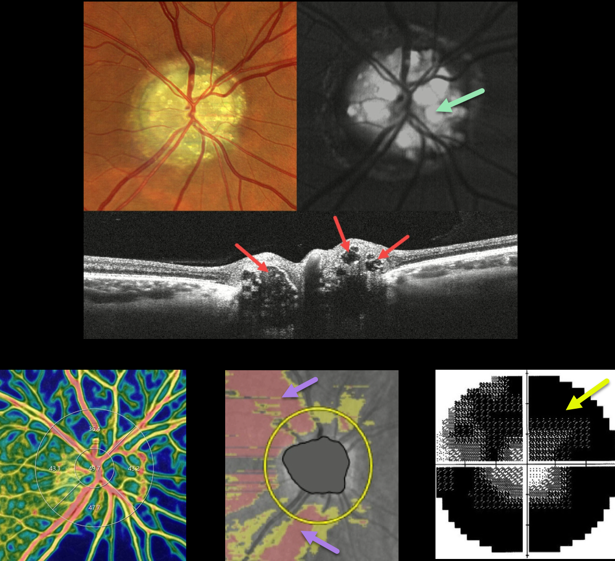
This 47YO female was examined for a routine eye examination. Although she initially denied any visual complaints, further questioning revealed a long history of peripheral vision loss. Vision was 20/20 in each eye, and the following findings were symmetrical and bilateral.
Color imaging of the right nerve shows severe optic disc drusen (ODD), which are hyper-autofluorescent. On OCT B-scan, the drusen appeared as relatively hyporeflective ovoid lesions within the substance of the nerve.
The drusen can be appreciated as blue flow voids on the OCT angiography density map. Diffuse retinal nerve fiber layer thinning is seen, and there is a severely constricted and depressed visual field. Her father was examined the same day and was discovered to also have ODD.
Learning Points:
ODD are refractile calcific concretions that form anterior to the lamina cribrosa. They are thought to be extruded, extracellular, calcified mitochondria and may be inherited as an autosomal dominant trait. Most patients are asymptomatic, but some can develop progressive visual field defects or central vision loss.
There is no treatment for the drusen themselves, although symptomatic macular neovascularization can be treated with thermal laser, photodynamic therapy, or anti-VEGF injections.
OPTIC NERVE DRUSEN
Will Gibson
Originally posted on @retina.rocks September 29, 2022
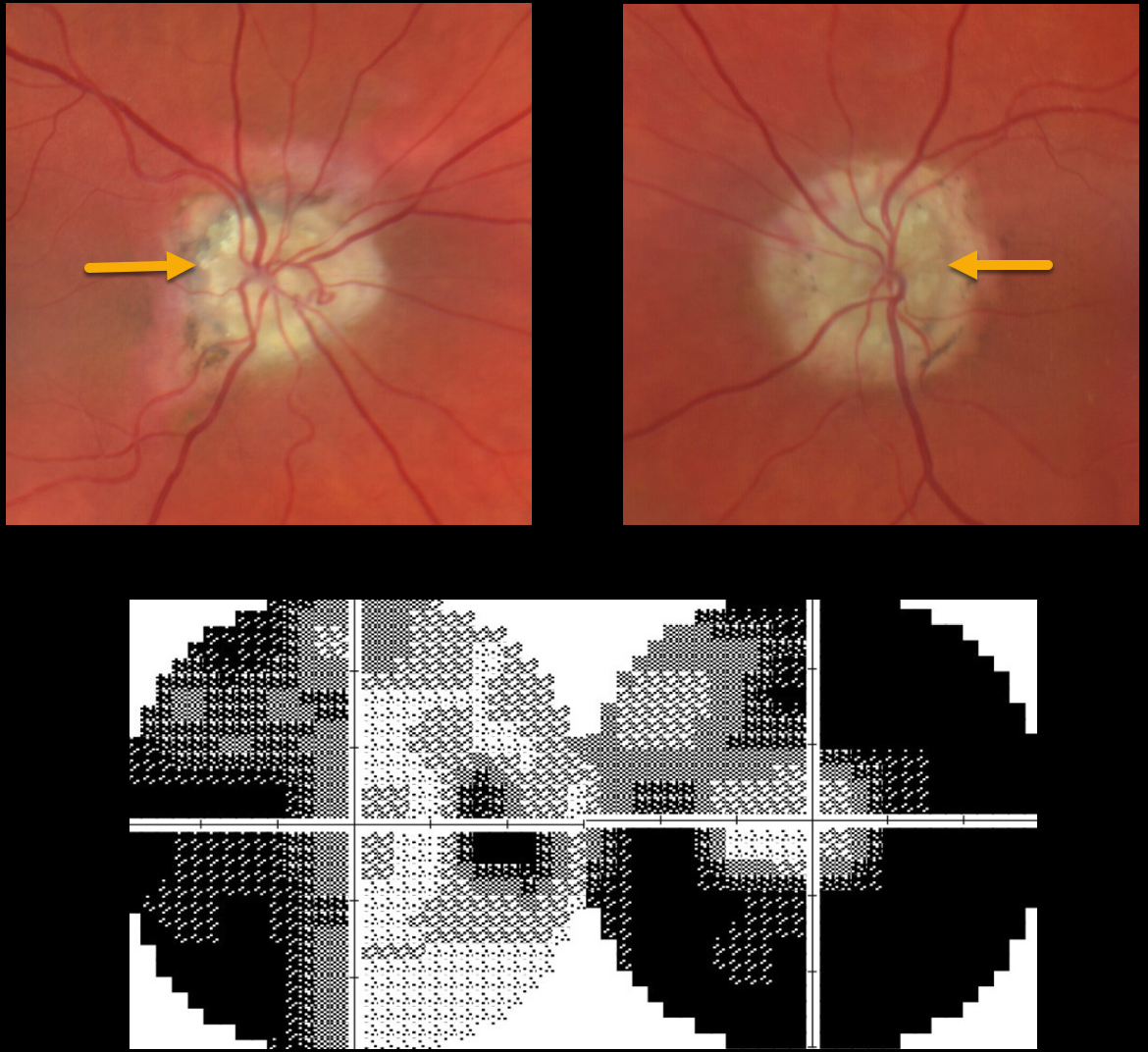
This 65YO female presented with dramatic bilateral optic nerve drusen with severe visual field defects. Vision was 20/20 OU.
Learning Points:
Optic disc drusen are refractile calcific concretions that form anterior to the lamina cribrosa. They are felt to be extruded extracellular calcified mitochondria and may be inherited as an autosomal dominant trait.
Most patients are asymptomatic, but some can develop progressive visual field defects or central vision loss.
There is no treatment for the drusen themselves, although symptomatic macular neovascularization can be treated with thermal laser, photodynamic therapy, or anti-VEGF injections.
OPTIC NERVE DRUSEN
Originally posted on @retina.rocks October 8, 2021
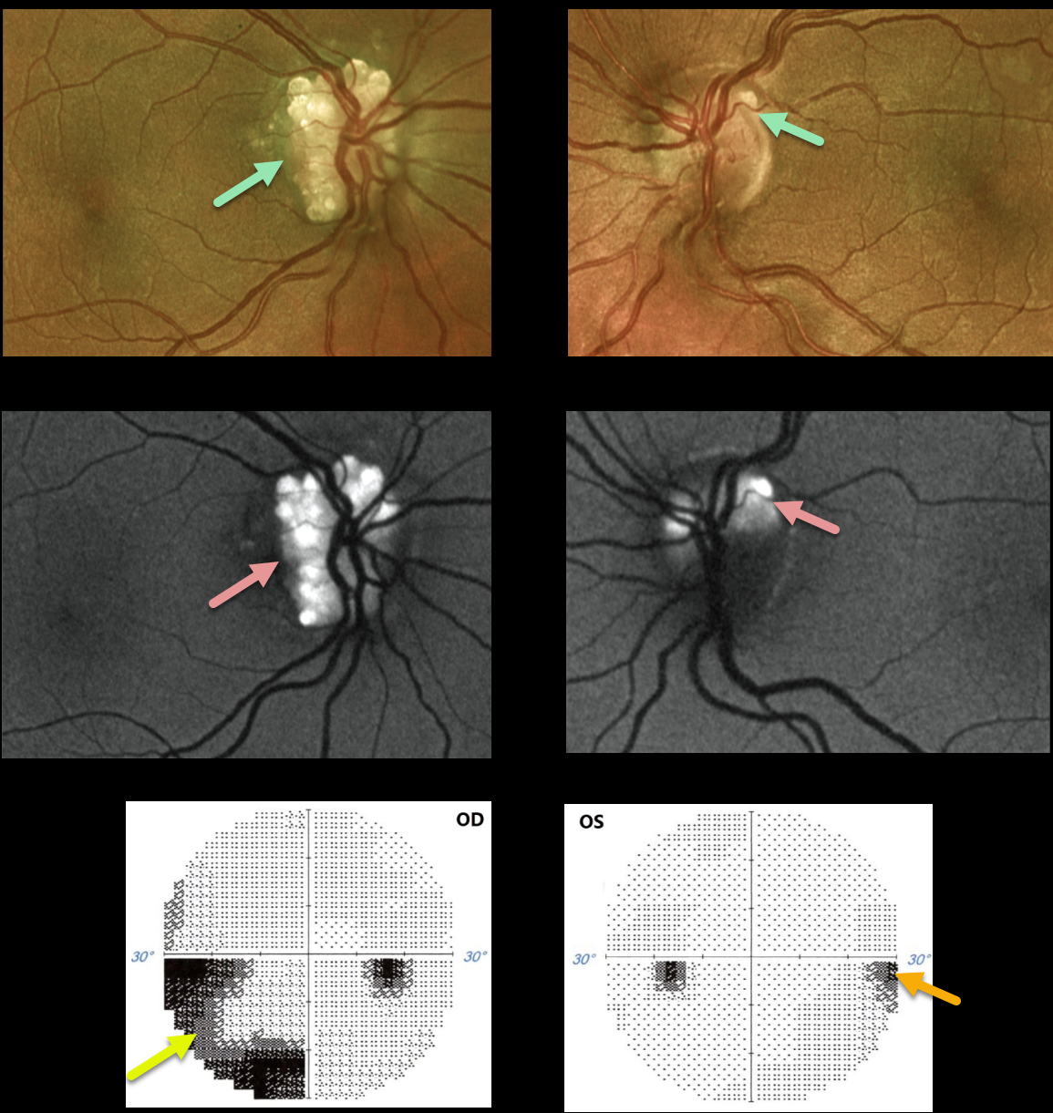
This 38YO female presented with bilateral optic disc drusen (ODD). Vision was 20/80 OD and 20/40 OS.
The drusen are markedly hyper-autofluorescent. The retinal nerve fiber is thinned, particularly in the right eye, due to the more extensive ODD. Humphrey visual fields show significant visual field defects inferonasally OD and mild nasal depression OS. Observation was recommended.
Learning Points:
Optic disc drusen (ODD) are refractile calcific concretions that form anterior to the lamina cribrosa. They are thought to be extruded, extracellular, calcified mitochondria and may be inherited as an autosomal dominant trait.
Porphyrins, organic molecules composed of four interconnected pyrrole subunits, are present within mitochondria. When these compounds break down, they become autofluorescent, which is likely why drusen autofluoresce.
Porphyrins are also part of the heme molecule in hemoglobin, accounting for why devitalized blood autofluoresces.
OPTIC NERVE DRUSEN
Originally posted on @retina.rocks July 22, 2021
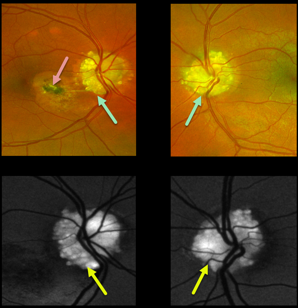
This 30YO male presented with dramatic bilateral optic nerve drusen. Vision was 20/20 OD and 20/200 in his amblyopic OS.
A spontaneously involuted macular neovascularization (MNV) is noted in the nasal macula. The drusen are markedly hyper-autofluorescent.
Observation was recommended.
Learning Points:
Optic disc drusen (ODD) are refractile calcific concretions that form anterior to the lamina cribrosa. They are thought to be extruded, extracellular, calcified mitochondria and may be inherited as an autosomal dominant trait.
MNV is a known complication of ODD. These can spontaneously involute, as in our patient, or can be treated with thermal laser, photodynamic therapy, or anti-VEGF injections.
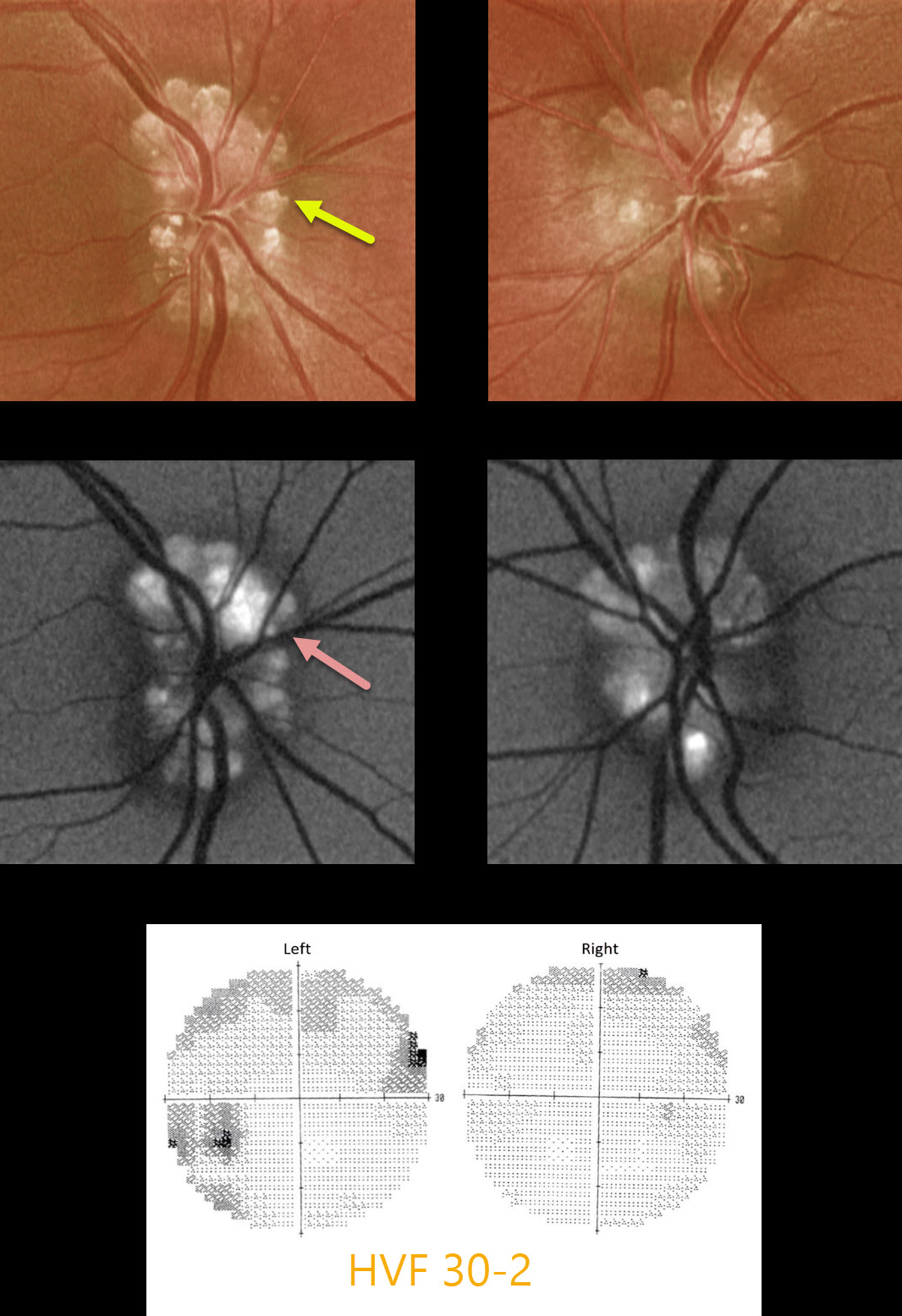
OPTIC NERVE DRUSEN
Originally posted on @retina.rocks January 4, 2021
This 29yo male presented with asymptomatic bilateral optic nerve drusen. Vision was 20/40 OD and 20/30 OS.
The drusen hyperautofluoresce, and visual fields show some non-specific depression and defects.
Learning Points:
Optic disc drusen are small hyaline bodies within the optic nerve that progressively calcify over time. They can be buried or superficial, giving the nerve a “lumpy-bumpy” appearance with normal overlying vessels. These nerves can often be confused with papilledema, especially in younger patients.
We have found ultrasonography to be totally unhelpful in younger patients, since drusen don’t calcify until later in life.
The drusen often hyperautofluoresce on fundus autofluorescence imaging. Swept-source OCT can occasionally visualize buried drusen, which can be either hypo- or hyperreflective.
When autofluorescence is negative, the best clinical test is fluorescein angiography, which will show optic nerve leakage with papilledema. Eyes with drusen will show either no nerve leakage or nodular staining.
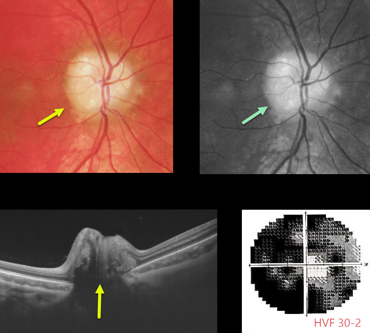
OPTIC NERVE DRUSEN
Originally posted on @retina.rocks September 28, 2020
Our patient is a 15YO boy with quite pronounced bilateral optic nerve drusen visible clinically, as well as on OCT (only right eye pictured). ODD will often hyperautofluoresce on fundus autofluorescence (FAF).
Although central vision was good, he complained of increasing peripheral vision loss due to drusen compressing the surrounding nerve fibers.
Learning Points:
Optic disc drusen (ODD) are refractile calcific concretions that form anterior to the lamina cribrosa. They are thought to be extruded, extracellular, calcified mitochondria and may be inherited as an autosomal dominant trait.
It is often difficult to differentiate ODD from true optic nerve edema. This is often the dilemma when seeing a child with a possible swollen nerve.
We have found ultrasonography to be unhelpful, since ODD don’t calcify until later in life.
B-scan OCT imaging is often unhelpful, although newer swept-source machines can directly visualize drusen. The RNFL will be variably thinned with ODD and thickened with true disc edema.
Fluorescein angiography (FA) is probably the best way to differentiate the two, since a swollen nerve will show late leakage, whereas ODD will not leak and often shows staining of the drusen.
Unfortunately, there is no proven treatment, although some have tried pressure-lowering medications for neuroprotection.
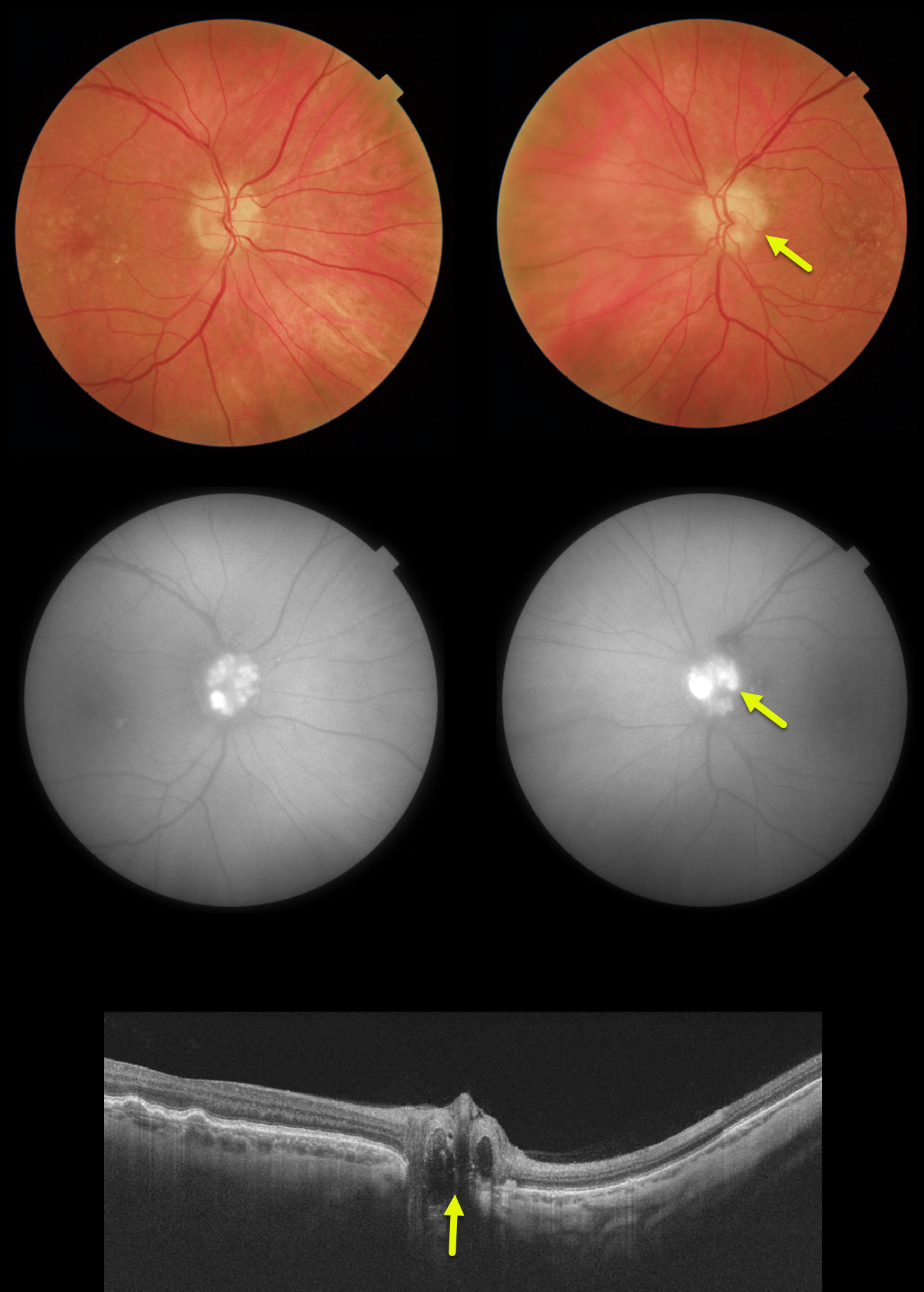
OPTIC NERVE DRUSEN
Originally posted on @retina.rocks March 16, 2020
Our patient’s optic disc drusen were obvious clinically, on FAF and on OCT. In younger patients they are usually much more difficult to see and can be confused with papilledema.
We have found ultrasonography in younger patients cases to be totally unhelpful since drusen don’t calcify until later in life.
If fundus autofluorescence (FAF) is negative, the best clinical test in these cases is fluorescein angiography, which will show optic nerve leakage with papilledema. Eyes with drusen will show either no nerve leakage or nodular staining.
Learning Points:
Optic disc drusen are small hyaline bodies within the optic nerve that progressively calcify over time. They can be buried or superficial giving the appearance of pseudopapilledema. The nerve usually has a “lumpy-bumpy” appearance with normal overlying vessels. Retinal nerve fiber layer will usually show variably thinning.
FAF can show drusen hyperautofluoresence. Swept source OCT can occasionally visualize buried drusen.which can be either hypo- or hyperreflective.


