

Optic Nerve Hypoplasia
<< Back to Cases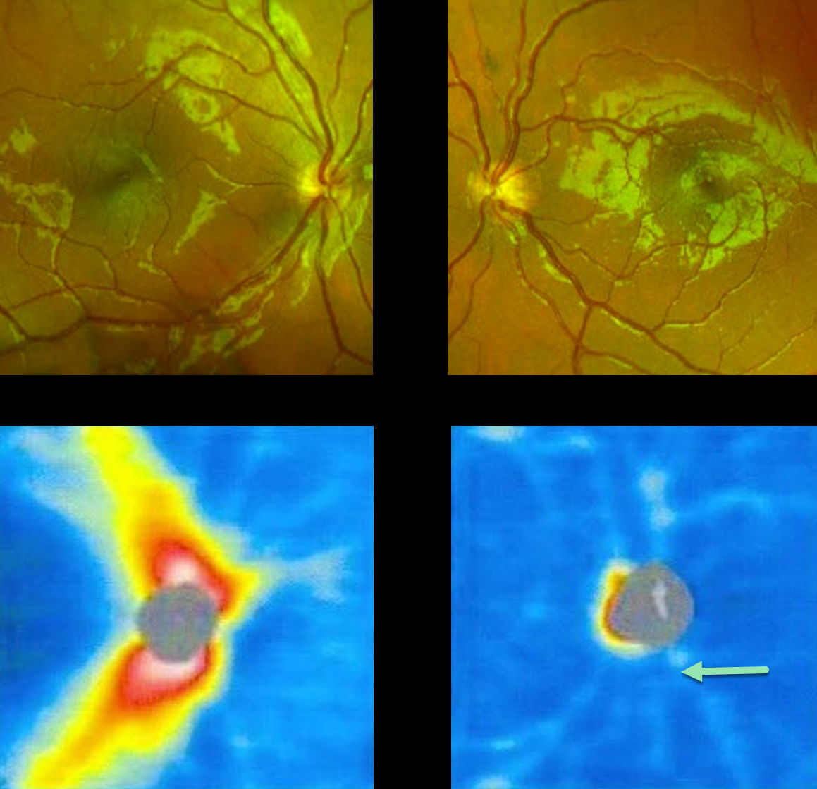
OPTIC NERVE HYPOPLASIA
Originally posted on @retina.rocks December 23, 2022
This 11YO adopted boy presented with a history of poor vision in his left eye since birth. He was referred for an abnormal-appearing left optic nerve.
Vision was 20/20 OD and hand motion OS. Although the left nerve clinically is hypoplastic with a surrounding scleral ring, color imaging failed to capture these disc findings. However, marked loss of the nerve fiber is noted compared to that seen in the right fundus. Optic nerve OCT is normal OD, but shows marked nerve fiber layer thinning OS.
MRI scanning showed an absent septum pellucidum with bilateral hypoplastic nerves, consistent with septo-optic dysplasia. He was referred to pediatric endocrinology.
Learning Points:
Hallmarks of optic disc hypoplasia include a decreased disc diameter, low disc/artery ratio, and the peripapillary double ring sign, a yellow/gray peripapillary halo delineated by an outer ring (the normal junction between the lamina cribrosa and sclera) and an inner pigmented ring (the abnormal extension of the retina and RPE over the outer portion of the lamina cribrosa).
Major risk factors include maternal diabetes and young maternal age. Vision is usually 20/200 or worse. Moderate or severe hypoplasia of the optic disc may
cause visual field defects. The condition may be associated with brain malformations, pituitary disorders, cerebral palsy, and seizures.
OPTIC NERVE HYPOPLASIA
Originally posted on @retina.rocks December 7, 2021
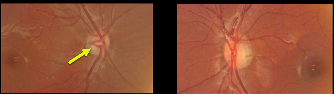
This healthy 20YO female presented with vision of 20/400 OD and 20/20 OS. Vision in her right eye was unchanged and poor since birth. The right nerve was hypoplastic, while the left nerve appeared fairly normal.
Learning Points:
Hallmarks of optic disc hypoplasia include a decrease in disc diameter, low disc/artery ratio, and the peripapillary double ring sign – yellow/gray peripapillary halo delineated by an outer ring (the normal junction between the lamina cribrosa and sclera) and an inner pigmented ring (the abnormal extension of the retina and RPE over the outer portion of the lamina cribrosa).
Major risk factors include maternal diabetes and young maternal age. Vision is usually 20/200 or worse. Moderate or severe hypoplasia of the optic disc may cause visual field defects.
The condition may be associated with brain malformations, pituitary disorders, cerebral palsy, and seizures.


