

Optic Nerve Pit and No Pit Retinoschisis
<< Back to Cases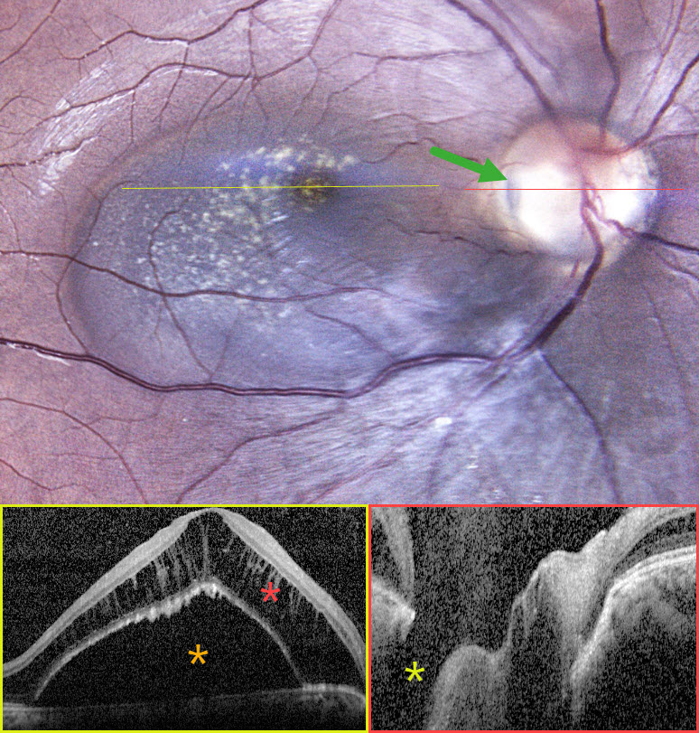
OPTIC NERVE PIT
Rohan Suresh Ninan
Originally posted on @retina.rocks August 28, 2026
This healthy 24YO female presented with asymptomatic retinal changes in her right eye, which was 20/20 one year earlier. Vision was 20/80 OD and 20/20 in her normal OS.
Fundus photography shows a serous macular detachment connected to a yellow-grey optic nerve pit along the temporal edge of the disc. OCT scanning shows an elevated serous detachment with marked overlying cystic schisis in the outer nuclear layer. The pit is noted on the OCT.
Learning Points:
An optic nerve pit is a rare (about 0.1% prevalence) congenital anomaly thought to be caused by incomplete closure of the optic fissure during gestation. Pits are most often located within or along the inferior-temporal disc margin and usually appear as a gray depression. While most optic pits are asymptomatic, they can cause vision loss by forcing fluid into the inner retina, outer retina, and, ultimately, the subretinal space (optic pit maculopathy). The source of the fluid remains a subject of ongoing debate, with opinions ranging from the vitreous to the subarachnoid space.
Given the lack of symptoms, continued observation was recommended for our patient.
OPTIC NERVE PIT
Shraddha Raj Shrivastava and Manish Nagpal
Originally posted on @retina.rocks July 24, 2026
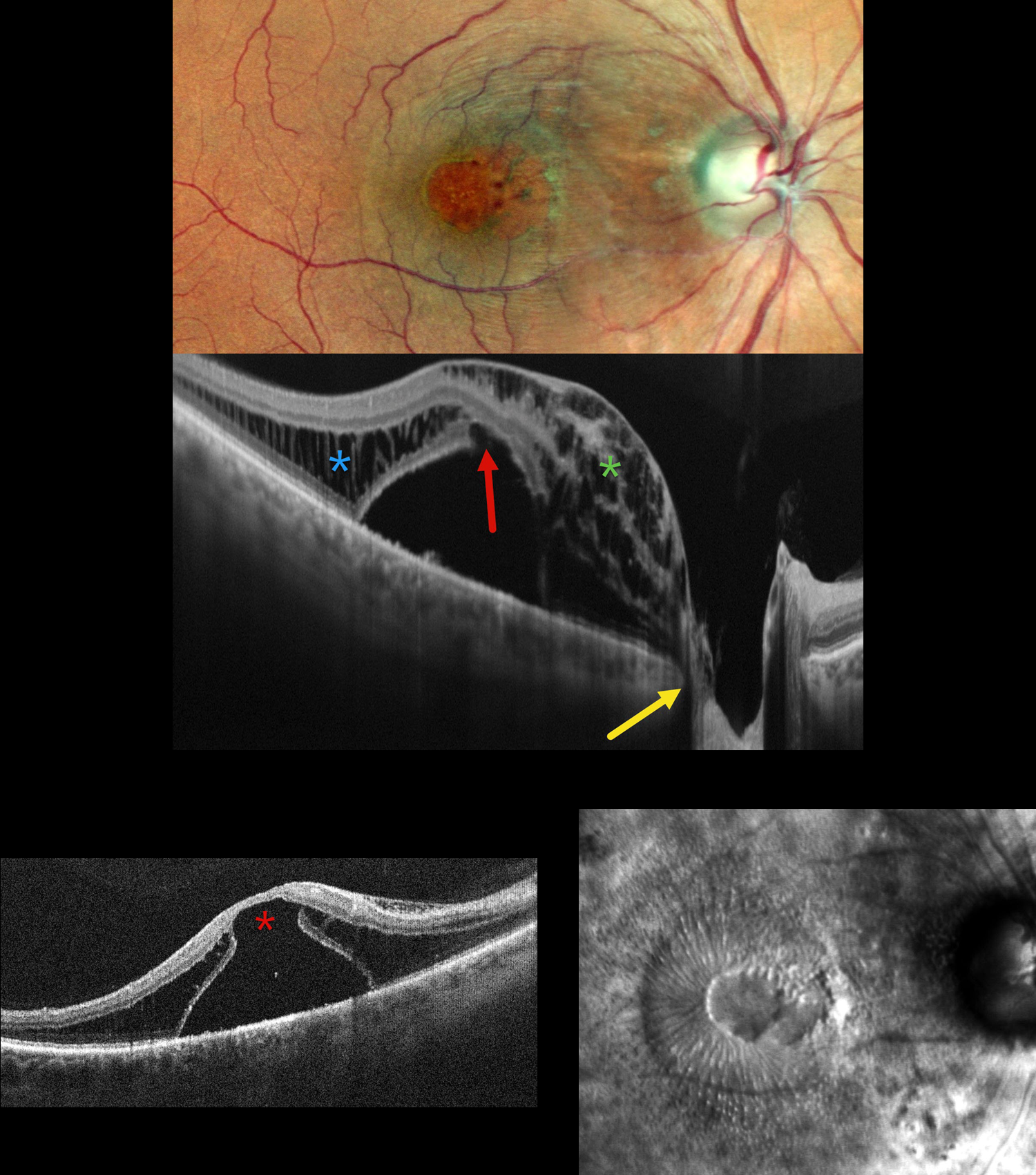
This 50YO male presented with 1 year of decreased vision in his right eye. He had been treated elsewhere with intravitreal injections for cystoid macular edema. Vision was 20/200 OD and 20/20 in his normal OS.
Pseudocolor SLO imaging shows a possible serous macular detachment with a large macular pseudohole. A possible pit is noted along the temporal aspect of the optic nerve. OCT scanning shows an elevated serous detachment. An outer-layer macular hole is noted along the posterior aspect of the fovea, along with full-thickness cystic retinal edema nasally and outer nuclear layer cystic edema temporally. The subretinal fluid directly communicates with the optic nerve pit. Retro mode reveals a unique view of the macular pathology, with the central outer macular hole and radiating fluid.
Learning Points:
An optic nerve pit is a rare (about 0.1% prevalence) congenital anomaly thought to be caused by incomplete closure of the optic fissure during gestation. Pits are most often located within or along the inferior-temporal disc margin and usually appear as a gray depression. While most optic pits are asymptomatic, they can cause vision loss by forcing fluid into the inner retina, outer retina, and, ultimately, the subretinal space (optic pit maculopathy). The source of the fluid remains a subject of ongoing debate, with opinions ranging from the vitreous to the subarachnoid space.
Vitrectomy surgery was discussed, and the patient elected for observation unless his symptoms worsened.
OPTIC NERVE PIT
Malvika Singh and Manish Nagpal
Originally posted on @retina.rocks May 26, 2026
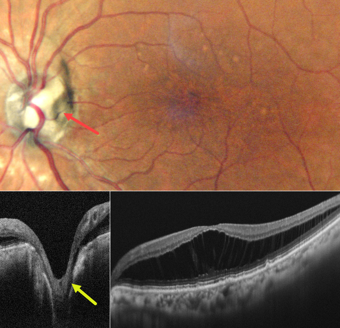
This 72YO female presented with 7 months of stable decreased vision in her left eye. Vision was 20/20 OD and 20/30 OS.
Pseudocolor SLO imaging shows irregular foveal pigment loss with more distal drusen-like changes. There is diffuse outer nuclear layer macular schisis on OCT. An optic pit is noted along the temporal edge of the nerve, which directly communicates with the schisis cavity. Observation was recommended.
Learning Points:
An optic nerve pit is a rare (about 0.1% prevalence) congenital anomaly thought to be caused by incomplete closure of the optic fissure during gestation. Pits are most often located within or along the inferior-temporal disc margin and usually appear as a gray depression. While most optic pits are asymptomatic, they can cause vision loss by forcing fluid into the inner retina, outer retina, and, ultimately, the subretinal space (optic pit maculopathy). The source of the fluid remains a subject of ongoing debate, with opinions ranging from the vitreous to the subarachnoid space.
OPTIC NERVE PIT WITH VITREOUS PLUG
The European VitreoRetinal Society (EVRS) and Miguel Rechichi
Originally posted on @retina.rocks January 9, 2026
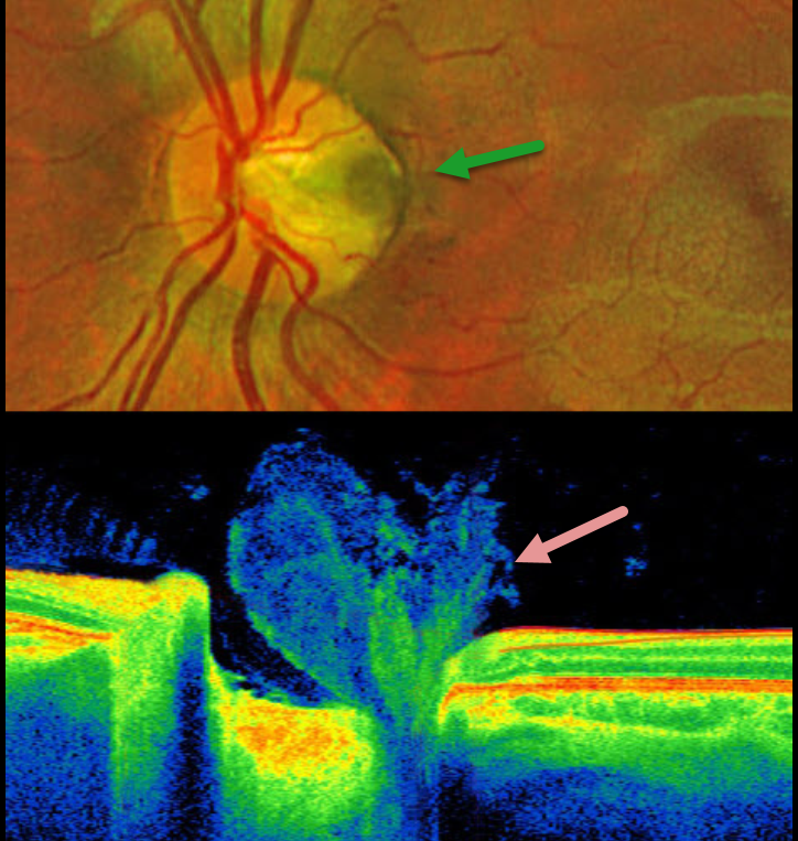
This healthy 6YO boy presented for a routine examination. Vision was 20/20 in his normal right eye and 20/20 in his left eye. OCT scanning shows a vitreous plug extending into the depth of the pit.
Optos color RG imaging shows a temporal optic nerve pit without associated macular abnormalities.
Learning Points:
An optic nerve pit is a rare (about 0.1% prevalence) congenital anomaly thought to be caused by incomplete closure of the optic fissure during gestation. Pits are most often located within or along the inferior-temporal disc margin and usually appear as a gray depression. While most optic pits are asymptomatic, they can cause vision loss due to fluid being forced into the inner retina, outer retina, and the subretinal space (optic pit maculopathy).
Utilizing high-resolution swept-source OCT, Spaide defined the optic pit complex as a broader means to characterize the structural abnormalities found with optic disc pits (Retina 2026;46:15-24). The spectrum of abnormalities includes lamina cribrosa defects (all eyes) with associated tissue cavitations extending a mean of 1855 microns posterior to a modified Bruch membrane opening. One of his patients had OCT findings similar to our patient’s, with vitreous extending into the pit. Sources of intra- and subretinal fluid included direct communication from a cavitation, perivascular hyporeflective spaces, intraneural channels extending toward cystoid spaces, and isolated retinal cysts. These concepts support the “dynamic and multifactorial process involving abnormal structural anatomy, disrupted tissue boundaries, potential misrouting of glymphatic flow, vitreous or cerebral spinal fluid (CSF)-related gradients, and mechanical forces exerted during eye movements.”
GLAUCOMATOUS PERIPAPILLARY RETINOSCHISIS
Vaibhav Sethi
Originally posted on @retina.rocks September 5, 2025
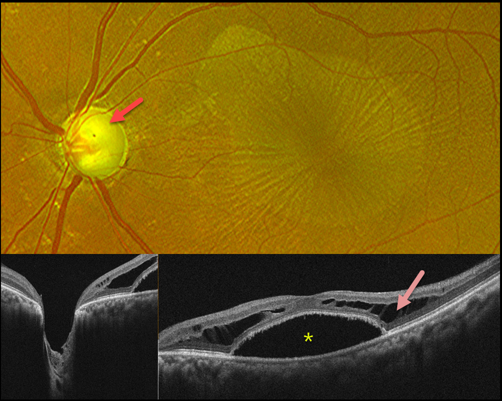
This 45YO male presented with blurred vision in his left eye. The right eye was phthisical due to absolute glaucoma. He was taking topical dorzolamide and travoprost OU. Vision was 20/400 OS. Intraocular pressure was 12 mm Hg.
Optos color RG imaging shows a nearly completely cupped glaucomatous nerve. There is an irregular oval discoloration of the outer macula with overlying radiating retinal striae. OCT scanning shows Henle’s layer schisis with a central macular detachment. An optic pit could not be identified clinically or on OCT.
Learning Points:
While peripapillary retinoschisis (PPRS) secondary to congenital optic disc cavitary abnormalities is a well-known phenomenon, similar findings in patients with glaucomatous cupping but without nerve head cavitation have more recently been described and are less common (Fujimoto et al, Ophthalmology Retina 2023;7:811-818). The retinal nerve fiber layer is most commonly involved in glaucoma, whereas in optic pit maculopathy, outer and inner nuclear layer schisis, foveal involvement, and subretinal fluid are more frequently noted.
Most cases of glaucoma-related PPRS are asymptomatic and spontaneously resolve. However, in fovea-involving or symptomatic cases, surgical intervention may improve vision and decrease time to resolution. There is no consensus regarding the optimal treatment approach, including whether to perform pars plana vitrectomy alone or with the addition of juxtapapillary laser treatment, ILM peeling, inner retinal fenestration, and/or autologous platelet concentrate.
We recommended adding brimonidine and timolol drops to further lower his IOP. We are not planning any surgical intervention.
OPTIC NERVE PIT
Seif Anwar
Originally posted on @retina.rocks July 23, 2025
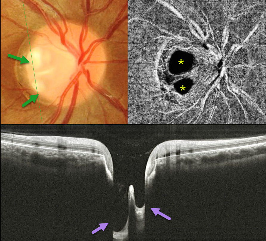
This healthy 20YO female presented with asymptomatic unilateral optic nerve findings in her right eye. Vision was 20/20 OD and 20/20 in her normal OS.
Color photography shows two independent optic nerve pits along the temporal edge of the nerve. OCT angiography dramatically illustrates the pits due to completely absent flow signals. The pits are also well imaged on OCT scans. Observation was recommended.
Learning Points:
An optic nerve pit is a rare (about 0.1% prevalence) congenital anomaly thought to be caused by incomplete closure of the optic fissure during gestation. Pits are most often located within or along the inferior-temporal disc margin and usually appear as a gray depression. Double pits are exceedingly rare (Babu et al, Indian J Ophthalmol 2020;68:663-665).
While most optic pits are asymptomatic, they can cause vision loss by forcing fluid into the inner retina, outer retina, and, ultimately, the subretinal space (optic pit maculopathy). The source of the fluid remains a subject of ongoing debate, with opinions ranging from the vitreous to the subarachnoid space.
NO OPTIC PIT RETINOSCHISIS
Originally posted on @retina.rocks March 13, 2025
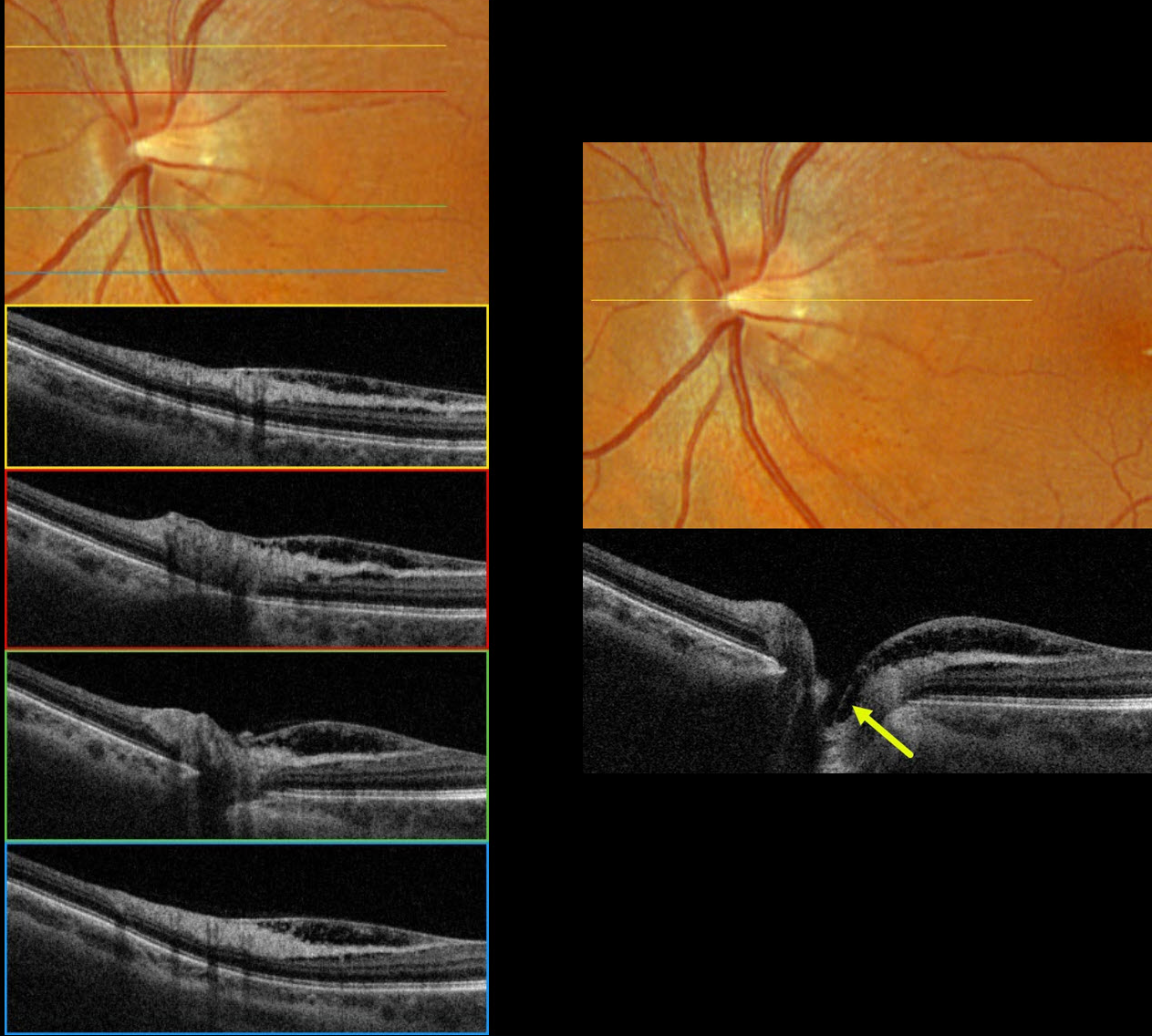
This 55YOF was referred for asymptomatic macular changes in her left eye. Vision was 20/30 bilaterally. The right eye was normal.
Optos color RGB imaging shows a normal appearing optic nerve with a small central cup. Very subtle inner retinal telangiectasia is seen inferotemporal to the disc. Triton swept-source OCT shows nasal macular inner retinoschisis, which extends into the central depression of the optic cup, but with no definite pit. This inner retinal schisis was variably present along the temporal disc margin, extending into the nasal macula. Fluorescein angiography was normal without leakage (not pictured).
Learning Points:
An optic nerve pit is a rare (about 0.1% prevalence) congenital anomaly thought to be caused by incomplete closure of the optic fissure during gestation. Pits are most often located within or along the inferior-temporal disc margin and usually appear as a gray depression. While most optic pits are asymptomatic, they can cause vision loss by forcing fluid into the inner retina, outer retina, and, ultimately, the subretinal space (optic pit maculopathy).
No optic pit retinoschisis (NOPIR) is a rare entity that causes pit-like findings in the absence of a pit or severe glaucomatous cupping (Fujimoto et al, Ophthalmology Retina 2023;7:811-818). As with optic pit maculopathy, eyes with good vision should be observed. Persistent vision loss from foveal involvement can be managed with vitrectomy, peeling of the internal limiting membrane, and fluid-gas exchange.
NO OPTIC PIT RETINOSCHISIS
Anand Temkar and Manish Nagpal
Originally posted on @retina.rocks December 12, 2024
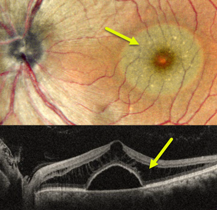
This 48YO male presented with one month of vision loss in his left eye. He was diagnosed elsewhere with idiopathic central serous chorioretinopathy. Vision was 20/20 in his normal OD and 20/40 OS.
Pseudocolor SLO imaging shows a round serous macular detachment, which is confirmed on OCT. The outer nuclear layer has a schisis-like thickening. Fluorescein angiography was normal (not shown). There was no optic pit clinically or on OCT.
Learning Points:
No optic pit retinoschisis (NOPIR) is a rare entity that presents with pit-like findings in the absence of a pit or severe glaucomatous cupping (Fujimoto et al, Ophthalmology Retina 2023;7:811-818). Somewhat similar findings can be observed in paraproteinemia (Mansour et al, Ophthalmology 2014;121:1925-1932), but our patient’s serum protein electrophoresis was normal.
As with optic pit maculopathy, eyes with good vision should be observed. Persistent vision loss from foveal involvement can be managed with vitrectomy, internal limiting membrane peeling, and fluid-gas exchange.
OPTIC NERVE PIT AND COLOBOMA
Kanwaljeet Harjot Madan
Originally posted on @retina.rocks June 13, 2024
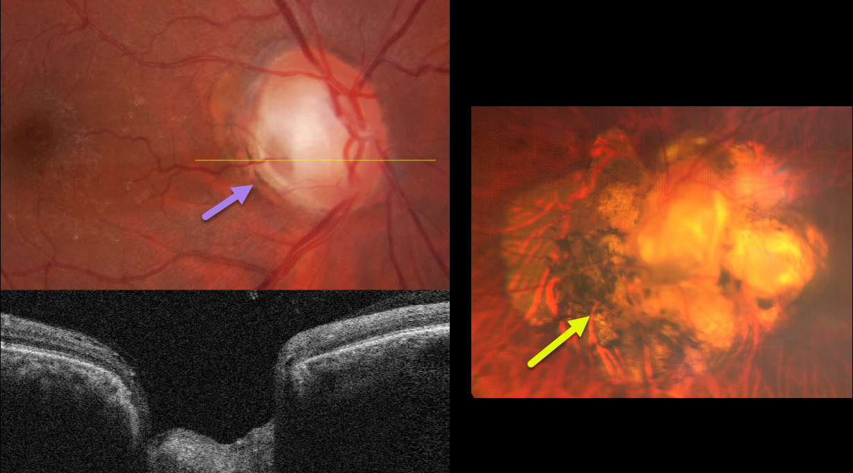
This healthy 63YO male was referred for cataract surgery in his right eye. He had a lifelong history of poor vision in his left eye. Vision was 20/60 OD and counting vision OS.
Color fundus imaging and OCT of the right eye show a grey inferotemporal optic nerve pit. Marked peripapillary scarring surrounds the left nerve, which is anomalous with an inferotemporal disc coloboma.
Learning Points:
An optic nerve pit and coloboma are cavitated abnormalities that can be a unilateral or bilateral condition and are caused by incomplete closure of the embryonic fissure. While most lesions are asymptomatic, they can cause vision loss by forcing fluid into the inner and outer retina, and finally into the subretinal space (optic pit maculopathy). Our patient currently shows no evidence of this.
Cataract surgery was performed on his right eye, and vision improved to 20/20. Observation for the disc abnormalities was recommended.
IDIOPATHIC CENTRAL SEROUS CHORIORETINOPATHY WITH INCIDENTAL OPTIC NERVE PIT
Originally posted on @retina.rocks May 30, 2024
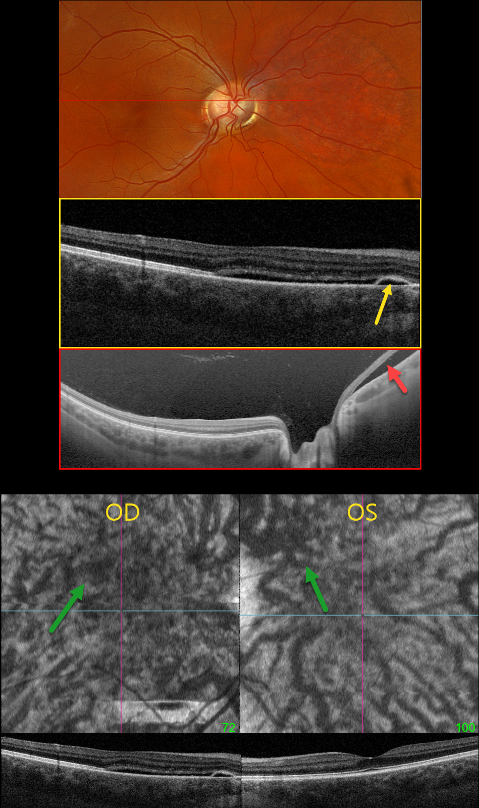
This healthy pregnant 32YO female presented with recent mild central vision loss in her right eye. Vision was 20/40 OD and 20/30 OS.
Optos color RGB imaging shows an anomalous right optic disc with an optic pit both nasally and temporally. Triton swept-source OCT shows both pits and subretinal fluid extending from the nerve nasally. OCT through the macula shows shallow nasal subretinal fluid extending into the macular center, along with a tiny nasal RPE detachment. This fluid does not communicate with the nerve or temporal pit. Optos Silverstone swept-source OCT through the posterior pole shows the subretinal fluid nasal to the nerve. Macular en face and B-scans show a thickened choroid with anomalous, thickened, and tortuous vessels.
Our patient’s symptoms are from idiopathic central serous retinopathy (ICSC), most likely related to her pregnancy. The macular fluid does not communicate with the temporal disc pit, and her macular OCT findings are classic for central serous. Fluorescein angiography was not performed due to her pregnancy. Although she has optic pit ‘maculopathy,’ this is in the nasal retina from her temporal disc pit. Observation was recommended.
Learning Points:
An optic nerve pit is a rare (about 0.1% prevalence) congenital anomaly thought to be caused by incomplete closure of the optic fissure during gestation. Pits are most commonly located within or along the inferior-temporal disc margin and usually appear as a gray depression. While most optic pits are asymptomatic, they can cause vision loss by forcing fluid into the inner and outer retina, and ultimately into the subretinal space (optic pit maculopathy). The source of the fluid remains a topic of ongoing debate, with the fluid originating either from the vitreous or the subarachnoid space.
OPTIC NERVE PIT
Barbara Parolini, Veronika Matello, Giulia Freschi, and Roberta Penzani
Originally posted on @retina.rocks May 14, 2024
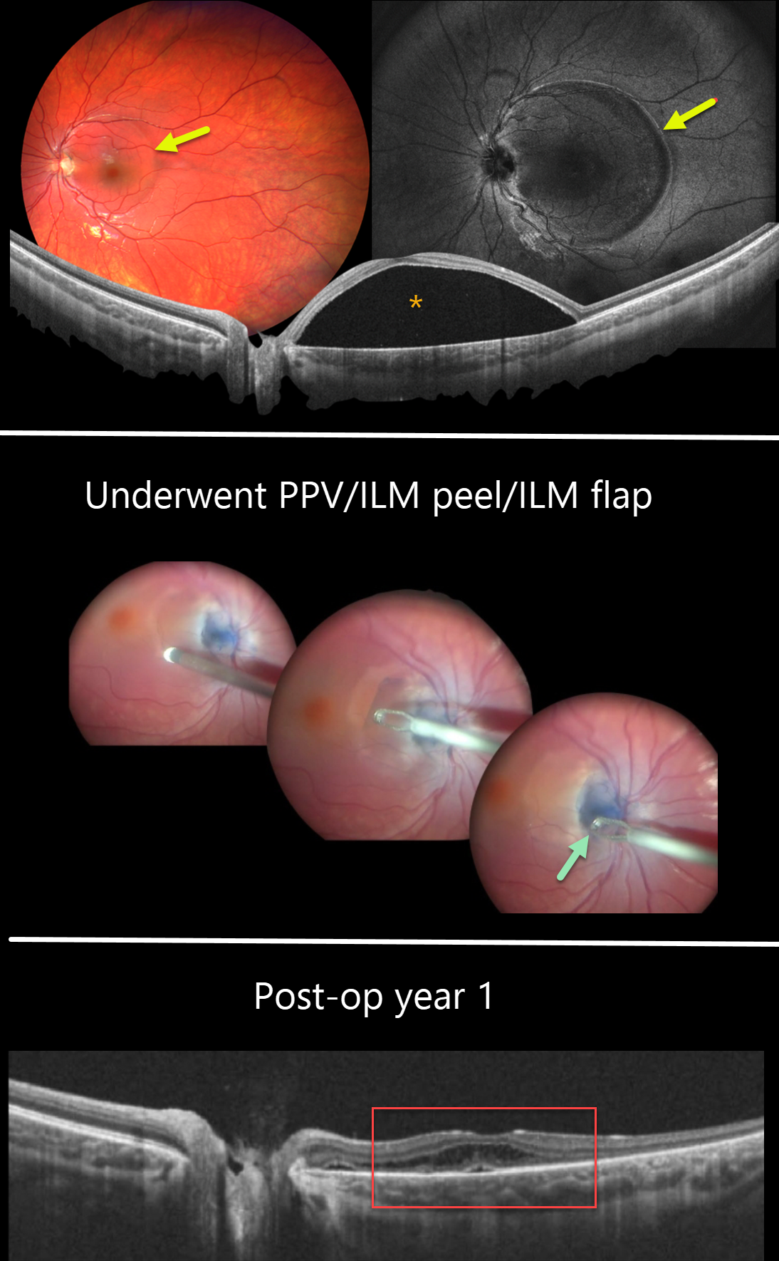
This 10YO female was referred for vision loss in her left eye from a macular detachment and optic pit. Vision was 20/20 in the normal OD and 20/80 OS.
Color imaging shows a round elevation of the entire macula, which is confirmed on widefield OCT. An optic nerve pit is noted along the inferotemporal edge of the nerve.
The macular detachment was initially treated with micropulse laser, resulting in partial reabsorption. Due to persistent fluid, one year later, pars plana vitrectomy with ILM peeling in the inter-papillomacular area and ILM flap into the pit was performed. One year postoperatively, the macular detachment resolved, and vision improved to 20/50.
Learning Points:
An optic nerve pit is a rare (about 0.1% prevalence) congenital anomaly thought to be caused by incomplete closure of the optic fissure during gestation. Pits are most often located within or along the inferior-temporal disc margin and usually appear as a gray depression.
While most optic pits are asymptomatic, they can cause vision loss by forcing fluid into the inner and outer retina, and ultimately into the subretinal space (optic pit maculopathy). The source of the fluid remains a topic of ongoing debate, with the fluid originating either from the vitreous or the subarachnoid space.
A host of treatments for optic pit maculopathy have been suggested over the years. However, a recent meta-analysis suggested that a plain-vanilla vitrectomy without gas tamponade is as successful as other procedures (Zheng et al, Ophthalmology Retina 2020;4:289-299).
OPTIC PIT MACULOPATHY
Asma Samsudeen and Ashish Sharma
Originally posted on @retina.rocks August 31, 2023
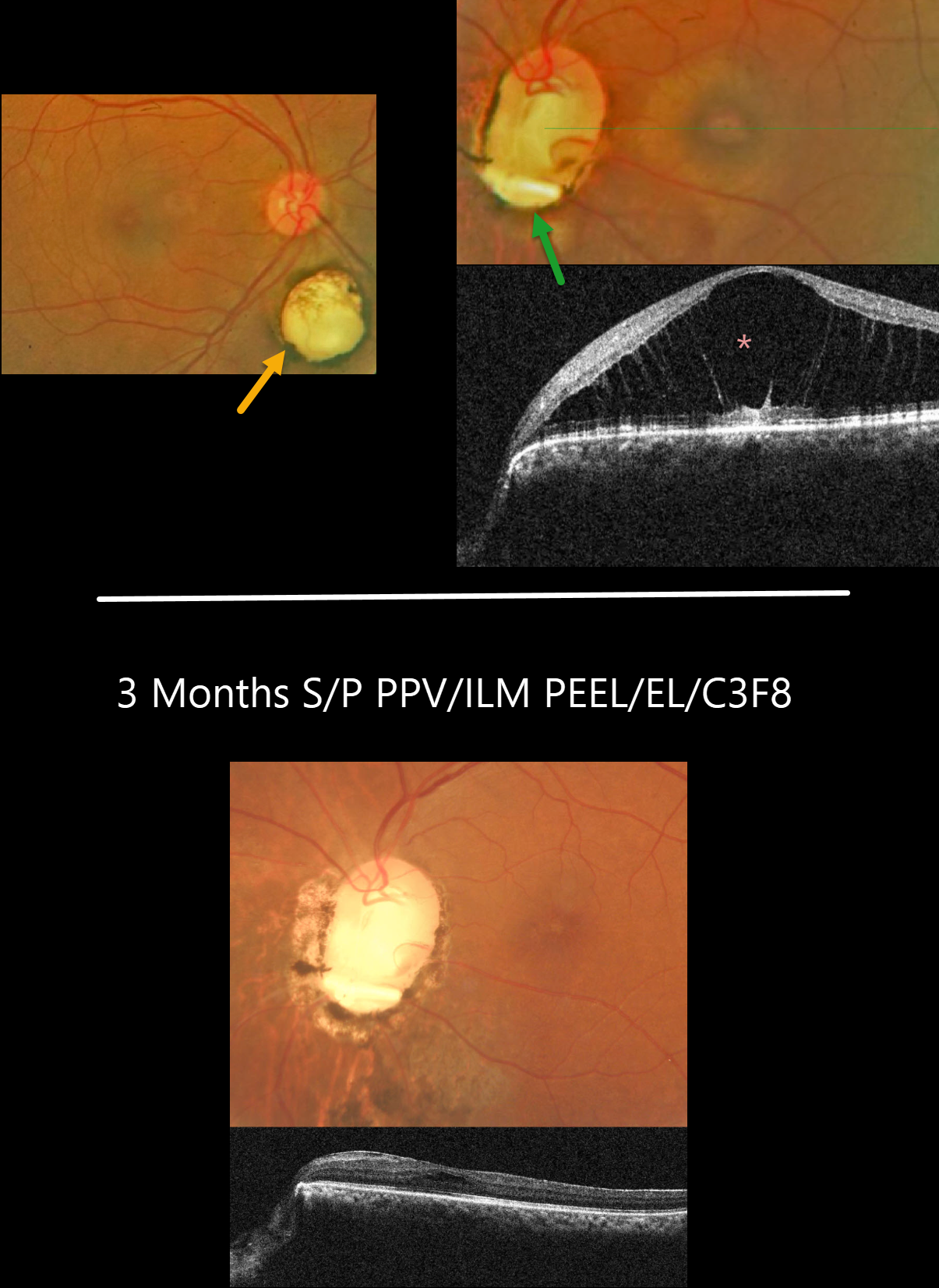
This 51YO female presented with 1 month of blurred vision in her left eye. There was no past ocular history. Vision was 20/30 OD and 20/80 OS. Anterior segments were normal.
Fundus photography of her right eye shows a small choroidal coloboma inferior to the nerve. The left nerve is anomalous with an inferior coloboma and optic pit. The central macula was elevated, with severe foveoschisis noted on OCT.
Vitrectomy with ILM peeling, endolaser along the disc margin, and C3F8 was performed, followed by some additional thermal laser around the nerve postoperatively.
Three months following vitrectomy, vision improved to 20/40 with complete flattening of the macular schisis.
Learning Points:
An optic nerve coloboma can be unilateral or bilateral and is caused by incomplete closure of the embryonic fissure. It has a pathogenesis similar to optic nerve pits, and our patient’s left nerve shows features of both.
While most optic pits are asymptomatic, they can cause vision loss by forcing fluid into the inner retina, outer retina, and, ultimately, the subretinal space (optic pit maculopathy).
The source of the fluid remains a point of ongoing debate, with the fluid originating from either the vitreous or subarachnoid space.
A host of treatments for optic pit maculopathy have been suggested over the years. However, a recent meta-analysis suggested that a plain-vanilla vitrectomy without gas tamponade is as successful as other procedures (Zheng et al., Ophthalmology Retina 2020;4:289-299).
OPTIC NERVE PIT
Emma Oreskovic and Natasha Draca
Originally posted on @retina.rocks January 27, 2023
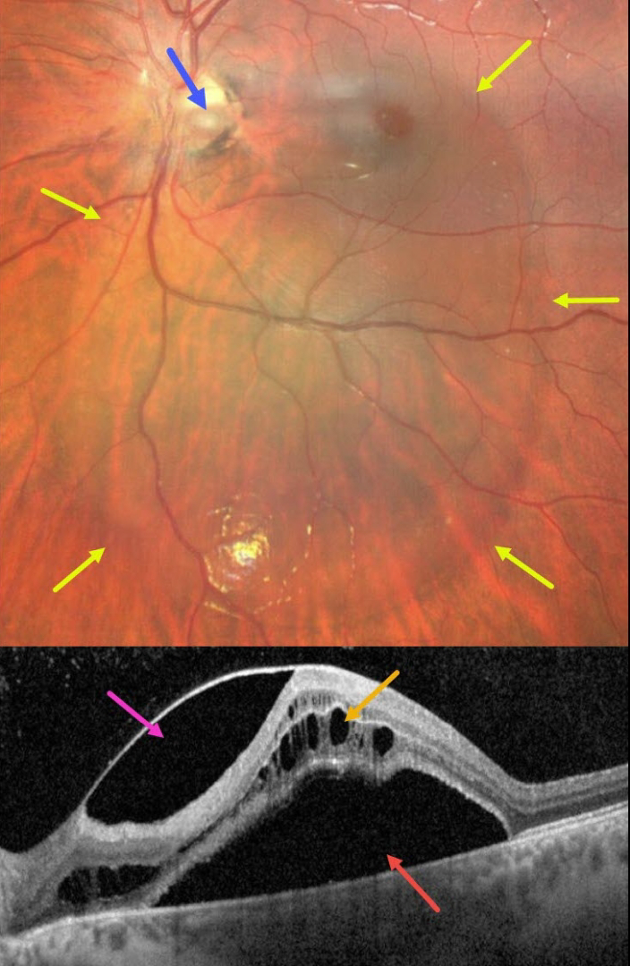
This 14YO boy presented with these asymptomatic and unilateral findings in his left eye. Vision was 20/50.
Fundus imaging shows a large vertical oval of retinal elevation extending from the superior macula into the inferior midperiphery. This fluid connects to an inferotemporal optic nerve pit.
OCT scanning shows subretinal fluid, cystic fluid mostly in the outer nuclear layer, and fluid below the internal limiting membrane vs within a split nerve fiber layer. He was referred to a retinal specialist for further evaluation and possible treatment.
Learning Points:
An optic nerve pit is a rare (about 0.1% prevalence) congenital anomaly thought to be caused by incomplete closure of the optic fissure during gestation. Pits are most commonly located within or along the inferior-temporal disc margin and usually appear as a gray depression.
While most optic pits are asymptomatic (as in this case), they can cause vision loss by forcing fluid into the inner retina, outer retina, and, ultimately, the subretinal space (optic pit maculopathy). The source of the fluid remains a subject of ongoing debate, with opinions ranging from the vitreous to the subarachnoid space.
A host of treatments for optic pit maculopathy have been suggested over the years. However, a recent meta-analysis suggested that a plain-vanilla vitrectomy without gas tamponade is as successful as other procedures (Zheng et al., Ophthalmology Retina 2020;4:289-299).
OPTIC NERVE COLOBOMA AND PIT
Originally posted on @retina.rocks December 2, 2022
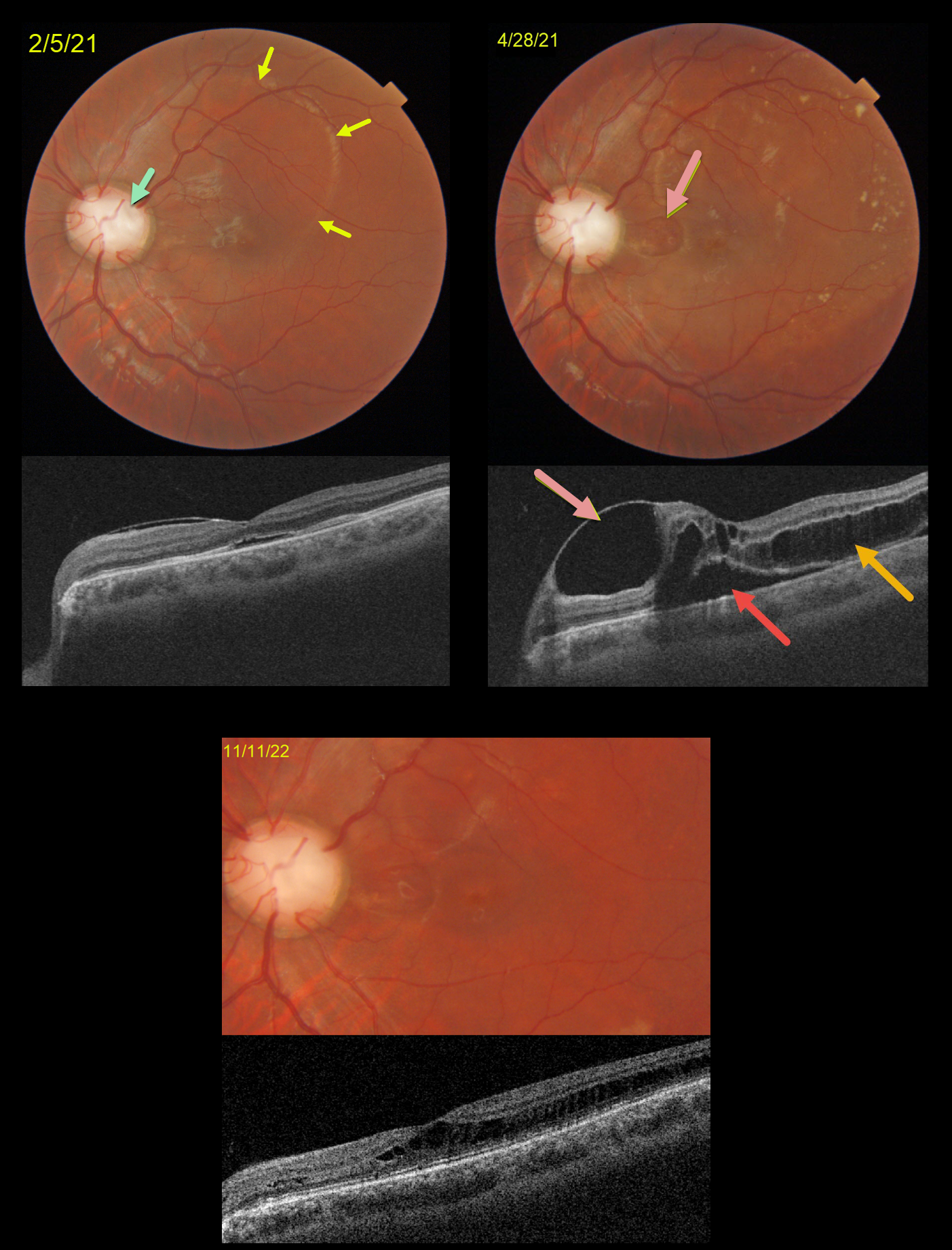
This is a follow-up to our 2/5/21 and 4/28/21 posts of a patient with bilateral optic nerve coloboma with optic pit maculopathy in his left eye.
On 2/5/21, vision was 20/70 with an optic pit and macular fluid. Despite the mostly peripheral fluid, the macular center was normal except for a trace amount of subretinal fluid (SRF).
Two months later, the peripheral fluid was better, but the central OCT findings were markedly different, with a nasal inner cyst, temporal outer schisis, and increased SRF. Although vision had decreased to 20/200, he subjectively felt his vision was better and elected continued observation.
When examined 1.5 years later on 11/11/22, the OCT findings had spontaneously improved. Temporal outer retinal schisis extends through the macular center, which is now free of SRF. Vision was 20/100, and he remained visually asymptomatic.
Learning Points:
An optic nerve coloboma can be a unilateral or bilateral condition caused by incomplete closure of the embryonic fissure.
While most optic pits are asymptomatic, they can cause vision loss due to fluid being forced into the inner retina, outer retina, and finally the subretinal space (optic pit maculopathy). The source of the fluid continues to be a point of ongoing debate, coming from either the vitreous or subarachnoid space.
A host of treatments for optic pit maculopathy have been suggested over the years. However, a recent meta-analysis suggested that a plain old vanilla vitrectomy without gas tamponade is as successful as any other procedure (Zheng et al Ophthalmology Retina 2020;4:289-299).
Our case illustrates how this fluid can spontaneously wax and wane, and that sometimes simple observation is the best option for some patients.
OPTIC NERVE PIT
Originally posted on @retina.rocks October 20, 2022
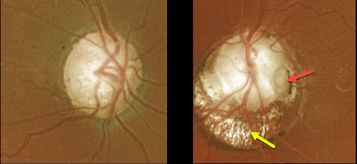
This 66YO female presented with an asymptomatic left optic nerve coloboma with a large temporal pit. The mildly colobomatous portion of the nerve is enlarged and slightly excavated inferiorly. Vision was 20/25 OD and 20/200 OS. Observation was recommended.
Learning Points:
Optic pits are most commonly located within or along the inferior-temporal disc margin and usually appear as a gray depression. They are thought to be caused by incomplete closure of the optic fissure during gestation.
While most optic pits are asymptomatic (as in this case), they can cause vision loss by forcing fluid into the inner and outer retina and, ultimately, the subretinal space (optic pit maculopathy).
The source of the fluid remains a subject of ongoing debate, with origins in either the vitreous or the subarachnoid space.
OPTIC NERVE COLOBOMA
Originally posted on @retina.rocks May 9, 2022
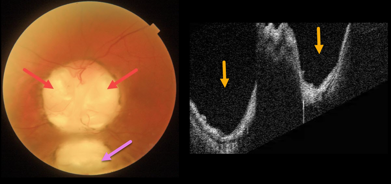
This 66YO male has mild lifelong decreased vision in his left eye. Vision was 20/70.
Triton imaging of the optic nerve coloboma shows optic nerve pits temporally and nasally, as well as a more inferior choroidal coloboma. Swept-source OCT dramatically reveals the pits.
We are following him yearly.
Learning Points:
Both optic nerve and choroidal colobomas can be unilateral or bilateral and are caused by incomplete closure of the embryonic fissure.
Patients with choroidal colobomas are at increased lifetime risk of rhegmatogenous retinal detachment due to breaks in the overlying thinned retina.
Optic nerve pits are also associated with serous macular detachment and macular neovascularization.
For a great review of cavitary optic nerve abnormalities, see Jain and Johnson, AJO 2014;158:423-435.
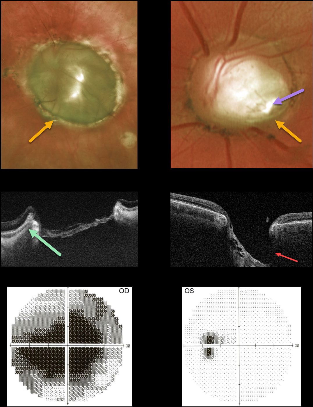
OPTIC NERVE COLOBOMA
Originally posted on @retina.rocks April 4, 2022
This 45YO female presented with bilateral optic nerve colobomas. Vision was hand motion OD and 20/25 OS.
Triton swept-source OCT shows a relatively featureless retina spanning the coloboma. The much smaller left nerve coloboma also contains an inferotemporal optic disc pit. The pit is also quite evident on OCT. The left nerve has a morning glory appearance with multiple vessels radiating outward from the peripheral disc margin, although there is no central glial tissue.
Visual fields show a large central defect OD with an enlarged blind spot OS.
Learning Points:
Optic nerve cavitary abnormalities can be unilateral or bilateral and result from incomplete closure of the embryonic fissure.
Our patient shows a spectrum of these abnormalities, including a huge optic disc coloboma OD, a much smaller disc coloboma OS with morning glory-like features, and an optic pit.
For a great review of cavitary optic nerve abnormalities, see Jain and Johnson, AJO 2014;158:423-435.
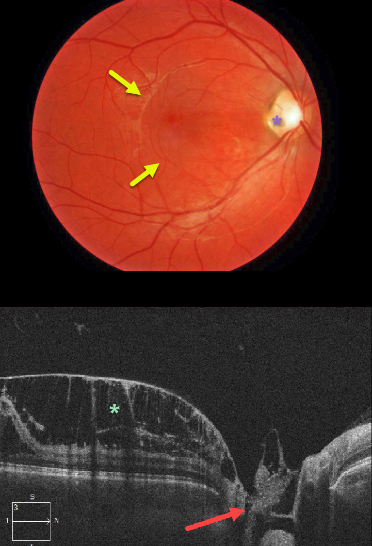
OPTIC NERVE PIT
Originally posted on @retina.rocks November 19, 2020
This patient presented with asymptomatic 20/30 vision OD and subtle macular thickening coming from an optic nerve pit along the temporal aspect of the optic nerve.
OCT shows a direct communication between the pit and the prominent inner macular schisis. Patients can also have subretinal fluid, which our patient did not. The left eye was completely normal.
Our patient opted for no treatment due to the lack of symptoms and fairly good vision.
Learning Points:
Optic pits are most commonly located within or along the inferior-temporal disc margin and usually appear as a gray depression. They are thought to be caused by incomplete closure of the optic fissure during gestation.
While most optic pits are asymptomatic, they can cause vision loss by forcing fluid into the inner and outer retina and, ultimately, the subretinal space (optic pit maculopathy). The source of the fluid remains a subject of ongoing debate, with origins in either the vitreous or the subarachnoid space.
A host of treatments have been suggested over the years. However, a recent meta-analysis suggested that a plain old vanilla vitrectomy without gas tamponade is as successful as any other procedure (Zheng et al Ophthalmology Retina 2020;4;289-299).
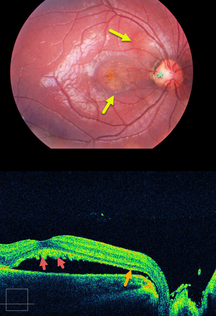
OPTIC NERVE PIT
Originally posted on @retina.rocks September 2, 2020
This patient has optic pit maculopathy, in which areas of subretinal fluid can be traced back to the optic nerve pit via an abnormal communication between the subretinal and subarachnoid spaces.
Shed photoreceptor outer segments accumulate on the back surface of the detached retina since the RPE cells can’t perform their normal function of phagocytosis. This shaggy vitelliform material gives the central macula a yellow appearance.
Learning Points:
Optic pits are congenital defects caused by the incomplete closure of the fetal fissure during embryogenesis. Macular detachment and schisis occur in up to 30-40% of eyes.
When symptomatic, a myriad of surgical procedures have been described. As with any problem with too many solutions, there is usually no best option.
A recent meta-analysis concluded that just a plain, vanilla vitrectomy is probably as successful as anything else (Zheng et al, Ophthalmology Retina 2020;4:389-299).


