

Optic Nerve Pseudo-Duplication
<< Back to Cases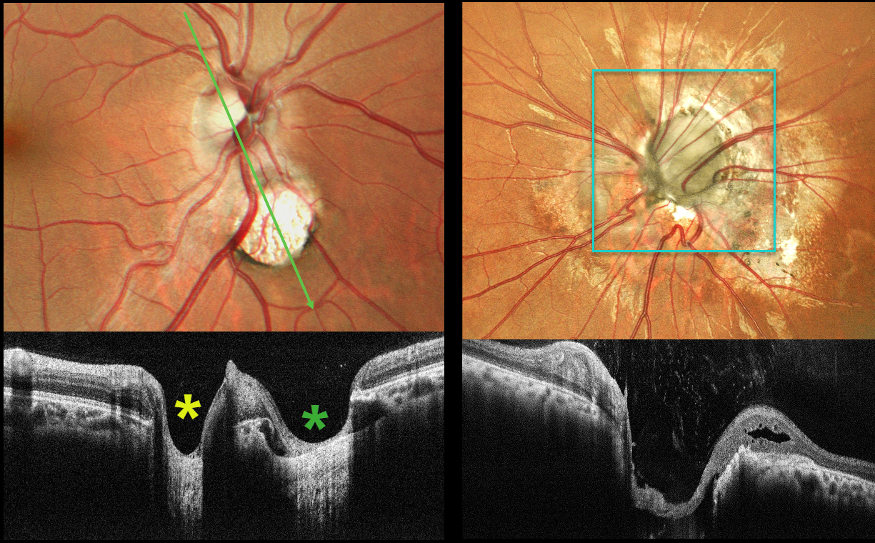
MORNING GLORY DISC AND CHOROIDAL COLOBOMA WITH PSEUDODUPLICATED OPTIC NERVE
Shraddha Raj Shrivastava and Manish Nagpal
Originally posted on @retina.rocks June 30, 2026
This 26YO male presented with a history of stable poor vision and inward deviation of his left eye since childhood. Vision was 20/20 OD and counting fingers in his esotropic, microphthalmic OS.
Color photography of the right posterior pole shows a round coloboma inferonasal to the nerve, giving the appearance of a pseudo-duplicated disc. OCT scanning through the nerve and coloboma reinforces the illusion of a second nerve head. The anomalous left nerve is enlarged and excavated, with numerous vessels radiating outwards from its inferonasal aspect.
Morning glory syndrome is typically unilateral and shares some similarities with optic disc coloboma and juxtapapillary staphyloma. This abnormality is named for its striking resemblance to the Morning Glory flower. Visual prognosis is usually poor. The condition can be associated with serous macular detachment. Neuroimaging should be considered to rule out transsphenoidal encephalocele and intracranial carotid artery dysgenesis, and fortunately, our patient’s imaging was negative. For a great review on the pathogenesis and treatment of maculopathy associated with cavitary optic disc anomalies, see Jain and Johnson, AJO 2014;158:423-435.
The coexistence of morning glory disc anomaly with microphthalmos, and contralateral chorioretinal coloboma in this case, may represent a continuum of optic fissure developmental defects with asymmetric expression, leading to a spectrum of cavitary optic disc anomalies in the same individual.
Pseudoduplication of the optic nerve occurs when a round choroidal coloboma or chorioretinal scar about the size of the optic nerve is associated with overlying radiating blood vessels (Bloom et al, Retinal Cases 2022;16:174-176). In our experience, this fake-out is most common from toxoplasmosis scars located near the optic nerve.
FULL-THICKNESS EYEWALL TRANSPLANT WITH INADVERTENT GRAFT INVERSION
Originally posted on @retina.rocks April 1, 2026
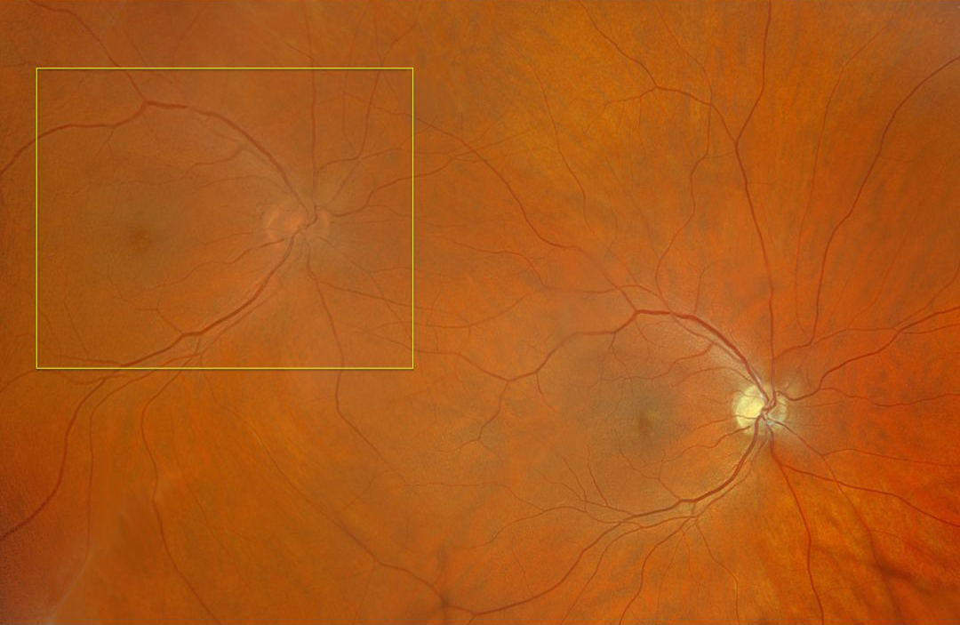
This healthy 43YO male fighter pilot developed a complex corneal laceration in his left eye. Despite multiple surgical procedures, he was left with an opaque cornea and an inability to fly due to the lack of normal binocular vision. Full-thickness eyewall transplantation from his normal left posterior segment into his right temporal posterior segment was therefore performed.
Intraoperatively, the dyslexic surgeon inadvertently inverted the allograft, resulting in the donor graft being inverted 180 degrees vertically. Optos color RGB imaging shows the donor nerve and macula in the right superotemporal midperiphery. Note that the disc should have been placed temporal to the donor macula, not nasal. Although surgically successful, this left the patient with monocular diplopia instead of the desired monocular binocular stereopsis. He remains grounded, and his case is still mired in litigation.
And happy April Fool’s Day!
CHOROIDAL COLOBOMA WITH OPTIC NERVE PSEUDODUPLICATION
Originally posted on @retina.rocks October 8, 2025
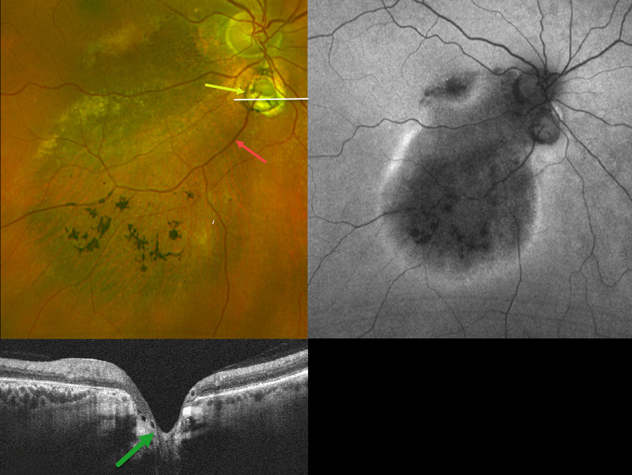
This 60YO female was referred for asymptomatic unilateral fundus findings in her right eye. Vision was 20/30 OD.
Optos color RG imaging shows a choroidal coloboma just inferior to the disc, giving the appearance of a pseudoduplicated optic nerve. Coarse pigmentary changes with some pigment migration extend from the macula into the inferior midperiphery. An inferotemporal retinal vein drains directly into the choroid through the coloboma. Triton swept-source OCT shows that the coloboma overlies an area of ectatic sclera. Fundus autofluorescence (FAF) shows variable hypo-FAF within the area of clinical pigmentary changes, with a surrounding rim of hyper-FAF.
Learning Points:
Choroidal colobomas are most often due to failure of the optic vesicle and choroidal fissure to close during fetal development. Lesions like this may also be from other causes, including toxoplasmosis. Choroidal coloboma can present as solitary or multiple lesions. Visual prognosis depends on the location and involvement of the optic nerve and macula.
Pseudoduplication of the optic nerve occurs when a round choroidal coloboma or chorioretinal scar about the size of the optic nerve is associated with overlying radiating blood vessels (Bloom et al, Retinal Cases 2022;16:174-176). In our experience, it is most common for toxoplasmosis scars to be located near the optic nerve.
Under normal conditions, the retinal and choroidal circulations remain separate and distinct. Rarely, the two circulations connect directly through a chorioretinal anastomosis. These can occur in chorioretinal scars (typically from inactive toxoplasmosis lesions), in macular telangiectasia, and in disciform scars.
The prominent gutter of inferior pigmentary changes indicates a prior retinal detachment, most likely related to spontaneously resolved fluid associated with the coloboma (Tanaka et al, Ophthalmology Retina 2021;5:702-710). Similar pigmentary changes are seen in central serous retinopathy, although our patient’s macular OCTs showed no pachychoroid features (not shown). Finally, this may represent previously resolved pit-like macular fluid, which is rarely found in the absence of a pit or severe glaucomatous cupping (Fujimoto et al, Ophthalmology Retina 2023;7:811-818).
CHOROIDAL COLOBOMA WITH OPTIC NERVE PSEUDODUPLICATION
Originally posted on @retina.rocks March 25, 2024
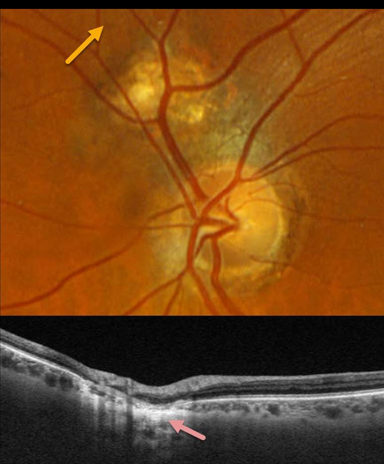
This 66YO male was referred for an asymptomatic lesion in his left fundus. Vision was 20/25 in his normal right eye and 20/25 in his left eye.
Optos color RGB imaging of the left eye shows a normal optic nerve. Just superonasal to the nerve is a round, colobomatous scar.
Triton swept-source OCT through this lesion shows disorganized neurosensory thinning with an underlying choroidal coloboma.
Learning Points:
Pseudoduplication of the optic nerve occurs when a round choroidal coloboma or chorioretinal scar about the size of the optic nerve is associated with overlying radiating blood vessels (Bloom et al, Retinal Cases 2022;16:174-176). In our experience, it is most common for toxoplasmosis scars to be located near the optic nerve.
DUPLICATED OPTIC NERVE
Originally posted on @retina.rocks March 31, 2023
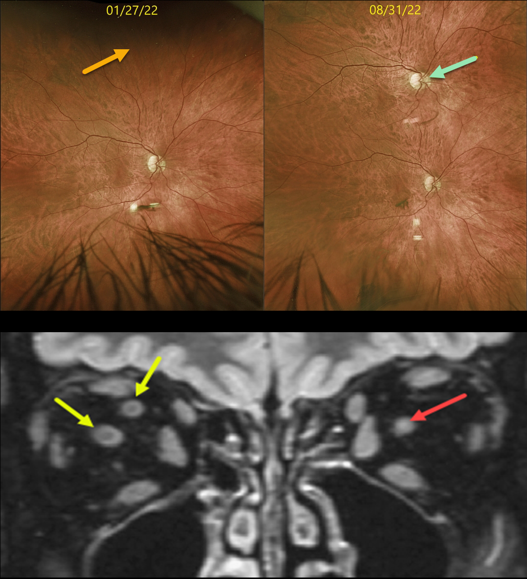
This 61YO diabetic male has been followed yearly for routine retinopathy screenings. On 1/27/22, vision was 20/20 with a normal right fundus. Note that the superior periphery is also normal.
On 8/31/22, he returned complaining of a new inferotemporal peripheral scotoma with monocular vertical diplopia in this eye. Vision was 20/20 centrally and 20/20 inferiorly. A new optic nerve and macula had developed in the interim. These findings were confirmed on MRI scanning, which revealed a single nerve OS and two nerves OD in the retro-orbital space.
Learning Points:
Duplication of the optic nerve is a non-heritable developmental abnormality with an extremely low prevalence of about 1 per 23.5 million live births, and is a rare cause of monocular diplopia. To the best of our knowledge, ours is the first such known de novo case.
AND HAPPY APRIL FOOL’S DAY 2023!
OPTIC NERVE PSEUDO-DUPLICATION WITH NVD ELSEWHERE
Originally posted on @retina.rocks December 18, 2020
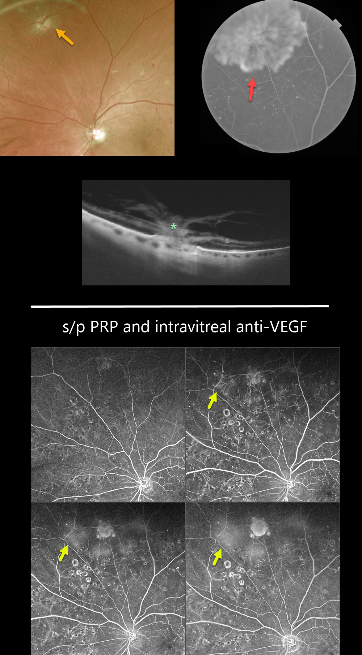
Is that another optic nerve?
This is a pseudo-duplicated optic disc from peripheral neovascularization emanating from a disc-sized mid-peripheral chorioretinal scar.
The OCT reveals the area of choriovitreal neovascularization.
Learning Points:
The neovascularization regressed following panretinal photocoagulation (PRP) supplemented with intravitreal Eylea injections.
Fluorescein angiography following PRP and anti-VEGF injections shows virtually no leakage from the neovascularization.
OPTIC NERVE PSEUDO-DUPLICATION
Originally posted on @retina.rocks December 18, 2019
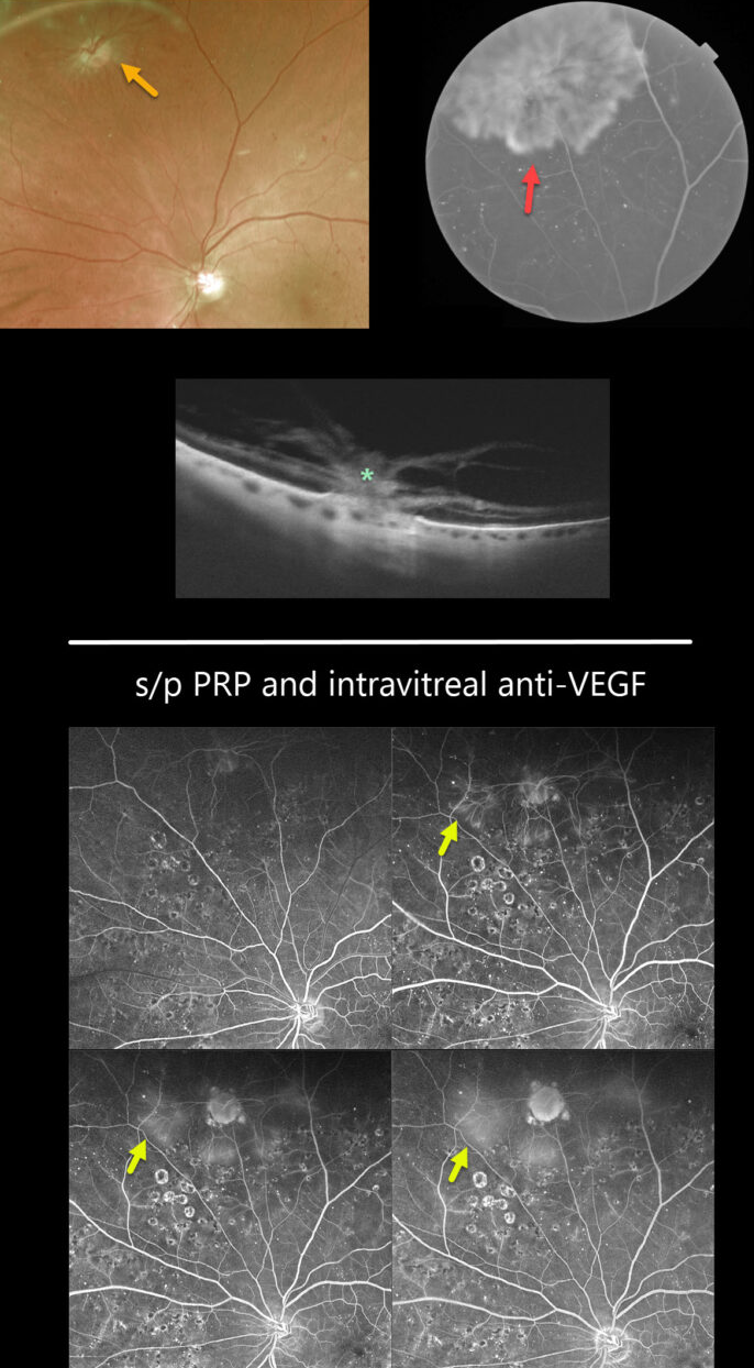
Is that another optic nerve????
Pseudoduplicated optic disc from peripheral neovascularization emanating from disc-sized mid peripheral chorioretinal scar. The neovascularization regressed following panretinal photocoagulation with intravitreal eylea injections. The OCT showing the choriovitreal neovascularization is amazing!


