

Optic Neuropathy
<< Back to Cases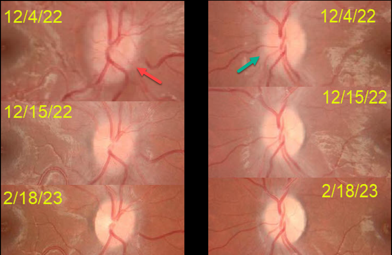
MOG-ASSOCIATED OPTIC NEURITIS
Asma Samsudeen and Ashish Sharma
Originally posted on @retina.rocks April 27, 2023
This 10YO boy presented on 12/4/22 with acute blurred vision in his right eye. He was hospitalized 2 weeks earlier for a viral illness with musculoskeletal pain and fever.
Vision was counting fingers OD and 20/20 OS. An afferent pupillary defect was noted OD. The right disc was moderately swollen, and milder disc swelling was noted OS. MRI was suggestive of bilateral optic neuritis along with a suspicious cervical cord lesion. He was diagnosed with possible parainfectious demyelination and started on high-dose intravenous steroids. Vision improved OD to 20/40 four days later.
On 12/15/22, vision was 20/20 OU, and the nerves were non-edematous. Despite oral mycophenolate mofetil (MMF) immunotherapy, he presented on 1/14/23 with counting vision OS, a new afferent pupillary defect OS, and new left disc edema (not shown).
Anti-NMO (neuromyelitis optica) testing was negative, and Anti-MOG (myelin oligodendrocyte glycoprotein, a protein located on the surface of central nervous system myelin) testing was positive, and was diagnosed with MOG antibody disease (MOG-AD).
Three days of intravenous pulse steroids were given in addition to continuing oral MMF.
Since he developed a relapse in his left eye while on MMF immunotherapy, Rituximab (RTX) will be added. When last examined on 2/18/23, vision was 20/20 OU.
Learning Points:
MOG-AD is an acquired immune-mediated demyelinating syndrome, distinct from multiple sclerosis, that causes inflammation of the optic nerve, spinal cord, and brain. Treatments include intravenous steroids, plasma exchange, and intravenous immunoglobulin. RTX reduces relapse frequency in the majority of patients (Bai et al., Multiple Sclerosis and Related Disorders, 2021;53:103044).
HYPERTENSIVE RETINOPATHY vs AION
Originally posted on @retina.rocks August 16, 2022
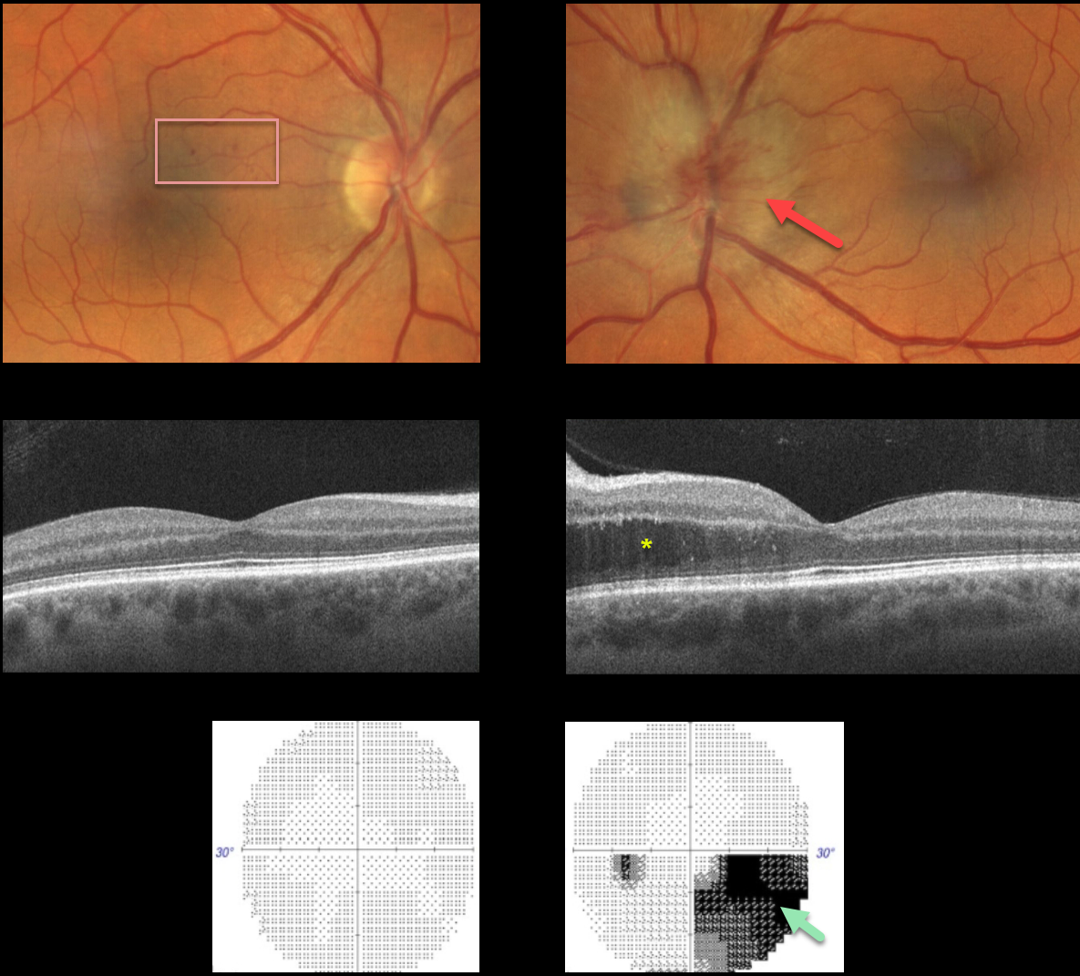
This 57YO male presented with unilateral optic nerve swelling in his left eye and a few faint nasal macular retinal hemorrhages in his right eye. Vision was 20/40 OD and 20/25 OS.
OCT was normal OD and thickened in the nasal outer nuclear layer OS. Visual fields were normal OD and inferiorly depressed, especially inferonasally, OS.
Blood pressure was 190/110, and he was immediately referred to the emergency room.
Learning Points:
Malignant hypertension, defined as blood pressure above 180 systolic and/or 120 diastolic, is a life-threatening medical emergency. Eye doctors are in a unique position to often diagnose this condition.
Patients will often present with bilateral optic nerve swelling, flame-shaped retinal hemorrhages, and, with more chronic disease, lipid precipitates in the nasal macular outer plexiform layer. However, rarely, the ocular findings may be asymmetric or even unilateral.
Since the macular fluid emanates from the optic nerve, the macular thickening is always worse nasally (as in our case), and is an important clinical clue for this diagnosis.
Our patient may also have had an isolated anterior ischemic optic neuropathy in his left eye and mild hypertensive retinopathy in his right eye. The partial altitudinal deffect is more common with this diagnosis than hypertension.
OPTIC NEUROPATHY
Originally posted on @retina.rocks November 20, 2020
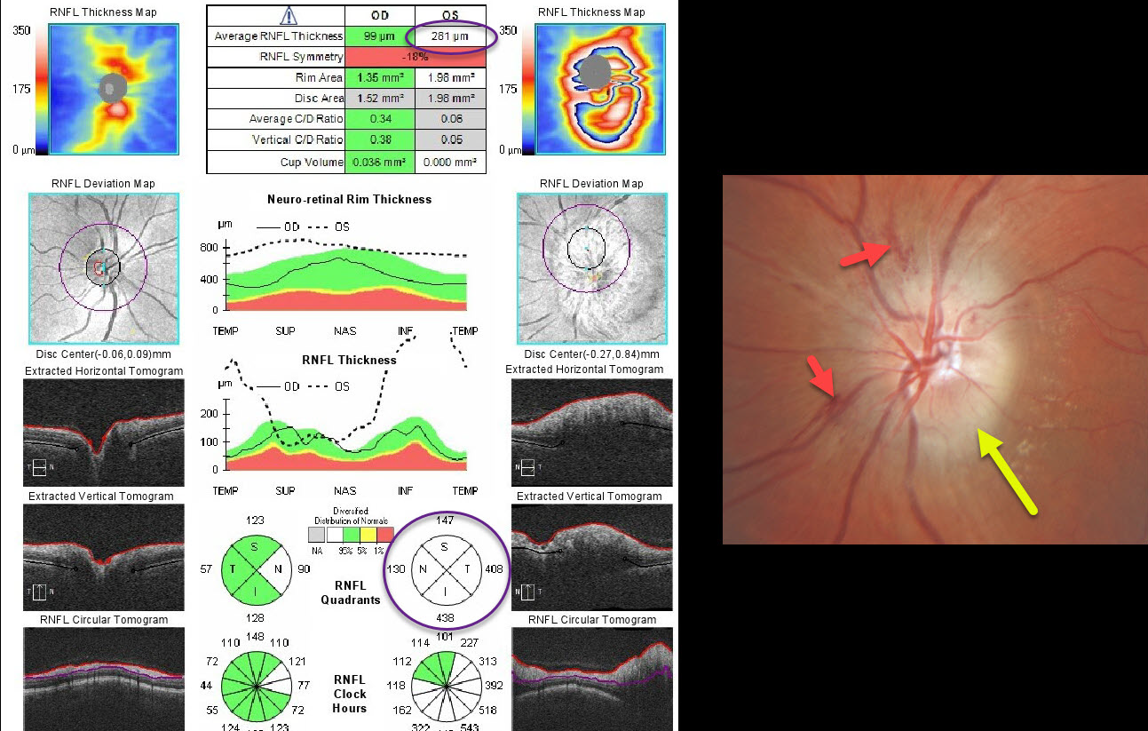
This patient presented with unilateral diffuse optic nerve edema with some splinter hemorrhages and possible inferotemporal lipid. Optic nerve OCT confirms markedly increased retinal nerve fiber layer thickness.
The differential diagnosis for a unilaterally acutely swollen nerve is quite long and includes arteritic (AION) and non-arteritic (NAION) anterior ischemic optic neuropathy, infectious causes (including syphilis and neuroretinitis), and inflammatory conditions (including sarcoidosis and optic neuritis). In this case, we diagnosed NAION.
Learning Points:
NAION presents with sudden painless unilateral central vision loss with an altitudinal defect. It is the most common acute optic neuropathy in patients over age 50.
It is thought to be caused by non-embolic occlusion of small branches of the short posterior ciliary arteries that supply the anterior optic nerve. It is associated with congenitally small and crowded nerves, optic disc drusen, medications (amiodarone and possibly phosphodiesterase inhibitors), hypertension, and diabetes. About 75% of patients have sleep apnea. Nocturnal hypotension or taking blood pressure pills at bedtime is also considered a risk factor.
Erythrocyte sedimentation rate and C-reactive protein should be ordered to rule out giant cell arteritis. There is no effective treatment.
Optic nerve edema resolves within a few months, often with secondary optic nerve pallor.


