

Outer Foveal Microdefect
<< Back to Cases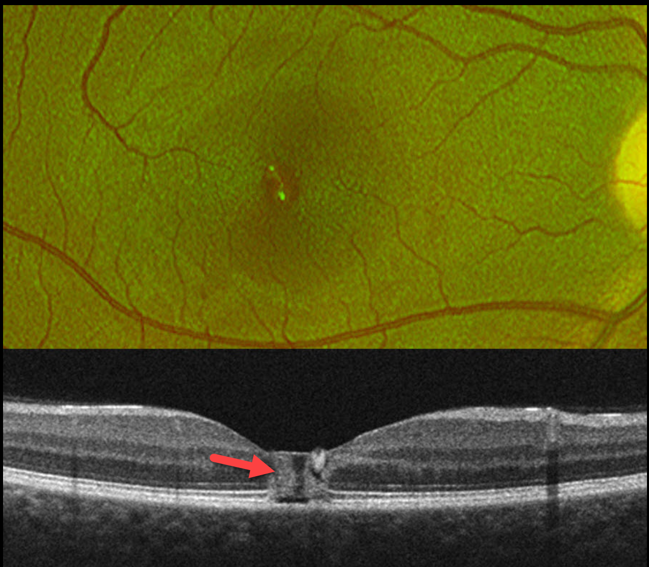
LASER MACULOPATHY
Vaibhav Sethi
Originally posted on @retina.rocks January 20, 2026
This healthy 19YO female noticed immediate blurred vision in her right eye after looking at a laser during a party. Her vision in this eye was 20/60 when she presented to us the following day.
Optos color RG imaging shows a foveal pseudohole. OCT scanning shows a central foveal column of full-thickness hyperreflectivity. There is loss of the outer segment bands overlying the RPE.
Learning Points:
Most cases of laser maculopathy reported in the literature are caused by intentional or inadvertent exposure to laser pointers (Bhavsar et al, Survey Ophthalmology 2021;66:231-260). However, more recently, there have been reports of macular injury from exposure to high-power lasers during concerts or from DJ laser machines (Perz-Montano et al, Doc Ophthalmol 2019;138:71-76). Most eyes recover fairly good vision with conservative management.
Our patient was immediately lost to follow-up.
WELDING ARC MACULOPATHY
Malvika Singh and Manish Nagpa
Originally posted on @retina.rocks December 24, 2025
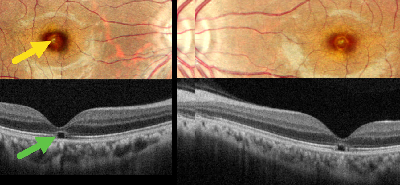
This 31YO male presented with 2 years of bilateral vision loss. He works as a welder and admits to rarely wearing his safety glasses. Vision was 20/30 OU.
Pseudocolor SLO imaging shows symmetrical, tiny yellow foveal scars. OCT shows outer foveal microdefects characterized by hyporeflectivity due to loss of all bands deep to the external limiting membrane.
Learning Points:
The welding process emits optical radiation across various wavelengths and intensities, including infrared, visible, and ultraviolet (UV) light. Exposure to this UV light without proper eye protection can result in keratoconjunctivitis (the most common clinical presentation) and foveal lesions similar to solar retinopathy. Findings include a small yellowish foveal spot or scar with an outer foveal microdefect on OCT (Singh et al, Surv Ophthalmology 2023;68:655-668). Vision is usually good despite the persistent clinical and OCT findings.
Our patient was lucky to have good vision despite his poor work habits. He was advised to use protective eyewear during welding.
WELDING ARC MACULOPATHY
Rohan Jain and Manish Nagpal
Originally posted on @retina.rocks May 1, 2025
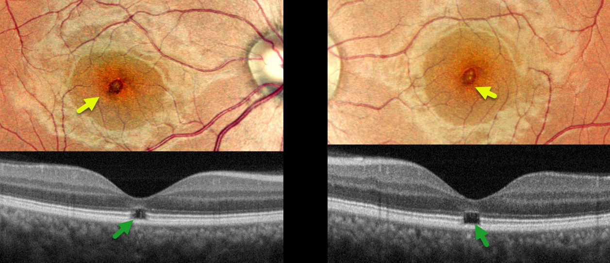
This 38YO male presented with 6-7 months of bilateral vision loss. He works as a welder and admits to rarely wearing his safety glasses. Vision was 20/25 OD and 20/20 OS.
Pseudocolor SLO imaging shows symmetrical, tiny yellow foveal scars. OCT shows outer foveal microdefects with hyporeflectivity due to loss of all bands deep to the external limiting membrane.
Learning Points:
The welding process emits optical radiation across various wavelengths and intensities, including infrared, visible, and ultraviolet (UV) light. Exposure to this UV light without proper eye protection can result in keratoconjunctivitis (the most common clinical presentation) and foveal lesions similar to those seen in solar retinopathy. Findings include a small foveal yellowish spot or scar with an outer foveal microdefect on OCT (Singh et al, Surv Ophthalmology 2023;68;655-668). Vision is usually good despite the persistent clinical and OCT findings.
Our patient was lucky to have good vision despite his poor work habits. He was advised to use protective eyewear during welding.
MACULAR PUCKER
Originally posted on @retina.rocks February 19, 2025
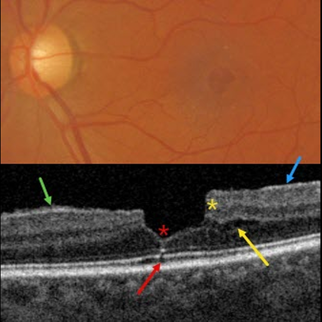
This patient presented with 20/30 vision due to a mildly symptomatic clinical macular pucker with a pseudohole; observation was recommended. Multimodal imaging reveals extensive pucker-related findings.
Triton color imaging shows an irregular macular pseudohole with a faint surrounding epiretinal membrane (ERM). True full-thickness macular holes are virtually always perfectly round, and these pucker-related pseudoholes are often irregularly round, as in this eye.
Swept-source OCT shows an adherent ERM with some shaggy-appearing adherent vitreous cortex, favoring epimacular proliferation. The fovea has a vertical contour, often present with ERM, due to the encircling membrane contracting towards the foveal center, dragging the inner retina centrally.
There are features of both a tractional lamellar macular hole (LMH) with splitting of the outer plexiform layer, along with a degenerative LMH with descending loss of tissue into the outer nuclear layer (Govetto et al, AJO 2016;164:99-109).
Finally, a tiny area of shadowing vs an outer foveal microdefect (Cohen et al, Ophthalmology Retina 2021;5:553-561) is seen.
LASER MACULOPATHY
Mattie Adams
Originally posted on @retina.rocks April 22, 2024
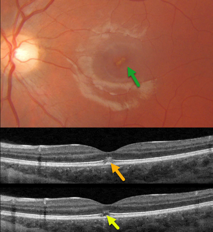
This 13YO girl presented with immediate vision loss in her left eye after looking at a laser pointer one week earlier. Vision was 20/20 in her normal right eye and 20/70 in her left eye.
Triton color photography shows an oblique, slightly linear area of foveal pigment loss. Swept-source OCT scans through this lesion show focal hyperreflectivity with variable loss of the ellipsoid zone and outer segments.
Learning Points:
Sun gazing is thought to cause acute damage to the RPE and photoreceptors via photochemical mechanisms involving reactive oxygen species. Similar photochemical damage can follow laser pointer exposure, although the clinical findings can be different (Bloom and Singal, Retinal Cases 2022;16:
89-91).
Sun gazing acutely produces bilateral, symmetric, pinpoint subfoveal yellow-white lesions in the outer retina, followed by variable clinical depigmented foveal RPE and focal photoreceptor OCT defects extending from the external limiting membrane to the RPE.
Laser pointer maculopathy can cause a similar lesion, but presumed more prolonged viewing, saccades, and laser pointer movement can also lead to larger areas of macular damage.
Outer foveal microdefects (Cohen et al, Ophthalmology Retina 2021;5:553-561) have been described in numerous conditions, including macular telangiectasia, tamoxifen use, ABCA4 disorders (cone-rod dystrophies, Stargardt disease and fundus flavimaculatus), phototoxicity, trauma, and vitreomacular traction disorders.
WHIPLASH MACULOPATHY
Originally posted on @retina.rocks March 5, 2024
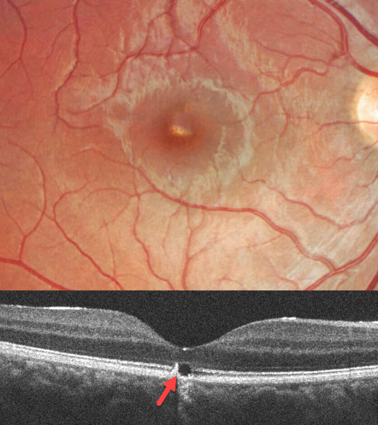
This 17YO male was in a car accident several weeks earlier with a whiplash injury. He noticed mild vision loss in his right eye since. Vision was 20/30.
Color imaging shows a somewhat linear area of foveal pigment loss. OCT scanning of this region shows an outer foveal microdefect with adjacent hyperreflectivity along its temporal edge.
Learning Points:
Initially described by Kelley et al (Arch Ophthalmol 1978;96:834-835), whiplash maculopathy is thought to occur from a vitreous coup-contrecoup injury following a motor vehicle accident. This can cause contusive RPE injury with transient macular edema (AJO 2007;143:348-350) or, more typically, a solar retinopathy-like defect in the outer retina.
Outer foveal microdefects (Cohen et al, Ophthalmology Retina 2021;5:553-561) have been described in numerous conditions, including macular telangiectasia, tamoxifen use, ABCA4 disorders (cone-rod dystrophies, Stargardt disease, and fundus flavimaculatus), phototoxicity, trauma, and vitreomacular traction disorders.
OUTER FOVEAL MICRODEFECT
Originally posted on @retina.rocks December 13, 2023
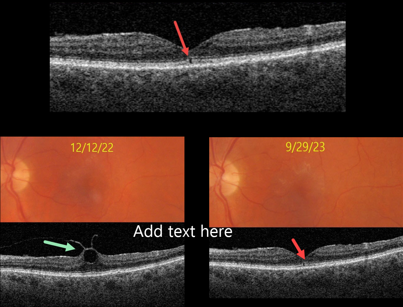
This 76YO female presented with no visual symptoms, 20/40 vision, and an abnormal OCT in her left eye. What caused this subtle OCT finding?
She was previously being followed for asymptomatic focal vitreomacular traction (VMT) in this eye. On 12/12/22, Triton color imaging showed a subtle foveal cyst with focal VMT on swept-source OCT.
We recommended observation due to the absence of symptoms, and when she returned on 9/29/23, these findings spontaneously resolved as the vitreous separated, leaving a tiny outer foveal microdefect.
Learning Points:
Outer foveal microdefects (Cohen et al, Ophthalmology Retina 2021;5:553-561) have been described in numerous conditions, including macular telangiectasia, tamoxifen use, ABCA4 disorders (cone-rod dystrophies, Stargardt disease, and fundus flavimaculatus), phototoxicity, trauma, and vitreomacular traction disorders.
The prevalence of VMT increases with age, and is present in about 1% of people over 40YO. The natural history is still being defined, but most patients have stable traction and vision. VMT can also resolve spontaneously in about 20% (Errera et al, Ophthalmology 2018;125:701-707).
Many patients with VMT remain completely asymptomatic despite impressive OCT findings. We therefore almost always observe VMT, as in this case, unless the patient has significant symptoms.
TAMOXIFEN RETINOPATHY
Aaron McNulty
Originally posted on @retina.rocks June 19, 2023
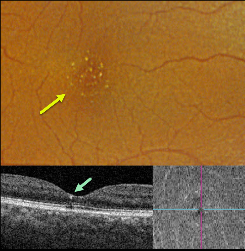
This 56YO female was referred for asymptomatic macular findings. The referring doctor mentioned that she may have retinal changes from the Tamoxifen that she was taking for over 3 years for breast cancer. Vision was 20/50 OD and 20/40 OS.
Optos color RGB imaging shows numerous tiny inner retinal crystalline deposits scattered throughout each fovea. These deposits appeared as tiny hyperreflective dots beneath the ILM on OCT B-scan and en face.
Our patient had already spoken with her oncologist before seeing us and had already stopped the Tamoxifen. The crystalline changes will likely remain and, hopefully, not progress going forward.
Learning Points:
Tamoxifen retinopathy shares phenotypes very similar to those of macular telangiectasia type 2 (MacTel 2, Lee et al., Ophthalmology Retina 2020;3:681-689), especially in the early stages, including retinal cavitations, right-angle venules, and inner retinal crystals.
The changes in Tamoxifen are confined to the central macula, whereas in MacTel2 they are present in a slightly larger area with an epicenter temporal to the foveal center (Hess et al, Ophthalmology Retina 2023;7:101-110). The retinal changes for both disorders likely share Muller cell dysfunction as the common cause.
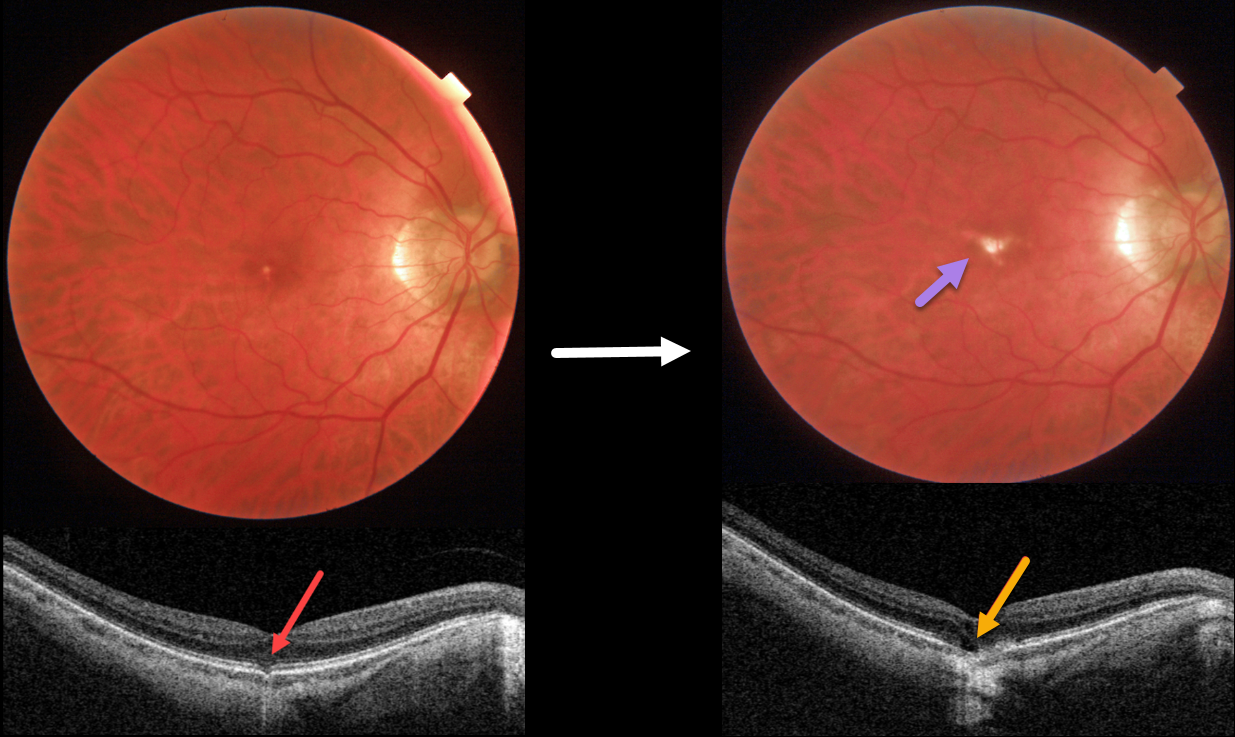
OUTER FOVEAL MICRODEFECT
Originally posted on @retina.rocks April 13, 2023
This 58YO male was referred for an asymptomatic foveal lesion in his right eye. There was a history of high myopia prior to LASIK surgery years earlier. Vision was 20/40 OD.
Color photography shows a small atrophic foveal scar. OCT shows a focal EZ defect with a possible tiny choroidal excavation. He returned about 2 years later for a routine follow-up visit.
Although he was binocularly without a new complaint, his vision had decreased to 20/200. There was now a definite, much enlarged foveal lacquer crack. OCT scanning shows a new outer foveal microdefect.
Learning Points:
Outer foveal microdefects (Cohen et al, Ophthalmology Retina 2021;5:553-561) have been described in numerous conditions, including macular telangiectasia, tamoxifen use, ABCA4 disorders (cone-rod dystrophies, Stargardt disease, and fundus flavimaculatus), phototoxicity, trauma, and vitreomacular traction disorders.
We are not aware of a prior report showing these lesions in association with myopic lacquer cracks. These defects, caused by focal loss of the outer retinal layers, leave an optically empty space that can be mistaken for subretinal fluid but is more of an optical gap within the outer retina.
Focal choroidal excavation (FCE), originally described by Jampol et al in 2006, is likely part of the pachychoroid spectrum since it is usually associated with a thickened choroid and pachyvessels. The FCE can be observed, although symptomatic manifestations, including active central serous retinopathy or macular neovascularization, often require treatment. An excellent review is by Verma et al, Br J Ophthalmol 2021;105:1043-1048.
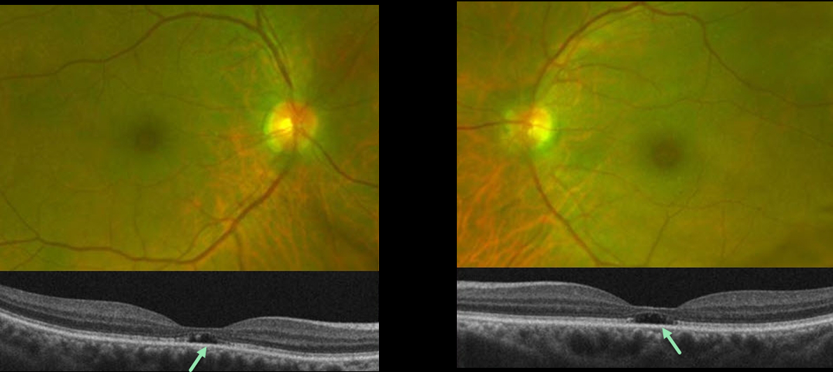
OUTER FOVEAL MICRODEFECTS FROM POSSIBLE IRD
Emily Shepherd
Originally posted on @retina.rocks March 10, 2023
This 65YO presented complaining of difficulty seeing at a distance for some time. She gave a history of radial keratotomy in both eyes about 20 years prior. There was no significant past medical history or family history of eye disease. Vision was 20/60 OD and 20/70 OS.
Optos color imaging shows mild symmetrical pericentral pigmentary change. Fundus autofluorescence and fluorescein angiography were unremarkable (not shown). However, OCT B-scans show bilateral subfoveal loss of the outer retinal tissue external to the external limiting membrane.
An ERG showed rod receptor dysfunction and abnormal dark adaptation with delayed adaptation, while a multifocal ERG was consistent with normal cone function.
Our patient underwent genetic testing and was found to have a heterozygous mutation for FLVCR1. This gene is associated with autosomal recessive posterior column ataxia with retinitis pigmentosa.
A recent report by Kuehlewein et al (Graefe’s Arch Clin Exp Ophthalmol 2019;257:629-638) showed a variety of phenotypes, none of which matched our patient. It is therefore difficult to say whether our patient’s findings are isolated (given the lack of funduscopic, autofluorescence, and angiographic findings beyond the OCT outer defects) or represent an atypical manifestation (given the abnormal electrophysiology) of FLVCR1 disease.
She is scheduled to return for follow-up imaging and visual field testing.
Learning Points:
Outer foveal microdefects (Cohen et al, Ophthalmology Retina 2021;5:553-561) have been described in numerous conditions, including macular telangiectasia, tamoxifen use, ABCA4 disorders (cone-rod dystrophies, Stargardt disease, and fundus flavimaculatus), phototoxicity, trauma, and vitreomacular traction disorders.
In our experience, in patients with inherited retinal diseases (IRD), these findings are most common with a pathogenic ABCA4 mutation (Leng et al, Retina 2012;32:1411-1419 and Wang et al Invest Ophthalmol Vis Sci 2022;53(5):28). These defects, caused by focal loss of the outer retinal layers, leave an optically empty space which can be confused with subretinal fluid but are more of an optical gap within the outer retina.
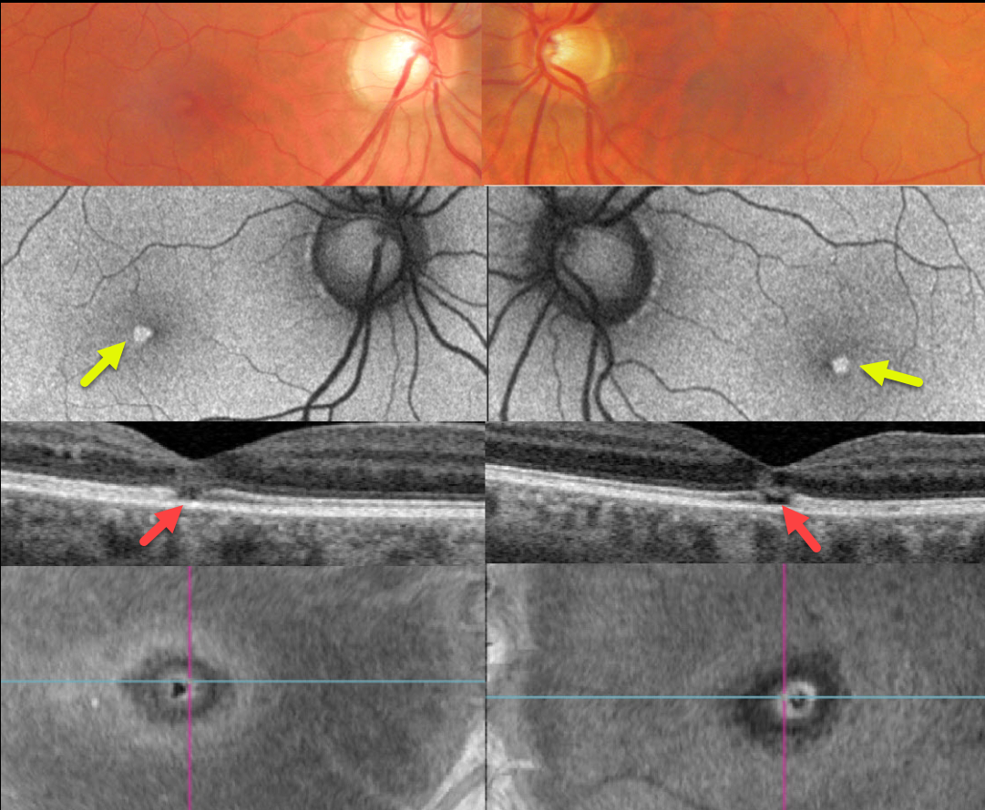
SOLAR RETINOPATHY
Originally posted on @retina.rocks February 27, 2023
This healthy 46YO male complained of mild bilateral central vision loss for about 5 years. Vision was 20/25 OD and 20/30 OS.
Optos color imaging shows subtle central foveal pigment loss. Fundus autofluorescence (FAF) shows these lesions to be hyper-FAF.
Swept-source OCT shows small central foveal outer retinal defects, and en face OCT of the outer retina shows a unique perspective of these lesions.
On further questioning, he noted that his visual symptoms immediately followed his viewing the Great Eclipse of 2017 without proper eye protection.
Learning Points:
The outer retinal damage in solar retinopathy is thought to be caused by photochemical outer retinal and RPE damage instead of a thermal burn. Vision is usually fairly good despite the outer retinal and RPE findings.
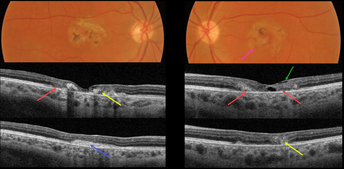
MACULAR TELANGIECTASIA
Originally posted on @retina.rocks November 8, 2022
This 49YO female presented with several weeks of vision loss in her left eye. A macular neovascularization (MNV) in her right eye was treated 4 years earlier with intravitreal Avastin. Vision was 20/200 OD and 20/70 OS.
Both maculas had central pigment loss with intraretinal pigment migration. A small dot of blood was noted inferonasally in the left macula.
OCT scanning shows outer retinal atrophy, pericentral retinal disorganization, a fibrosed, inactive MNV in the right macula, and hyporeflective loss of retinal tissue (cavitation) in the left outer macula. Faint loss of the temporal inner retinal tissue is highlighted by intact internal limiting membrane (ILM drape sign).
Anti-VEGF therapy was started for a presumed MNV in her left eye.
Learning Points:
Originally described by Gass (Arch Ophthalmology 1982;100:769-780), MacTel2 is a neurodegenerative disorder, most likely originating from Muller cell dysfunction.
The term, telangiectasia, is misleading in that the funduscopic findings are mostly non-vascular. The refractile inner retinal ‘crystals’ are thought to be Muller cell footplates. The foveal retina shows a whitish discoloration, most likely due to retinal opacification caused by dysfunctional Muller cells.
Photoreceptor loss allows RPE cells to migrate along intraretinal capillaries, causing the coarse clinical pigment clumping.
All of these changes are usually in the temporal fovea, so our patient is atypical in that they involve the entire central macular regions.
SOLAR RETINOPATHY
Originally posted on @retina.rocks April 27, 2022
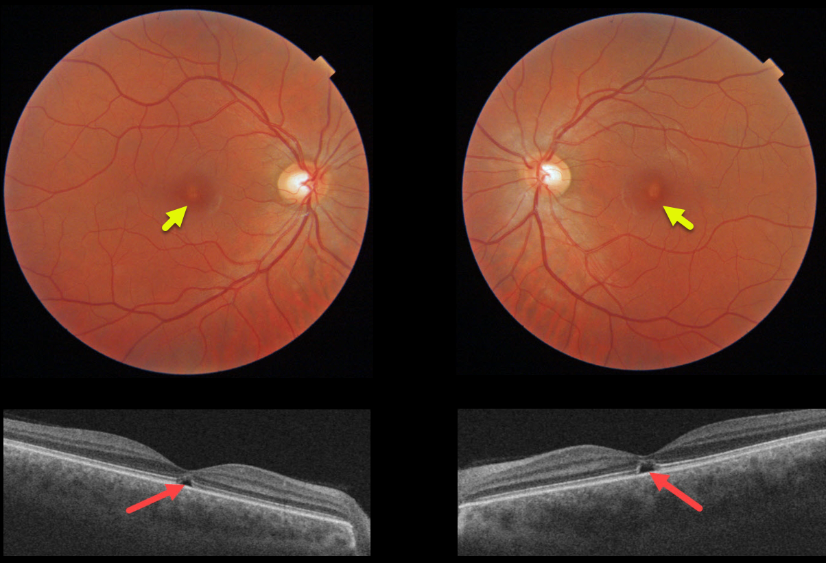
This 31YO male was referred for relatively asymptomatic central macular pigmentary changes. He gave a long history of twice-daily sun gazing, which always caused a very pleasurable sneezing episode. Vision was 20/30 OD and 20/40 OS.
Triton color imaging shows tiny foveal pigment loss. Swept-source OCT shows bilateral ellipsoid zone and outer segment defects.
We diagnosed him with the ACHOO reflex (Autosomal Dominant Compelling Helioophthalmic Outburst). And no, this isn’t an April Fools post, and we’re not talented enough to even make this stuff up if we wanted to! Patients with the ACHOO reflex sneeze when exposed to bright lights, typically sunlight.
We advised him to stop his sun gazing ritual, although we’re not sure this will be possible for him.
Learning Points:
The outer retinal damage in solar retinopathy is thought to be caused by photochemical outer retinal and RPE damage instead of a thermal burn. Vision is usually fairly good despite the outer retinal and RPE findings.
SOLAR RETINOPATHY
Originally posted on @retina.rocks February 24, 2022
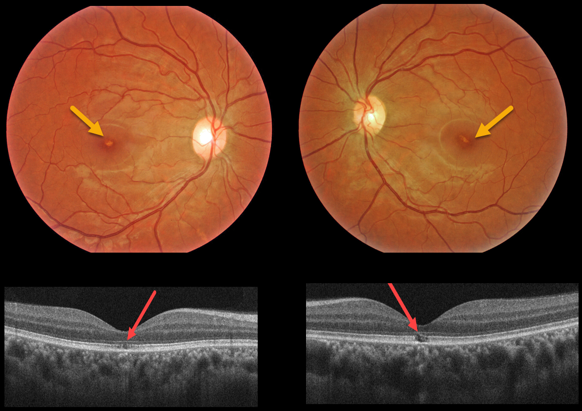
This 32YO female with schizophrenia reported regular sun gazing every morning for many years. Vision was 20/40 OU.
Triton color imaging shows tiny foveal pigment loss. Swept-source OCT shows bilateral ellipsoid zone and outer segment defects.
Learning Points:
The outer retinal damage in solar retinopathy is thought to be caused by photochemical outer retinal and RPE damage instead of a thermal burn.
Vision is usually fairly good despite the outer retinal and RPE findings.
MACULAR TELANGIECTASIA
Originally posted on @retina.rocks December 27, 2021
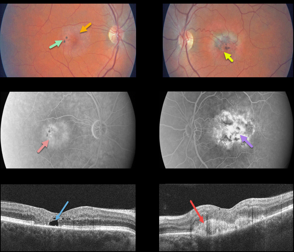
This patient presented with classic findings of macular telangiectasia type 2 (MacTel2), including an opaque perifoveal retina, inner retinal crystal, and black clumps of intraretinal pigment migration.
Deep retinal angiographic leakage is noted in the right eye with more diffuse staining in the left eye. OCT scanning shows an outer retinal cavitation OD and type 2 macular neovascularization (MNV) OS.
Learning Points:
Originally described by Gass (Arch Ophthalmology 1982;100:769-780), MacTel2 is a neurodegenerative disorder, most likely originating from Müller cell dysfunction.
The term telangiectasia is misleading because the funduscopic findings are mostly nonvascular.
The refractile inner retinal ‘crystals’ are thought to be Müller cell footplates. The foveal retina shows a whitish discoloration, most likely due to retinal opacification resulting from Müller cell dysfunction.
Photoreceptor loss allows RPE cells to migrate along intraretinal capillaries, causing the coarse clinical pigment clumping.
The majority of patients will also have subclinical chorioretinal anastomosis associated with right-angle venules (Spaide et al., Retina 2018;38:1920-1929).
Secondary MNV can be successfully treated per age-related macular degeneration protocols.
MACULAR PUCKER
Originally posted on @retina.rocks August 5, 2021
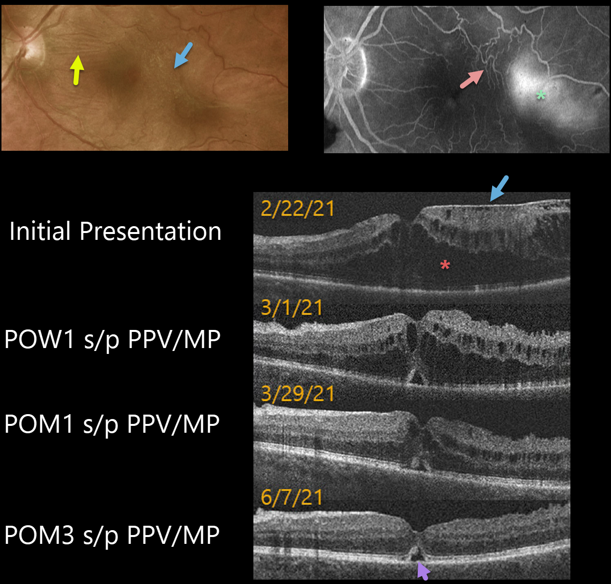
This 62YO male presented with 20/200 vision in his left eye. An epiretinal membrane (ERM) was noted in the temporal macula, associated with more nasal horizontal retinal striae.
Optos fluorescein angiography shows distortion of the temporal macular vessels with late leakage.
Vitrectomy with membrane peeling was performed the following day, and one week later, on 3/1/21, vision improved to 20/70 with a significant decrease in macular thickening.
When last examined on 6/7/21, about 3 months post-op, vision was still 20/70, but the macular thickening and contour were almost normal except for a small blister of foveal subretinal fluid.
Learning Points:
OCT scanning confirmed the temporal ERM, but the severe cystic macular thickening with possible tractional schisis that extended into the nasal macula seemed disproportionate to the imaged temporal ERM. Fortunately, both the vision and macular thickening significantly improved following vitrectomy with membrane peeling.
MACULAR TELANGIECTASIA
Originally posted on @retina.rocks January 22, 2021
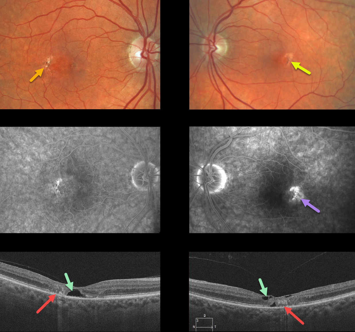
Our patient shows classic bilateral findings of macular telangiectasia type 2 (MacTel2) with temporal foveal intraretinal pigment migration and inner retinal crystals, as well as angiographic staining and leakage.
Our patient’s optical coherence tomography scans also show classic findings of mostly temporal foveal retinal atrophy, disorganization, and hyporeflective tissue loss (cavitations).
Learning Points:
MacTel type 2 is a neurodegenerative disorder, most likely originating from Müller cell dysfunction. The term telangiectasia is misleading because the funduscopic findings are mostly nonvascular.
It is thought that photoreceptor loss allows RPE cells to migrate along intraretinal capillaries, creating the pigment clumping. This is a similar mechanism to that found in other causes of intraretinal pigment migration, including photoreceptor loss or damage, such as retinitis pigmentosa, chronic retinal detachment, and blunt trauma.
The refractile inner retinal ‘crystals’ are thought to be Muller cell footplates, similar to the refractile dots found in retinoschisis.
Although much remains to be learned about its pathophysiology and there is no treatment for the underlying disease, secondary macular neovascularization can be successfully treated according to age-related macular degeneration protocols.
TAMOXIFEN RETINOPATHY
Originally posted on @retina.rocks September 9, 2020
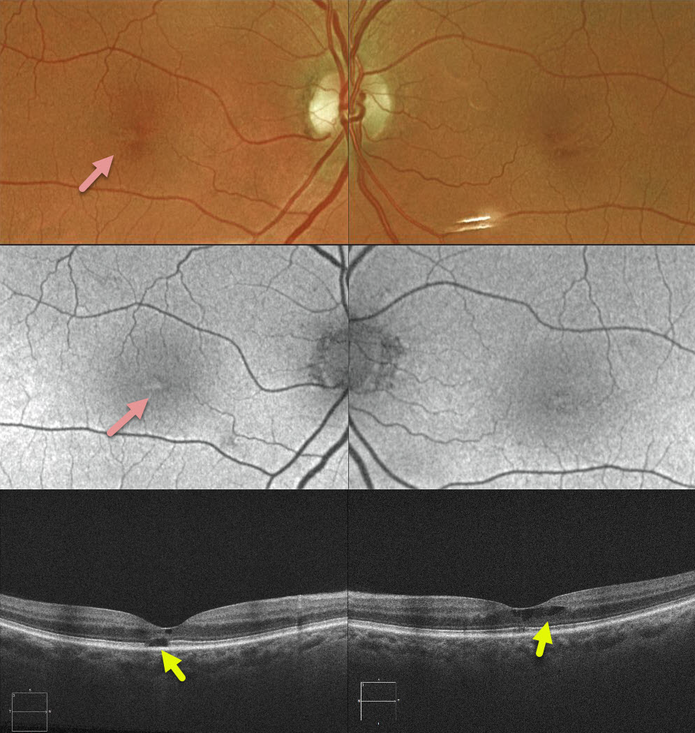
This 72YO female recently completed a 5-year course of Tamoxifen as adjunctive treatment for breast cancer. She had no visual symptoms with 20/30 vision bilaterally.
Clinically, there were subtle bilateral foveal pigmentary changes. B-scan and en-face OCT showed bilateral cavitary changes.
Learning Points:
Tamoxifen retinopathy shares findings very similar to those of macular telangiectasia type 2 (MacTel2), including retinal cavitations, right-angle venules, and inner retinal crystals. The changes in MacTel2 are usually confined to the temporal fovea, whereas with Tamoxifen, they seem more diffusely distributed throughout the entire central macula. The retinal changes for both disorders likely share Muller cell injury as the underlying cause.
The prevalence of Tamoxifen retinopathy may be as high as 12%. Although most oncologists do not require this, periodic ophthalmic screening, OCT, and examinations may be indicated since patients can lose central vision if toxicity develops.
SOLAR RETINOPATHY
Originally posted on @retina.rocks July 31, 2020
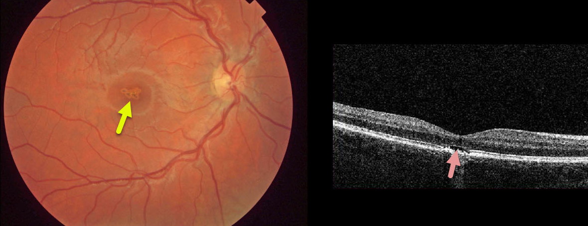
This asymptomatic 10YO boy presented with 20/40 vision OD and 20/20 vision OS.
There were multifocal small subretinal pigment clumps with surrounding pigment loss in the right central macula.
OCT shows foveal outer segment loss with ragged RPE loss. The left macula was normal.
He denied looking at a laser pointer but did admit to a history of sun-gazing. He likely closed his left eye each time he looked at the sun, which explains the unusual finding of unilateral solar retinopathy.
Learning Points:
The outer retinal damage in solar retinopathy is thought to be caused by photochemical outer retinal and RPE damage instead of a thermal burn. Vision is usually fairly good.
WHIPLASH MACULOPATHY
Originally posted on @retina.rocks July 1, 2020
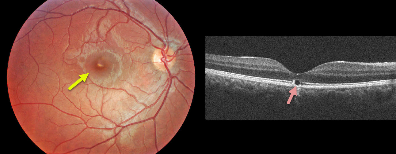
This 17YO male presented with sudden unilateral vision loss immediately following a whiplash from a motor vehicle accident.
Clinically, he has a yellowish linear subretinal foveal discoloration. OCT shows a focal loss of the outer subfoveal retina.
Learning Points:
These findings are classic for whiplash maculopathy, in which sudden acceleration/deceleration causes the vitreous to tug on or impact the retina, resulting in outer retinal and RPE damage.


