

Outer Retinal Tubulation
<< Back to Cases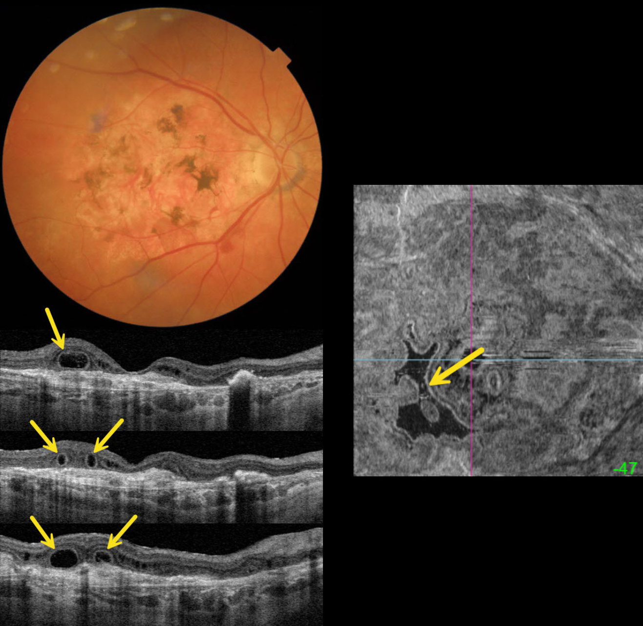
OUTER RETINAL TUBULATION IN NEOVASCULAR AMD
Originally posted on @retina.rocks November 18, 2025
This 89YO female is receiving ongoing intravitreal Eylea in her 20/40 left eye for neovascular AMD. She has a stable 20/400 atrophic disciform scar in her right eye.
Triton color imaging shows a variably pigmented flat disciform scar. Swept-source OCT shows variable outer retinal atrophy overlying a hyperreflective inactive macular neovascularization (MNV). A complex pattern of outer retinal tubulation (ORT) is seen on numerous B-scans. En face OCT shows another view of these meandering tubular structures.
Learning Points:
ORT is often noted overlying inactive MNV and should not be confused with exudative fluid or cysts, which lack a hyperreflective border. The outer hyperreflective band likely represents inner segment mitochondria undergoing fission and translocation toward the nucleus (Litts et al, Retina 2018;38:445-461). ORT, initially described by Zweifel et al (Arch Ophthalmol 2009;127:1596-1602), is a neurodegenerative condition of the photoreceptors and Muller cells associated with atrophy affecting the outer retina and retinal pigment epithelium, including advanced AMD and inherited retinal diseases.
BIETTI CRYSTALLINE DYSTROPHY
The European VitreoRetina Society (EVRS) and Shishir Verghese
Originally posted on @retina.rocks October 24, 2025
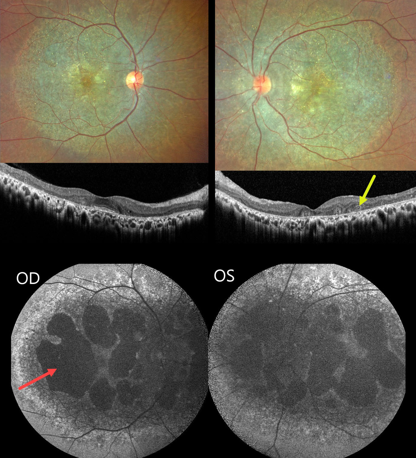
This healthy 36YO female presented with a long history of bilateral vision loss with night blindness. Family history was negative. Vision was 20/80 bilaterally. Anterior segments were normal.
Fundus imaging shows extensive pigmentary degeneration with subretinal crystalline deposits involving both posterior poles. OCT scanning shows hyperreflective dots overlying the RPE-Bruch’s membrane complex with variable outer retinal and RPE loss. A few areas of outer retinal tubulation are noted temporally OS. Fundus autofluorescence (FAF) shows diffuse hypo-FAF throughout each posterior pole, dense hypo-FAF within areas of discrete atrophy, and dots of more peripheral hyper-FAF.
Learning Points:
Bietti crystalline dystrophy is an autosomal recessive ocular disorder that affects both the anterior and posterior segments. Clinical features include crystalline deposits in the retina, cornea, and rarely the crystalline lens, as well as retinal pigment epithelial clumping and atrophy. Mutations in CYP4V2 that cause dysregulated lipid metabolism have been implicated in its pathogenesis. While there is no definitive treatment, anti-VEGF therapy can be administered for secondary macular neovascularization. See Saatci et al for a recent review (Clinical Ophthalmology 2023;17:953-967).
WET AMD WITH OUTER RETINAL TUBULATION
Originally posted on @retina.rocks April 12, 2023
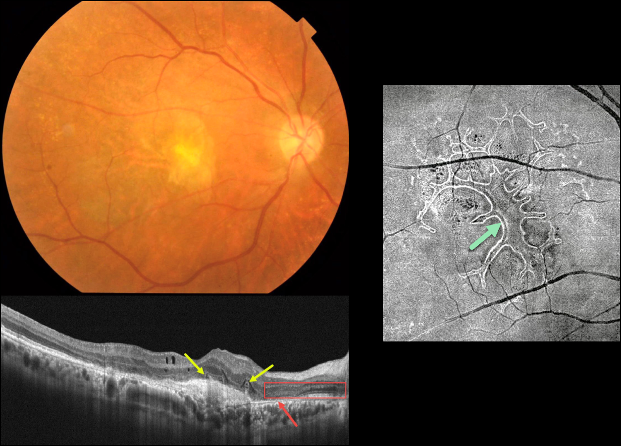
This 63YO male received several intravitreal anti-VEGF injections for neovascular AMD in his right eye, the most recent being 6 months earlier. Vision is counting fingers.
Color photography shows an inactive atrophic foveal scar. OCT scanning shows a hyperreflective macular neovascularization (MNV). The outer retina overlying the MNV is disorganized with several overlying hyporeflective lesions with surrounding hyperreflective rims. More nasally, there is complete RPE loss with bare Bruch’s membrane. The overlying retina is anomalous with additional hypo- and hyperreflective bands.
En face OCT scanning through the outer retina provides a more dramatic view of the extensive tubulation-branching network.
Learning Points:
Outer retinal tubulation (ORT) is often noted overlying inactive MNV with ongoing anti-VEGF therapy and should not be confused with exudative fluid or cysts, which lack a hyperreflective border. The outer hyperreflective band likely represents inner segment mitochondria undergoing fission and translocation toward the nucleus (Litts et al, Retina 2018;38:445-461).
ORT, initially described by Zweifel et al (Arch Ophthalmol 2009;127:1596-1602), is a neurodegenerative condition of the photoreceptors and Muller cells associated with atrophy affecting the outer retina and retinal pigment epithelium, including advanced AMD and inherited retinal diseases.
AMD MACULAR ATROPHY
Originally posted on @retina.rocks November 2, 2022
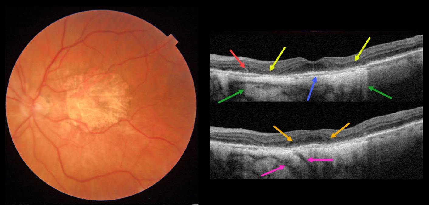
This 84YO female has stable advanced non-exudative AMD with bilateral macular atrophy (MA). There are numerous classic findings on multimodal imaging of her left eye.
Color imaging shows foveal MA. Swept-source OCT shows pericentral outer retinal and RPE atrophy. This atrophy allows for more light to illuminate the underlying choroid and sclera, as well as visualizing two short posterior ciliary arteries posterior to the sclera. The variably atrophic RPE allows for visualization of Bruch membrane, which is normally fused in the RPE-Bruch layer seen in normal eyes. There is a tiny area of outer retinal tubulation (ORT) nasally. Finally, hyporeflective outer plexiform layer wedge defects are noted along the descending outer retinal atrophy.
Wedge defects develop at the boundaries of the degenerating outer plexiform layer in about 75% of MA eyes (Mones et al, Ophthalmology 2012;119:1412-1419).
ORTs represent photoreceptor and Muller cell degeneration (Dolz-Marco et al, Ophthalmology 2017;124:1353-1367). These ORTs have a hyper-reflective border with a central hyporeflective core.
Learning Points:
In 1970, Dr. Gass originally described macular atrophy from AMD as “geographic areas of atrophy” in the setting of “senile macular choroidal degeneration” (Schmitz-Valkenberg, Retina 2016;36:2250-2264).
Today, the terminology is much more specific due to advances in technology and improved visualization of retinal and choroidal structures.
From Sadda et al, Ophthalmology 2018:537-548, the new classification is as follows: Complete RPE and Outer Retinal Atrophy (cRORA), Incomplete RPE and Outer Retinal Atrophy (iRORA), Complete Outer Retinal Atrophy (cORA), and Incomplete Outer Retinal Atrophy (iORA).
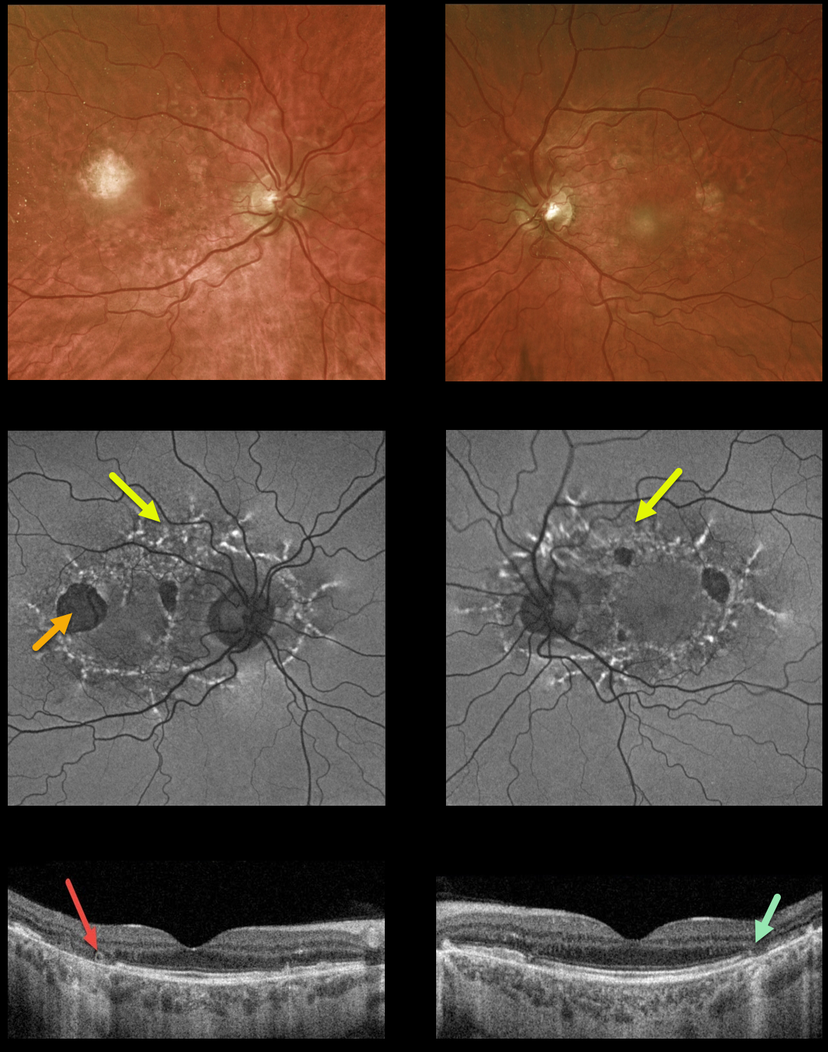
MATERNALLY INHERITED DIABETES AND DEAFNESS (MIDD) + OUTER RETINAL TUBULATION (ORT)
Originally posted on @retina.rocks October 3, 2022
This 59YO male presented with vision of 20/400 OD and 20/30 OS. He has severe lifelong hearing loss, and both he and his mother have type 2 diabetes.
Optos ultrawidefield imaging shows temporal macular atrophy. Fundus autofluorescence shows the true extent of the pathology, with linear interconnected subretinal streaks of hyper-FAF associated with areas of hypo-FAF macular atrophy.
Swept-source OCT shows variable, mostly temporal outer retinal and RPE atrophy, along with a small area of temporal outer retinal tubulation (ORT) OD.
Learning Points: Maternally inherited diabetes and deafness (MIDD) is responsible for up to 3% of all cases of diabetes, and results from a mutation of mitochondrial DNA A3243G. MIDD often masquerades as a pattern macular dystrophy.
Fundus autofluorescence (FAF) in our experience is the best way to visualize these changes. The FAF appearance somewhat resembles that seen with Elmiron toxicity.
Peripapillary hypoautofluorescence, more densely-packed macular autofluorescent changes, and earlier central macular involvement suggest Elmiron toxicity over other causes (see Barnes et al Ophthalmology Retina 2020;4:1196-1201). Note that our case spares the central macula and peripapillary retina, which is more consistent with MIDD.
ORT is often noted overlying inactive MNV with ongoing anti-VEGF therapy and should not be confused with exudative fluid or cysts which lack a hyperreflective border. The outer hyperreflective band likely represents inner segment mitochondria undergoing fission and translocation toward the nucleus (Litts et al, Retina 2018;38:445-461).
ORT, initially described by Zweifel et al (Arch Ophthalmol 2009;127:1596-1602), is a neurodegenerative condition of the photoreceptors and Muller cells associated with atrophy affecting the outer retina and retinal pigment epithelium, including advanced AMD and inherited retinal diseases.
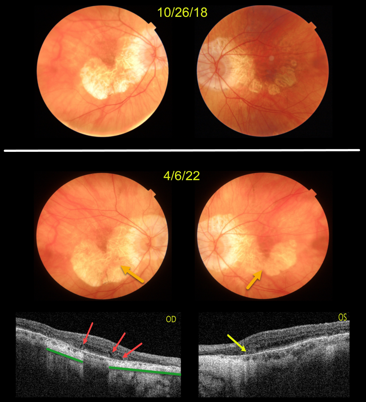
AMD MACULAR ATROPHY
Originally posted on @retina.rocks April 28, 2022
This patient originally presented in 2018 with vision of 20/40 OD and 20/50 OS. Areas of AMD-related macular atrophy (MA) skirted each macular center.
Four years later, vision had decreased to 20/100 OU due to progressive atrophy. Comparing the two images from 2018 to 2022, there is a bilateral linear enlargement for all margins of the atrophy, particularly in the left eye.
Triton swept-source OCT B-scans show classic findings for MA. The right eye has multiple wedge defects, hyporeflective wedges which develop at the boundaries of the degenerating outer plexiform layer in about 75% of MA eyes (Mones et al, Ophthalmology 2012;119:1412-1419). In addition, loss of the outer retina and RPE allows more OCT light to be transmitted into the choroid, resulting in distinct areas of choroidal and scleral hyperreflectivity.
There is a tiny area of outer retinal tubulation (ORT) nasally in the left eye, which represents photoreceptor and Muller cell degeneration (Dolz-Marco et al, Ophthalmology 2017;124:1353-1367). These ORTs have a hyper-reflective border with a central hyporeflective core.
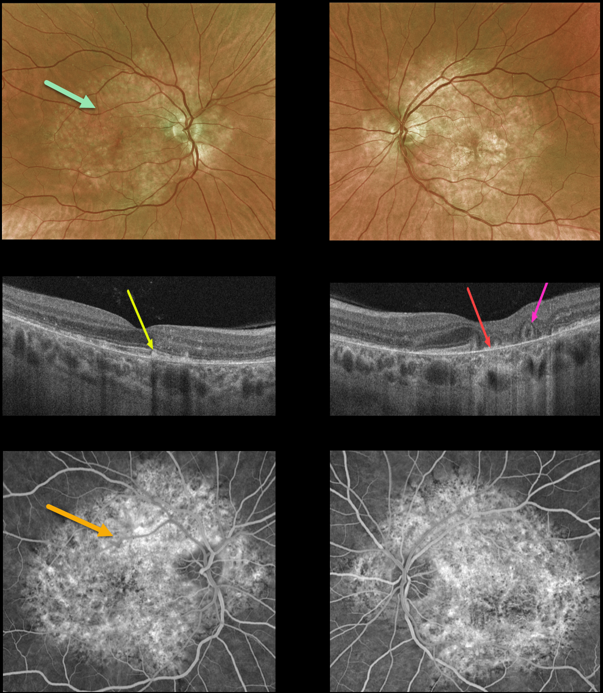
PENTOSAN POLYSULFATE SODIUM MACULOPATHY + OUTER RETINAL TUBULATION
Originally posted on @retina.rocks March 1, 2022
This 54YO female presented with a 9-month history of blurred vision in her left eye. She was on Elmiron (pentosan polysulfate sodium, PPS) for about 20 years due to interstitial cystitis, but stopped the medication before seeing us due to the widespread publicity about its potential retinal toxicity.
Optos color imaging shows variable yellow-orange atrophic pigmentary changes throughout the macular and peripapillary posterior poles.
OCT of the right macula shows scattered hyperreflective lesions mostly within the outer segment layers. The left macular OCT shows variable outer retinal and RPE loss, especially centrally, where bare Bruch’s membrane is visible. There is also an area of outer retinal tubulation (ORT) more temporally.
Fluorescein angiography shows window defects within these regions.
Learning Points:
Elmiron was approved by the FDA in 1996 for treating interstitial cystitis. Recently, a unique PPS retinopathy has been described, and our patient shows classic findings.
Toxicity seems to develop over many years and can mimic more common disorders, including age-related macular degeneration and macular dystrophies.
Peripapillary hypoautofluorescence, more densely packed macular autofluorescent changes, and earlier central macular involvement suggest PPS toxicity over other causes (see Barnes et al Ophthalmology Retina 2020;4:1196-1201), including maternally inherited diabetes and deafness (MIDD).
ORT is often noted overlying inactive macular neovascularization with ongoing anti-VEGF therapy and should not be confused with exudative fluid or cysts, which lack a hyperreflective border. The outer hyperreflective band likely represents inner segment mitochondria undergoing fission and translocation towards the nucleus (Litts et al, Retina 2018;38:445-461).
ORT, initially described by Zweifel et al (Arch Ophthalmol 2009;127:1596-1602), is a neurodegenerative condition of the photoreceptors and Muller cells associated with atrophy affecting the outer retina and retinal pigment epithelium, including advanced AMD and inherited retinal diseases.
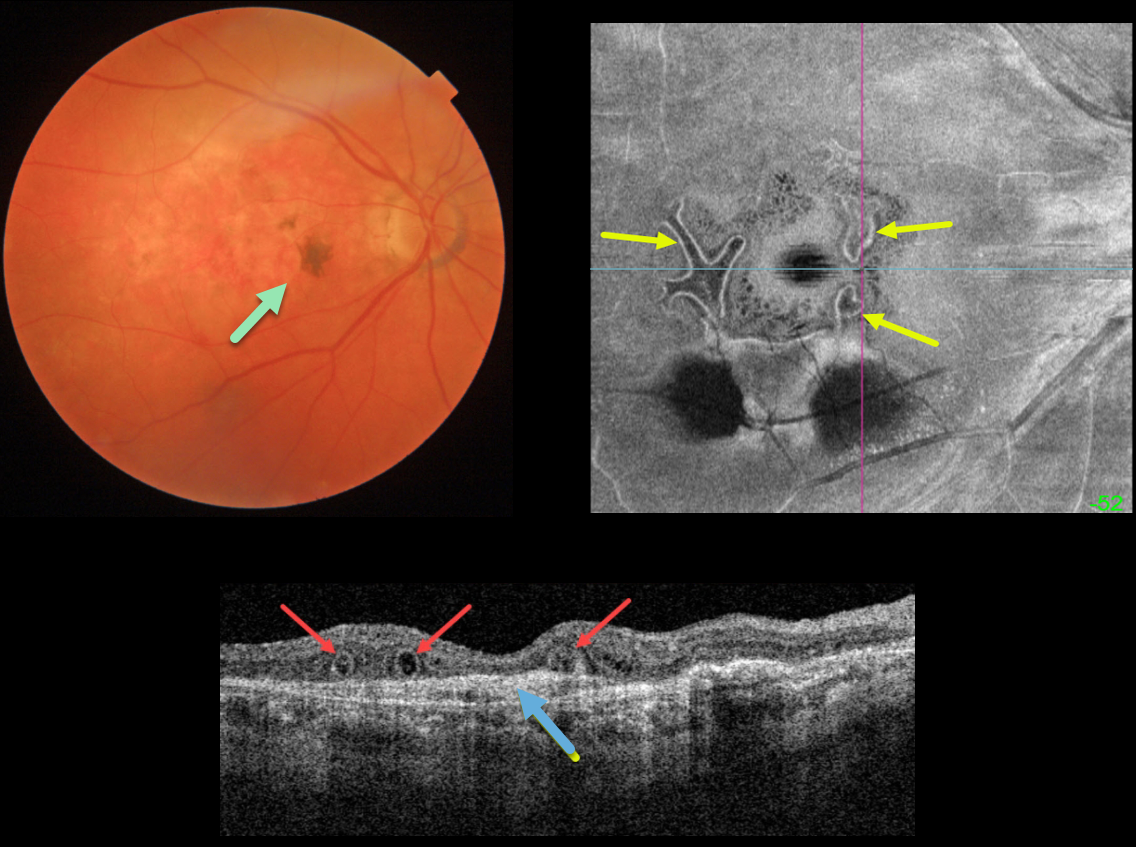
OUTER RETINAL TUBULATION FOLLOWING ONGOING ANTI-VEGF FOR NEOVASCULAR AMD
Originally posted on @retina.rocks September 23, 2021
This is an 86YO female who has stable dry macular scarring from wet age-related macular degeneration with ongoing anti-VEGF injections.
Triton swept-source OCT shows numerous outer retinal tubulations (ORT) overlying an inactive type 1 macular neovascularization (MNV). These tubules are more dramatically seen with en face imaging of the outer retina.
Learning Points:
Outer retinal tubulation is often noted overlying inactive MNV with ongoing anti-VEGF therapy and should not be confused with exudative fluid or cysts, which lack a hyperreflective border.
ORT, initially described by Zweifel et al (Arch Ophthalmol 2009;127:1596-1602), is a neurodegenerative condition of the photoreceptors and Muller cells associated with atrophy affecting the outer retina and retinal pigment epithelium, including advanced AMD and inherited retinal diseases.
The outer hyperreflective band likely represents inner segment mitochondria undergoing fission and translocation toward the nucleus (Litts et al, Retina 2018;38:445-461).
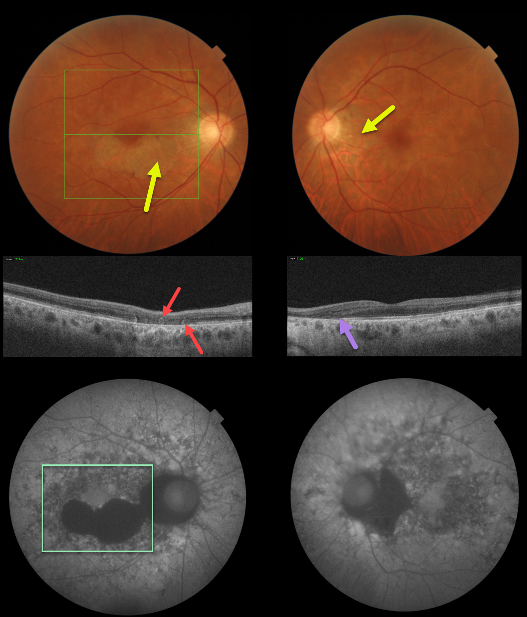
ACUTE POSTERIOR MULTIFOCAL PLACOID PIGMENT EPITHELIOPATHY (APMPPE) WITH OUTER RETINAL TUBULATION
Originally posted on @retina.rocks June 4, 2021
This patient has bilateral fundus findings consistent with resolved acute posterior multifocal placoid pigment epitheliopathy (APMPPE).
Triton fundus photos show placoid areas of atrophic pigmentary changes, which are variably hypo-autofluorescent.
Triton swept-source OCT in the right eye shows outer retinal atrophy with outer retinal tubulation (ORT) and nasal outer retinal atrophy in the left eye.
Learning Points:
ORT, initially described by Zweifel et al (Arch Ophthalmol 2009;127;1596-1602), is a neurodegenerative condition of the photoreceptors and Muller cells associated with atrophy affecting the outer retina and retinal pigment epithelium, including advanced AMD and inherited retinal diseases.
ORT is often seen overlying inactive macular neovascularization in the setting of ongoing anti-VEGF therapy and should not be confused with exudative fluid or cysts, which lack a hyperreflective border. The outer hyperreflective band likely represents inner segment mitochondria undergoing fission and translocation toward the nucleus (Litts et al, Retina 2018;38;445-461).
Acute posterior multifocal placoid pigment epitheliopathy (APMPPE) is a usually bilateral inflammatory vs ischemic disorder of the inner choroid, RPE, and outer retina.
Blurry vision and paracentral/central scotomas often follow a viral illness. Acutely multifocal creamy yellow or grayish-white subretinal placoid lesions are scattered throughout the posterior poles. The lesions and visual symptoms usually resolve within 4 to 8 weeks with secondary chorioretinal changes. Recurrent lesions are rare, and vision is usually fairly good.
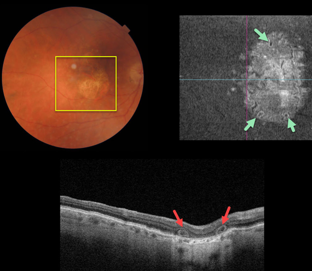
OUTER RETINAL TUBULATION
Originally posted on @retina.rocks February 16, 2021
This 80YO female was 20/200 in her right eye due to central macular atrophy from atrophic age-related macular degeneration (AMD).
Triton swept-source optical coherence tomography showed outer retinal tubulation (ORT) in the outer retinal layers. The extent of these tubular structures was best visualized on en face imaging.
Learning Points:
Outer retinal tubulation, initially described by Zweifel et al (Arch Ophthalmol 2009;127;1596-1602), is a neurodegenerative condition of the photoreceptors and Muller cells associated with atrophy affecting the outer retina and retinal pigment epithelium, including advanced AMD and inherited retinal diseases.
ORT is often seen overlying inactive macular neovascularization in the setting of ongoing anti-VEGF therapy and should not be confused with exudative fluid or cysts, which lack a hyperreflective border.
The outer hyperreflective band likely represents inner segment mitochondria undergoing fission and translocation toward the nucleus (Litts et al Retina 2018;38;445-461).
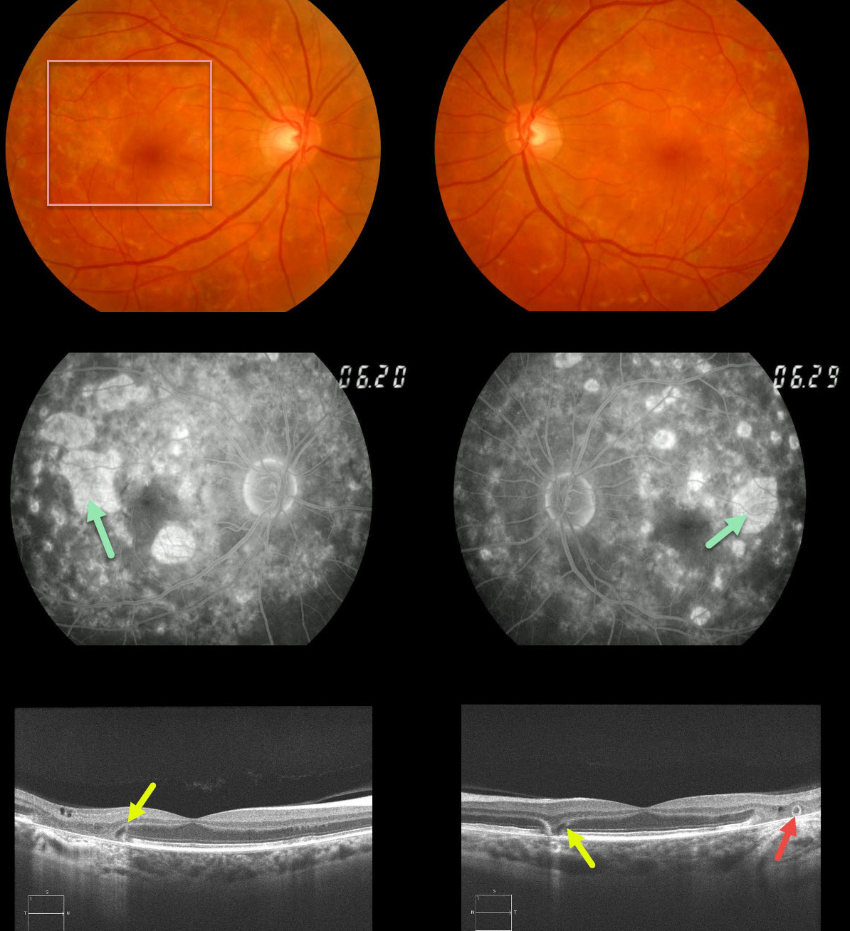
MATERNALLY INHERITED DIABETES AND DEAFNESS (MIDD)
Originally posted on @retina.rocks December 4, 2020
This 62yo female with a history of type 2 diabetes and mild hearing loss initially presented in 2012 with asymptomatic 20/30 vision OU and bilateral areas of focal macular atrophy and subretinal yellow fleck-like lesions.
The retinal pigment epithelial changes were much more pronounced on fluorescein angiography. OCT scanning shows classic wedge defects along the border of the macular atrophy as well as an area of outer retinal tubulation in the left eye.
Learning Points:
Maternally inherited diabetes and deafness (MIDD) accounts for up to 3% of all cases of diabetes and results from a mutation in mitochondrial DNA at position A3243G. MIDD often masquerades as a pattern macular dystrophy.
Fundus autofluorescence (FAF), in our experience, is the best way to visualize these changes, but it was not available at the office when we initially saw her. MIDD patients need to be screened for other potential co-existing medical conditions, including cardiac arrhythmias, myopathy, and renal disease.


