

Pachychoroid Spectrum
<< Back to Cases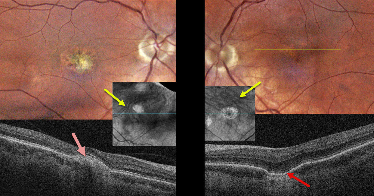
PACHYCHOROID SPECTRUM PREVIOUSLY MISDIAGNOSED AS OCULAR HISTOPLASMOSIS
Steven Bloom
Originally posted on @retina.rocks April 22, 2026
This healthy 39YO female gave a history 18 years earlier of prior anti-VEGF therapy OD for ocular histoplasmosis. She was visually asymptomatic, and vision was 20/40 OD and 20/30 OS.
Color photography of her left eye shows a patchy area of pigment loss in the superonasal fovea. OCT scanning through this area shows a choroidal excavation. The right macula has an overall mostly depigmented area of scarring, which appears as a mound of subretinal hyperreflectivity on OCT. Choroidal en face OCT shows dilated pachyvessels OU that cross the horizontal meridian (vortex anastomoses). There were no areas of peripheral chorioretinal scarring in either eye. Observation was recommended.
Learning Points:
Pachychoroid, meaning “thick” choroid, is secondary to large, dilated outer choroidal vessels. Increased oncotic pressure from these dilated vessels can cause RPE dysfunction, serous detachment, and macular neovascularization (MNV). Pachychoroid disease spectrum is a growing list of findings, including idiopathic central serous chorioretinopathy (ICSC), pachychoroid neovascularization, polypoidal choroidal vasculopathy, pachychoroid pigment epitheliopathy, pachydrusen, peripapillary pachychoroid syndrome, choroidal excavation, and pachyvitelliform maculopathy.
Our guess is that our patient had active ICSC when she was treated for her ‘ocular histoplasmosis.’ The lesion in the right macula is atypical for ICSC. But the other findings, including dilated choroidal vessels with vortex anastomoses and a choroidal excavation in her left eye, argue for pachychoroid as her unifying diagnosis.
PACHYVITELLIFORM MACULOPATHY
Originally posted on @retina.rocks April 7, 2026
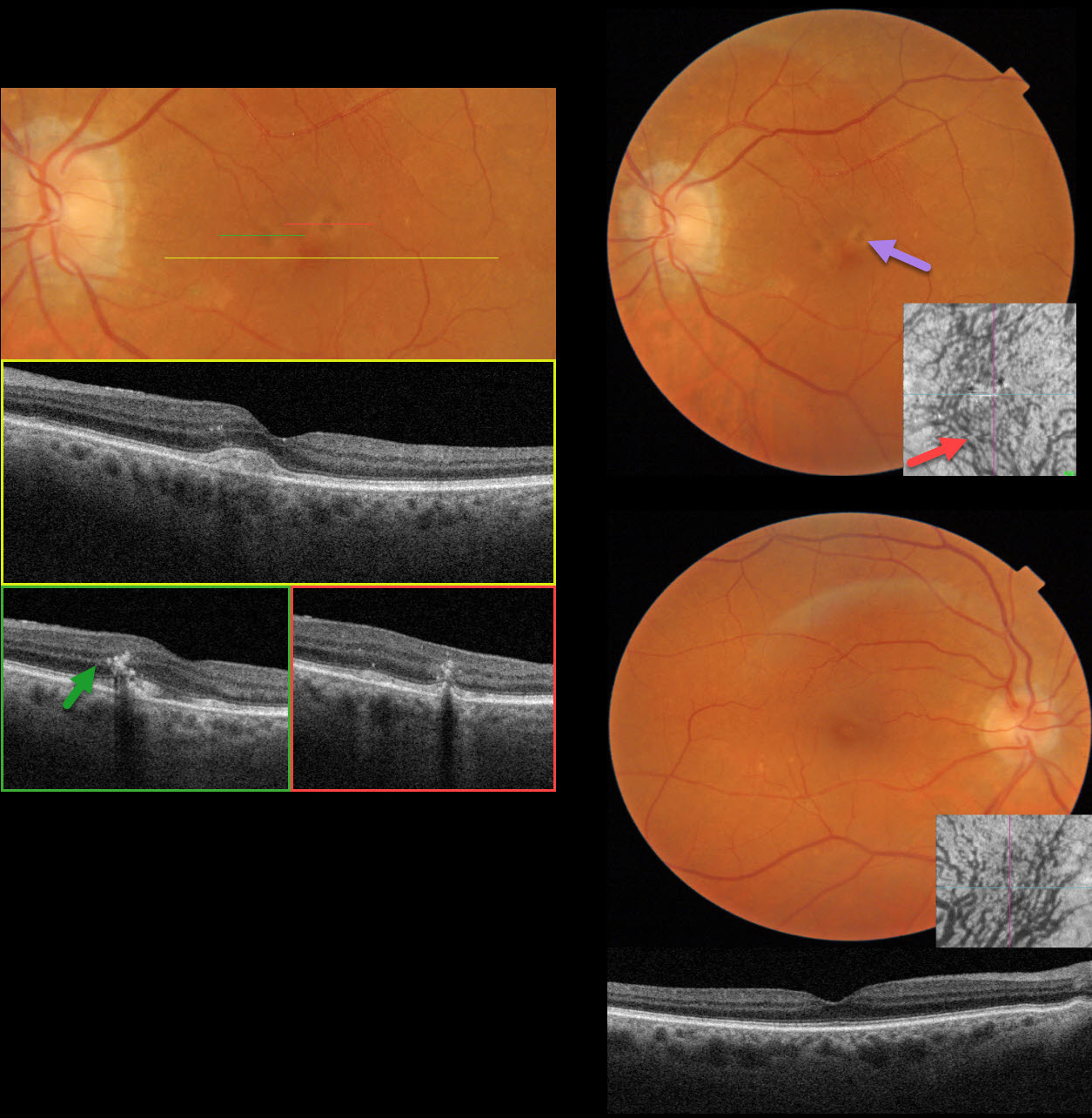
This 81YO female was referred for asymptomatic retinal findings. Vision was 20/30 OD and 20/30 OS.
Triton color imaging OS shows several yellowish subretinal pigment clumps in the superior fovea. Swept-source OCT through the central macula shows an acquired vitelliform lesion (AVL). Scans through each of the pigment clumps show coarse intraretinal hyperreflective foci. Some ectopic pachydrusen are noted in the inferotemporal right macula. OCT shows large outer choroidal vessels, and en face OCT shows the entire macula to be drained by the inferior vortex system with dilated vessels. The choroidal vessels are also dilated OS.
Learning Points:
Pachychoroid, meaning “thick” choroid, is secondary to large, dilated outer choroidal vessels. Increased oncotic or mechanical pressure from these dilated vessels can cause RPE dysfunction, serous detachment, and macular neovascularization (MNV). Our patient has pachychoroid findings despite having a ‘normal’ choroidal thickness on B-scan OCT. The choroidal thickness depends on numerous factors, including the patient’s age and refractive error. A high myope’s choroid should be quite thin, but if it appears as a ‘normal’ thickness, it is thick for that eye. We also often see pachychoroid disease in elderly patients whose choroid is abnormally thickened for their age, but appears ‘normally’ thick when not accounting for this.
Pachychoroid disease spectrum includes central serous chorioretinopathy, pachychoroid neovascularization, polypoidal choroidal vasculopathy, pachychoroid pigment epitheliopathy, pachydrusen, peripapillary pachychoroid syndrome, and focal choroidal excavation (Cheung et al, Eye 2025;39:819-834 ). Hilely et al recently described an additional entity, pachyvitelliform maculopathy (Br J Ophthalmol 2024;108:753-759), defined by AVL associated with pachychoroid features. The authors hypothesize that the thickened choroid causes RPE dysfunction, resulting in the accumulation of undigested photoreceptor outer segments with AVL formation.
We believe a better term for these disorders is venous overload choroidopathy, described by Spaide et al (Progress in Retinal and Eye Research 2022;86:100972). This hypothesis suggests scleral venous outflow as a unifying cause for this ever-expanding spectrum.
PACHYCHOROID SPECTRUM PREVIOUSLY MISDIAGNOSED AS AMD
Originally posted on @retina.rocks July 30, 2025
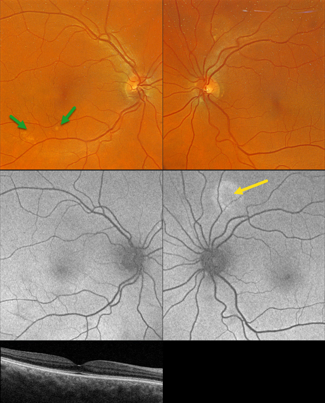
This healthy 65YO founder of Retina Rocks presented for his once-per-decade ‘eye examination’ which included an undilated slit lamp examination by his partner, Triton swept-source OCT, and Optos color RGB imaging. His mother had a history of neovascular AMD, and he has a known history of drusen. Given his family history, he has been taking AREDS2 supplementation for many years. There were no ocular symptoms, and vision was 20/20 bilaterally.
Triton color imaging shows some pachydrusen in the inferotemporal right macula. Swept-source OCT is normal except for a thickened choroid. Optos fundus autofluorescence (FAF) shows a round area of hyper-FAF superior to the left nerve.
Learning Points:
Pachydrusen were described by Spaide as a distinct pachychoroid phenotype (Retina 2018;38:708-716). Unlike typical AMD, where cone-based soft drusen are symmetrically localized to the foveal region, pachydrusen are eccentrically located, far less numerous, and have an irregular contour. Since choroidal thickness is influenced by numerous factors, including age and refractive status (Xie et al, IOVS 2022;63;2;34), these must always be considered when reviewing an individual patient’s scans. Eyes with AMD typically have age-related choroidal thinning, but eyes with pachydrusen, as in this case, are associated with a relatively thickened choroid.
Our patient became aware of his macular drusen well before pachychoroid was described by Freund in 2013 (Warrow et al, Retina 2013;33:1659-1672), and he attributed his early macular drusen to AMD. In 2025, he understands that his findings are consistent with the pachychoroid spectrum, as evidenced by the bilaterally thickened choroid, pachydrusen OD, and FAF findings suggestive of prior extramacular central serous OS. He does not have intermediate AMD, is at minimal risk for developing AMD, and thus doesn’t need AREDS2 supplementation. However, like the rest of us, he is only human and finds emotional comfort in continuing to take these supplements.
PACHYCHOROID NEVOASCULARIZATION
Originally posted on @retina.rocks March 31, 2025
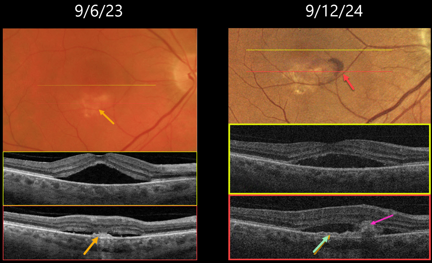
This 64YO male presented on 9/6/23 with several weeks of vision loss in his right eye. Vision was 20/40 OD.
Triton color imaging shows a faint round foveal serous detachment. Swept-source OCT confirms a classic idiopathic central serous chorioretinopathy (ICSC) foveal detachment. There is a small hyperreflective placoid elevation of the RPE along the inferior edge of the detachment corresponding to a yellow-white area of subretinal pigmentation. Due to significant symptoms, he elected for early photodynamic therapy (PDT), which successfully resolved the serous fluid two months later with 20/30 vision (not shown).
He did well until he returned on 9/12/24 with recurrent blurred vision OD, which had decreased slightly to 20/40. There is new subretinal blood within the region of the persistent placoid RPE elevation inferiorly. OCT shows recurrent serous foveal fluid centrally. There is an enlargement of the shallow inferior RPE elevation adjacent to the subretinal blood. Monthly intravitreal Avastin therapy was started.
Learning Points:
Initially described by Pang and Freund, pachychoroid neovasculopathy is a variant of type 1 (below the RPE) macular neovascularization (MNV) found as part of the pachychoroid spectrum, including ICSC and pachychoroid pigment epitheliopathy (Retina 2015;35:1-9). About one-third of chronic ICSC eyes with flat irregular RPE detachments will harbor an MNV on OCT angiography (Bousquet et al, Retina 2018;38:629-639), which is probably a form of pachychoroid neovascularization (Hwang et al, Retina 2020;40:1724-1733).
In retrospect, our patient most likely had a pachychoroid neovascularization, which initially responded to PDT. When he returned one year later with a definite active MNV, we elected to start anti-VEGF monotherapy. He was unfortunately immediately lost to follow-up.
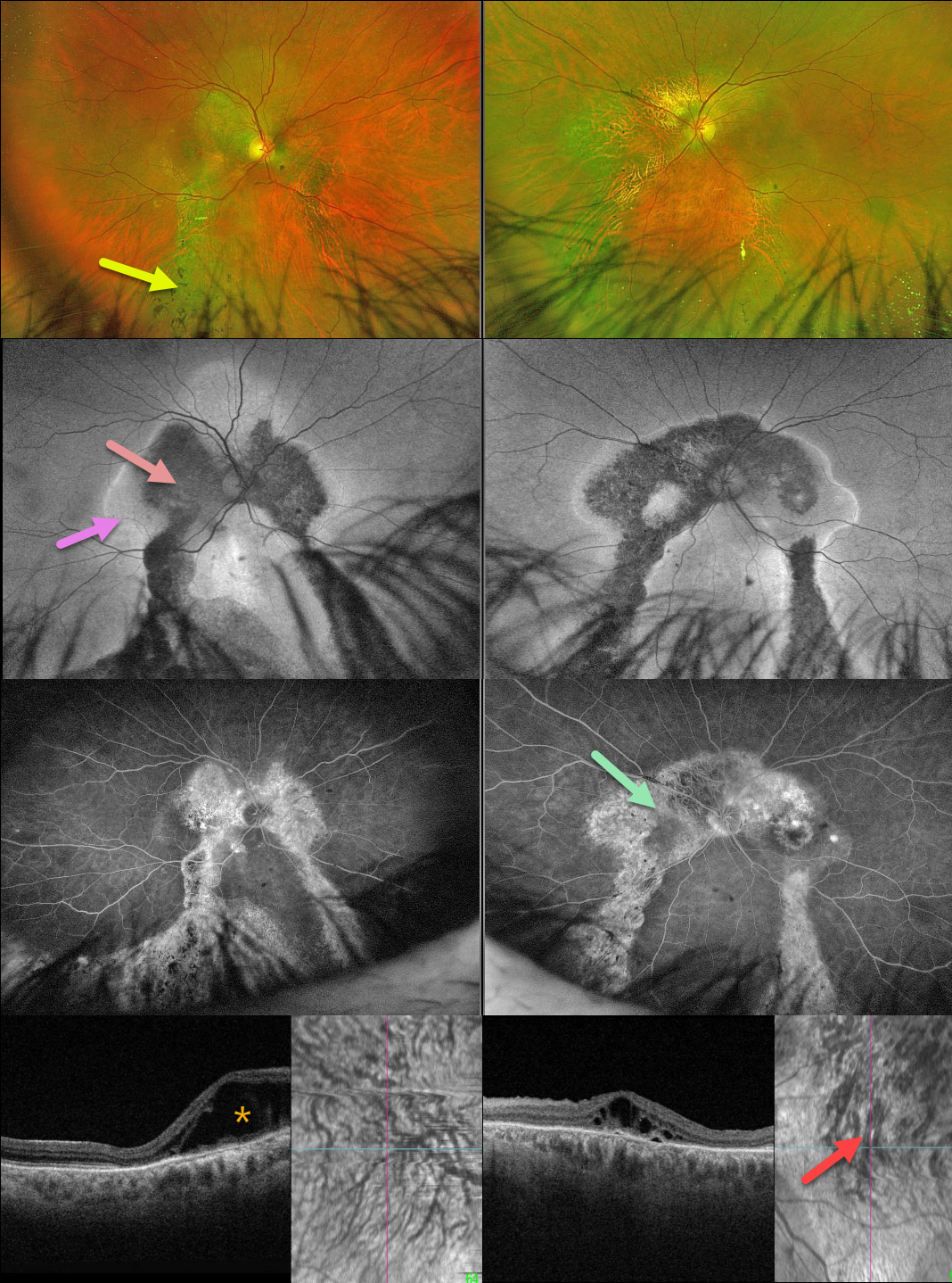
POSSIBLE PACHYCHOROID SPECTRUM
Originally posted on @retina.rocks June 11, 2024
This healthy 62YO male presented with a several-year history of bilateral vision loss. There was no family history of eye disease. Vision was 20/400 OU.
Optos color RG imaging shows bilateral, symmetrical peripapillary and macular pigmentary changes extending into the gutters towards the inferior peripheries. Intraretinal pigment migration (spicules) is noted inferiorly.
On fundus autofluorescence (FAF), these areas are hypo-FAF centrally with hyper-FAF along their margins, and exhibit window defects on fluorescein angiography.
Outer retinal fluid is noted nasally OD and centrally OS on Triton swept-source OCT. En face OCT shows bilateral choroidal pachyvessels with anastomoses. Genetic testing was completely negative.
Learning Points:
Gutters are commonly seen in pathology with chronic subretinal leakage, including central serous retinopathy, choroidal hemangioma, choroidal nevus, and uveal melanoma. These pigmentary changes are usually best imaged with FAF. Our patient’s OCT certainly shows pachychoroid features, but the symmetrical double gutters in each eye would be quite unusual for central serous retinopathy.
Our patient’s gutters are somewhat reminiscent of a patient we have previously posted with the Senior-Løken Syndrome, although this is associated with renal failure and an NPHP1 mutation, which our patient did not have.
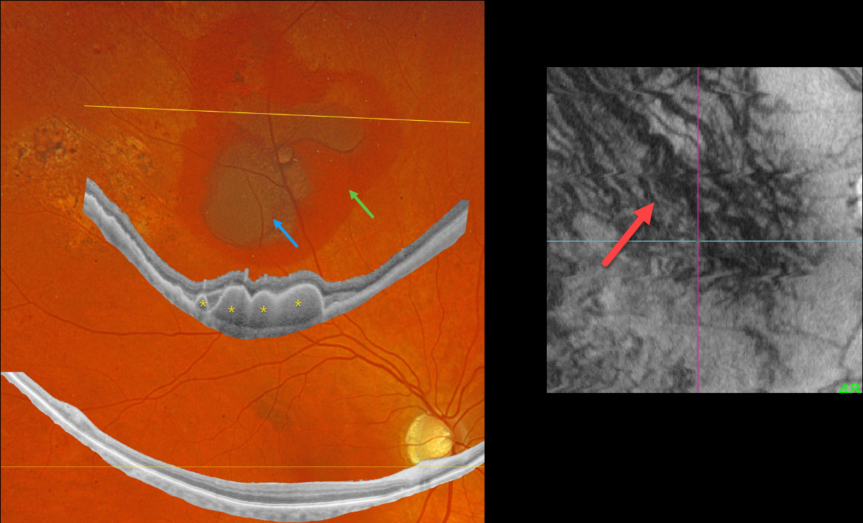
PERIPHERAL EXUDATIVE HEMORRHAGIC CHORIORETINOPATHY
Originally posted on @retina.rocks April 17, 2024
This 64YO diabetic male presented with asymptomatic retinal findings in his left eye. Vision was 20/40 OU.
Optos color RGB imaging shows superior subretinal and sub-RPE blood. Optos Silverstone widefield swept-source OCT through this lesion shows prominent multiple choroidal polyps. Subretinal pigmentary clumping temporal to the blood likely represents findings from absorbed subretinal blood. Moderate non-proliferative retinopathy is noted.
En face imaging is quite abnormal with vortex anastomoses and dilated choroidal vessels.
Observation was recommended.
Learning Points:
Pachychoroid, meaning “thick” choroid, is secondary to large, dilated Haller vessels. Increased oncotic/mechanical pressure from these dilated vessels can cause RPE dysfunction, serous detachments, and macular neovascularization.
What defines a ‘pachy’ choroid remains quite unclear (Spaide, Retina 2021;41:231-237), since pachychoroid-related findings can be present despite a ‘normal’ macular choroidal thickness as in our patient.
Spaide recently described venous overload choroidopathy, a unifying concept that better explains the underlying pathogenesis (Progress Retinal Eye Research 2022;86:100973).
The pachychoroid spectrum also includes ectopic neovascularization (aka peripheral exudative hemorrhagic chorioretinopathy), with the choroid showing progressive thickening towards the temporal periphery (Shroff et al, Retina 2021;41:1518-1525).
Our patient’s widefield OCT beautifully shows the choroidal polyps responsible for the peripheral subretinal bleeding.
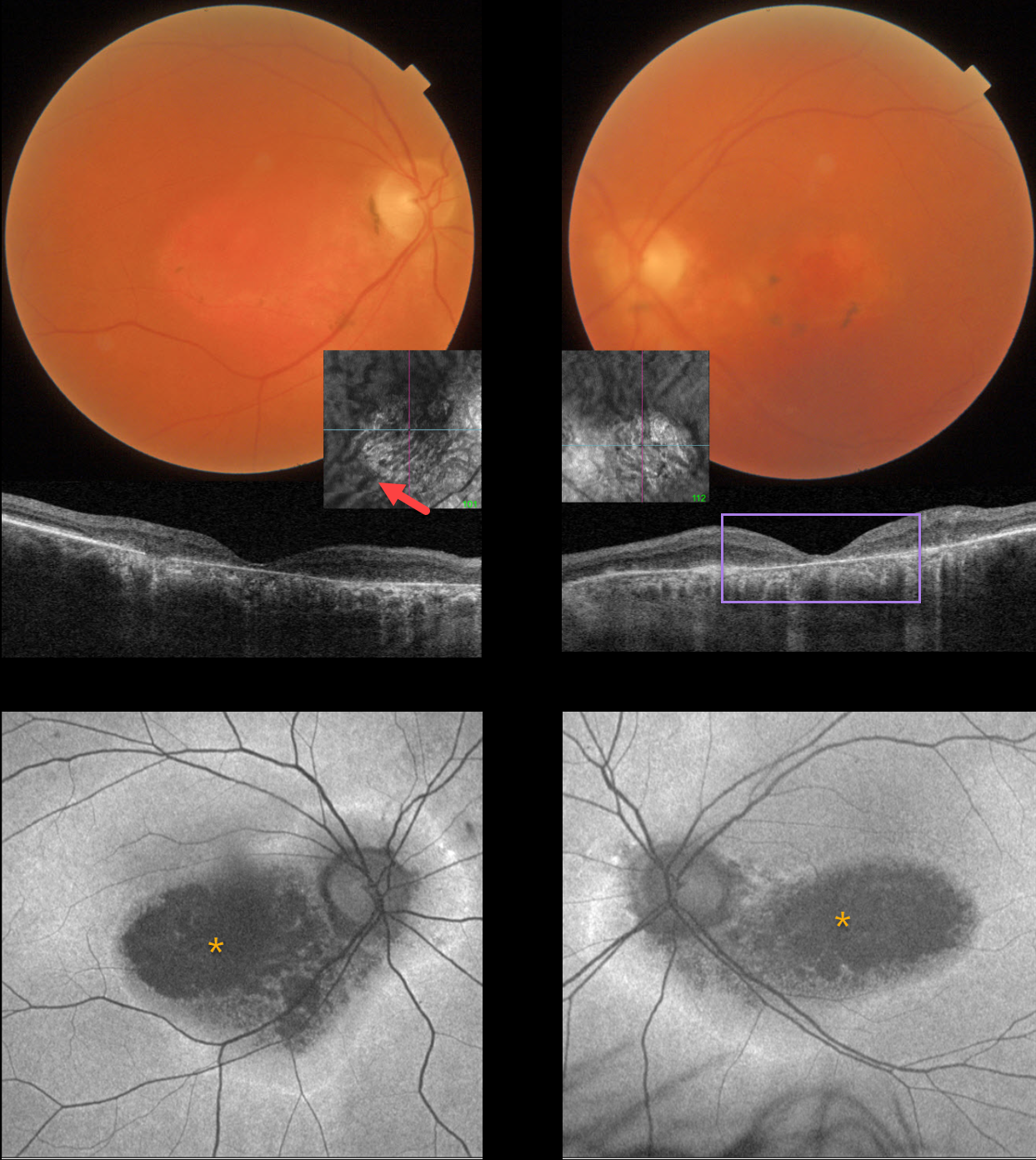
HYDROXYCHLOROQUINE TOXICITY + PACHYCHOROID
Originally posted on @retina.rocks March 11, 2024
This 67YO female has a known history of Plaquenil toxicity. Her bilateral 20/200 vision and macular findings have been stable since discontinuing this medication 30 years ago.
Triton color imaging and swept-source OCT show classic findings for severe hydroxychloroquine toxicity, including an oval area of foveal pigment loss and outer retinal atrophy.
Most patients will have preserved central foveal pigment giving a bulls-eye appearance, although our patient has loss of outer retina and RPE throughout. Interestingly, choroidal en face imaging shows bilateral dilated choroidal pachyvessels.
On fundus autofluorescence (FAF), the central maculas are hypo-FAF, although an atypical hyper-FAF ring extends around each optic nerve.
Learning Points:
Current screening guidelines should make Plaquenil toxicity, as found in our patient, a thing of the past. Last revised in 2016, the American Academy of Ophthalmology (AAO) recommends that a baseline exam be performed before starting the medication, with annual screenings beginning at least 5 years after initiating treatment, unless major risk factors are present. However, in practice, patients are usually screened yearly once they are placed on this medication.
Optical coherence tomography (OCT) is performed annually to assess outer retinal findings, including ellipsoid zone loss. Humphrey 10-2 visual field testing (24-2 for Asians, since their macular involvement is usually more peripheral) is also needed annually, as about 10% of patients will have field loss despite normal examinations and OCT testing.
Plaquenil dosing should also be based on real, not ideal, weight to better predict the optimal dose. See Marmor et al Ophthalmology 2016;123:1386-1394 for the full screening guidelines.
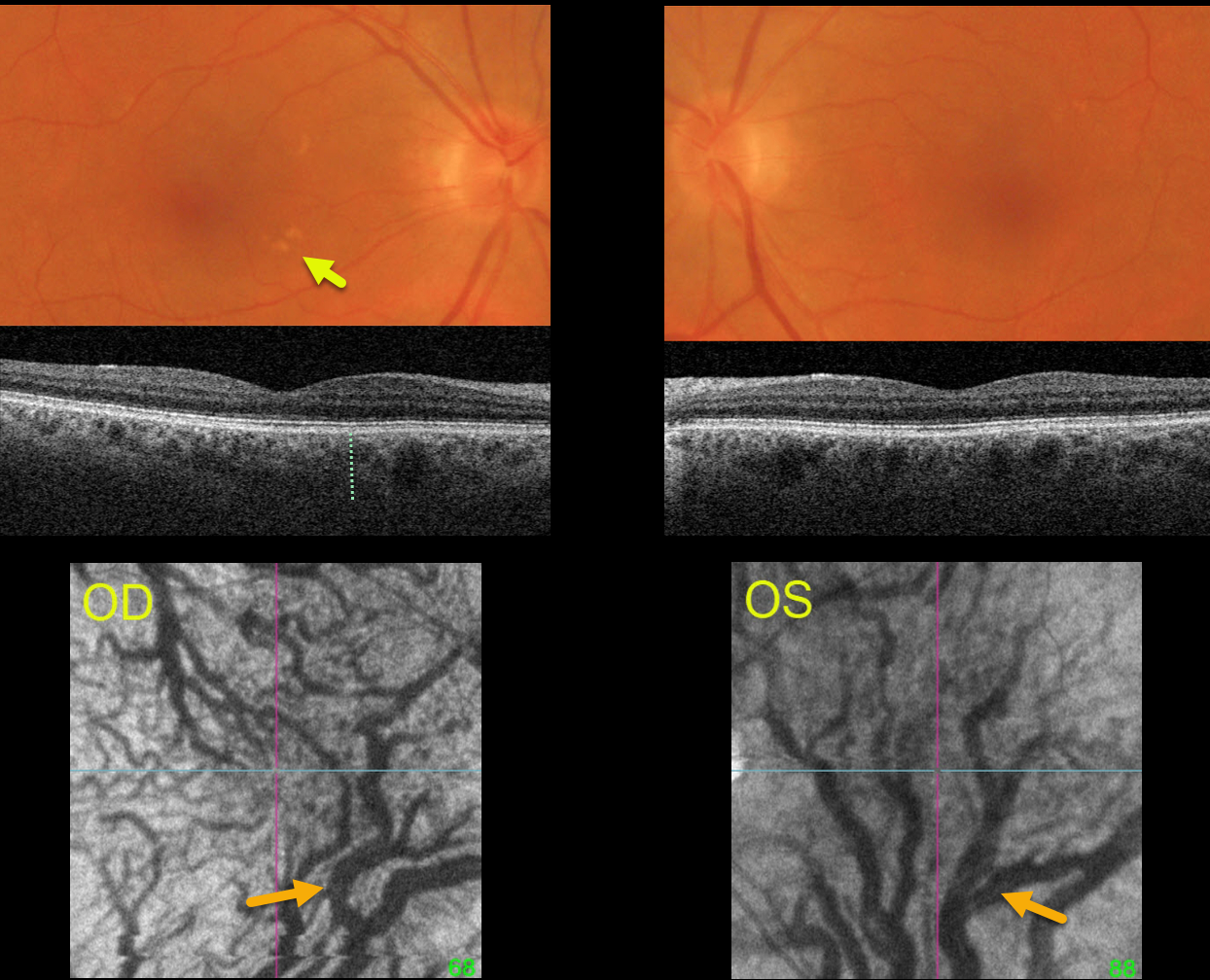
CHOROIDAL VENOUS ANASTAMOSIS
Originally posted on @retina.rocks October 5, 2023
This healthy 67YO female was examined for a symptomatic posterior vitreous detachment in her right eye. She was previously visually asymptomatic, and vision was 20/25 OU.
Optos color RGB imaging shows several large irregular drusen in the nasal right macula and faint drusen in the superotemporal left macula.
Triton swept source OCT shows a relatively thickened choroid for the patient’s age, along with large choroidal vessels. En face imaging through each choroid shows marked vortex dilation bilaterally. Each macula, particularly in her left eye, is drained by the inferior vortex system.
Learning Points:
Pachychoroid, meaning “thick” choroid, is secondary to large dilated Haller vessels. Increased oncotic pressure from these dilated vessels can cause RPE dysfunction, serous detachment, and macular neovascularization.
Another, more subtle finding is pachydrusen. Unlike typical age-related drusen that are pinpoint or round and centered in each macula, pachydrusen are fewer in number, more ectopic, and often have an irregular contour (Spaide, Retina 2018;38:708-716).
The choroid is typically thinned with AMD, but eyes with pachydrusen have thicker choroids. Although our patient’s macular clinical findings were quite subtle and the OCT B-scans were suggestive of pachychoroid, her en face images most dramatically show the choroidal pathology.
CHOROIDAL EXCAVATION
Originally posted on @retina.rocks August 15, 2023
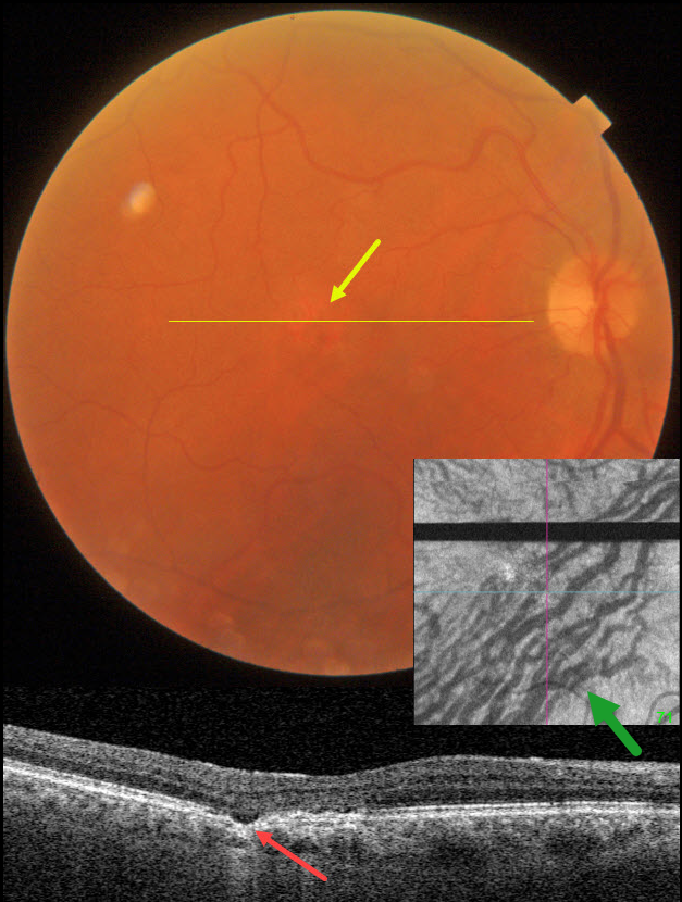
This 74YO male was seen for a routine eye examination. He told us he had a laser procedure on his right eye 18 years ago. Based on this image, what type of laser was it?
Fundus photography shows a normal macula except for an area of depigmentation in the temporal fovea.
Swept-source OCT through this area shows a small focal choroidal excavation. Although the choroid appears to have normal thickness, large outer choroidal vessels spanning virtually the entire choroid are noted nasally.
Choroidal en face OCT is markedly abnormal with dilated vessels and vortex anastomoses.
Our patient shows several pachychoroid-related findings, including a thick choroid for their age, large choroidal vessels, FCE, and vortex anastomoses. Our patient currently has asymptomatic pachychoroid despite having a ‘normal’ choroidal thickness on OCT.
So what type of laser did our patient have? Photodynamic therapy for central serous retinopathy, which has remained in remission since treatment in 2005
Learning Points:
Pachychoroid, meaning “thick” choroid, is secondary to large dilated Haller vessels. Increased oncotic pressure from these dilated vessels can cause RPE dysfunction, serous detachment, and macular neovascularization.
The choroidal thickness depends on numerous factors, including refractive error and age. A high myope’s choroid should be quite thin, but if it appears as a ‘normal’ thickness, it is thick for that eye. We also often see pachychoroid disease in elderly patients whose choroid is abnormally thickened for their age, but appears ‘normally’ thick when not accounting for this.
FCE, originally described by Jampol et al in 2006, is likely part of the pachychoroid spectrum, as it is usually associated with a thickened choroid and pachyvessels. The FCE can be observed, although symptomatic manifestations, including active central serous retinopathy or macular neovascularization, often require treatment. An excellent review is by Verma et al, Br J Ophthalmol 2021;105:1043-1048.
The choroidal venous system, as with the neurosensory retinal vasculature, usually respects the horizontal meridian. With pachychoroid disorders, these anastomoses develop, and in this case, the inferior vortex system drains nearly the entire macula.
For a wonderful review highlighting the concept of venous overload choroidopathy, see Spaide et al, Progress Retinal Eye Research 2022;86:100973.
MACULAR HOLE
Shilpi Narnaware and Prashant Bawankule
Originally posted on @retina.rocks August 8, 2023
This 60YO female presented with 1 month of vision loss in her right eye. Vision was 20/100 OD and 20/40 OS.
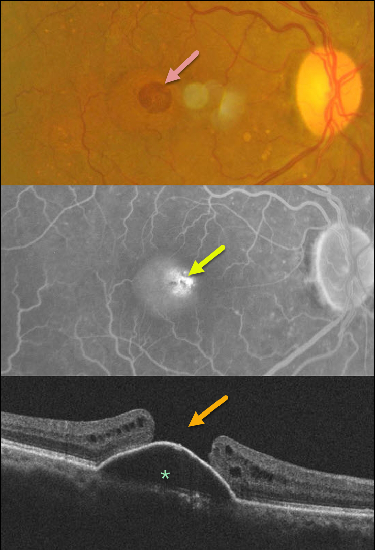
Fundus photography of her right eye shows a large macular hole with some nasal pachydrusen. There was an old branch retinal vein occlusion without macular drusen in her left eye (image not shown).
Fluorescein angiography shows central pooling beneath a retinal pigment epithelial detachment (PED) with increased transmission through the macular hole. OCT scanning shows a PED with an overlying large macular hole.
This case presents a unique and interesting challenge in that macular hole surgery likely would not close the hole, given the underlying PED. She received 3 monthly Lucentis injections in her right eye, with no change in the PED.
Our thought would be to try a different anti-VEGF agent, such as Eylea, or consider adding photodynamic therapy for a possible pachychoroid-related avascular PED.
A similar case showed spontaneous closure of a macular hole following anti-VEGF therapy (see Chakraborty and Sheth, Indian J Ophthalmol Case Rep 2023;3:439-440).
If our patient’s macular hole remained open once the PED flattened, then macular hole surgery would be performed.
PACHYCHOROID NEOVASCULARIZATION
Originally posted on @retina.rocks July 18, 2023
This 85YO female presented with recent vision loss in her right eye. Vision was 20/200 OD and 20/40 OS.
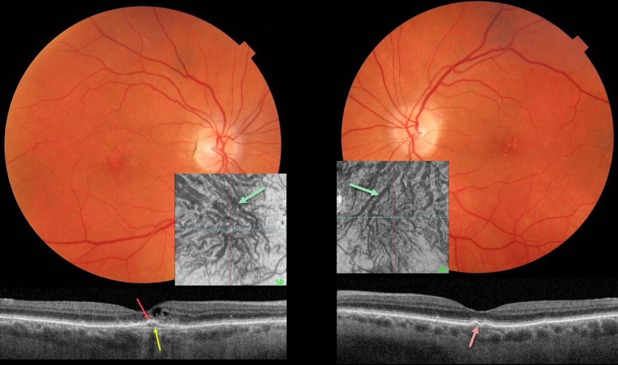
Fundus photography of her right eye shows granular foveal pigmentary changes with a few faint, medium-sized drusen in her left eye.
Swept-source OCT OD shows a shallow, irregular elevation of the foveal RPE with visible Bruch’s membrane. Some cuticular drusen are noted bilaterally. Although the choroid itself is not thickened, large choroidal vessels are seen bilaterally, and choroidal en face imaging shows abnormal, dilated superior anastomotic vortex vessels that drain the entirety of each macula.
Learning Points:
Pachychoroid, meaning “thick” choroid, is secondary to large dilated Haller vessels. Increased oncotic pressure from these dilated vessels can cause RPE dysfunction, serous detachment, and macular neovascularization (MNV).
Our patient has pachychoroid despite having a ‘normal’ choroidal thickness on B-scan OCT. The choroidal thickness depends on numerous factors, including the patient’s age and refractive error. A high myope’s choroid should be quite thin, but if it appears as a ‘normal’ thickness, it is thick for that eye.
We also often see pachychoroid disease in elderly patients whose choroid is abnormally thickened for their age, but appears ‘normally’ thick when not accounting for this.
Originally described by Pang and Freund (Retina 2015;35:1-9), pachychoroid neovasculopathy is a variation of type 1 MNV (located below the RPE). About one-fifth of patients diagnosed with neovascular AMD actually have pachychoroid neovasculopathy (Farvardin et al, Int J Retina Vitreous 2022;8:78), and about one-third of eyes will progress to polypoidal choroidal vasculopathy by 5 years (Siedlecki et al, Ophthalmology Retina 20222;6:807-813).
Although our 85YO patient also has macular drusen, the summation of her findings most likely points to pachychoroid disease as the cause of her MNV. Anti-VEGF therapy was begun.
RPE TEAR
Originally posted on @retina.rocks June 14, 2023
This 73YO female presented with acute vision loss in her right eye. Vision was 20/40 OD and 20/30 OS. There was a several-year history of type 2 diabetes with mild bilateral nonproliferative diabetic retinopathy.
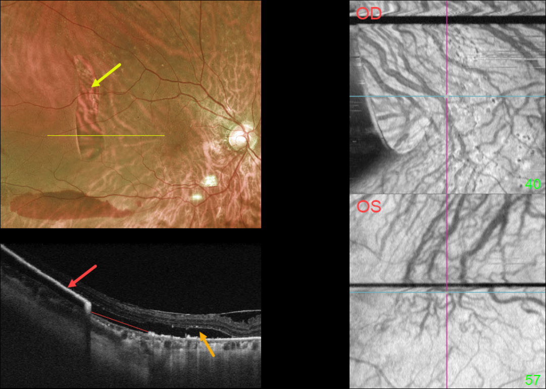
Optos imaging shows a large vertical area of depigmentation in the distal superotemporal macula. There is a serous detachment surrounding this region, along with a far inferior subretinal hemorrhage.
Swept-source OCT through this lesion shows bare Bruch’s membrane from an RPE tear. The retracted RPE is markedly hyperreflective more temporally. Subretinal fluid overlies this region.
Macular choroidal en face imaging shows bilateral anomalous vasculature with choroidal anastomoses.
Learning Points:
A tear of the RPE is most commonly seen in RPE detachments (PEDs) associated with neovascular AMD, but can also occur in numerous other etiologies, including central serous chorioretinopathy and polypoidal choroidal vasculopathy.
The tear leaves a depigmented region where the RPE was and is now absent, along with a pigmented, relatively straight subretinal lesion from the scrolled RPE edge.
The choroidal en face OCT findings are more consistent with the pachychoroid spectrum. RPE tears are found in polypoidal choroidal vasculopathy, another pachychoroid disorder (Zhao et al, Retina 2020;40:477-489).
See Ersoz et al for a great review on RPE tear classification, pathogenesis, predictors, and management (Surv Ophthalmology 2017;62:493-505).
PACHYCHOROID SYNDROME
Will Gibson
Originally posted on @retina.rocks March 22, 2023
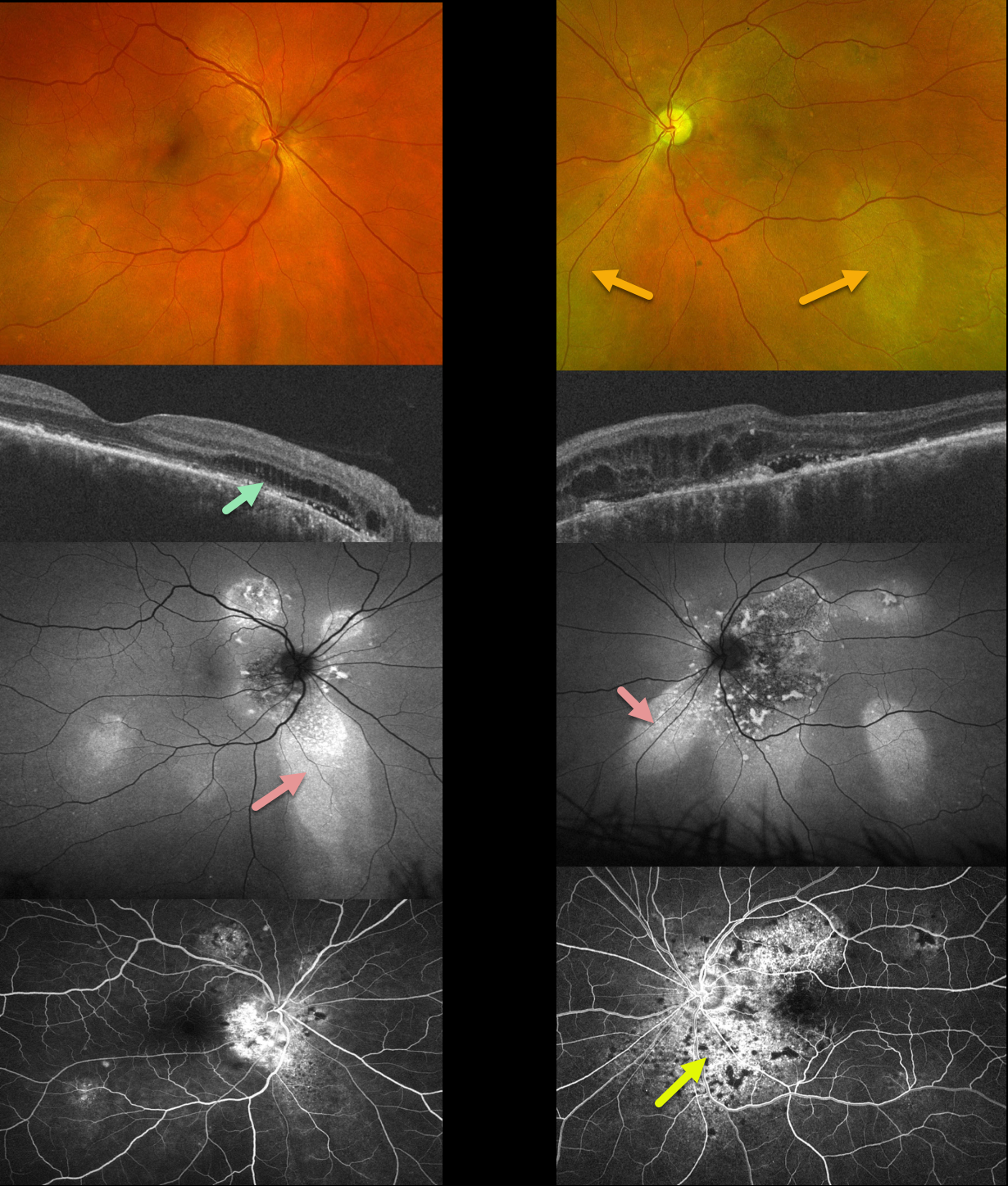
This 51YO male has been followed for years with self-limited episodes of idiopathic central serous retinopathy (ICSC).
Optos color imaging of both eyes shows regions of inferior pigmentary loss, most pronounced in the OS. These findings are much more dramatic on fundus autofluorescence. Swept-source OCT shows variable bilateral loss of the outer retinal bands. Outer retinal fluid with trace subretinal fluid is seen centrally and nasally OS. Fluorescein angiography shows variable staining.
Learning Points:
This patient has numerous features of the pachychoroid spectrum, including idiopathic central serous retinopathy (ICSC), pachychoroid neovasculopathy, polypoidal choroidal vasculopathy, pachychoroid pigment epitheliopathy, peripapillary pachychoroid syndrome, and choroidal excavation.
Our patient’s current findings are dominated by the peripapillary pachychoroid syndrome. Originally described by Phashukkuwatana et al (Retina 2018;1652-1667), findings include peripapillary choroidal thickening associated with nasal macular subretinal and/or intraretinal fluid. Most patients also have chorioretinal folds.
The fluid waxes and wanes, and is responsive to photodynamic therapy if treatment is needed for persistent symptomatic foveal fluid (Iovino et al, Eye 2022;36:716-723). Our patient is being observed for now.
PACHYCHOROID PIGMENT EPITHELIOPATHY
Originally posted on @retina.rocks March 6, 2023
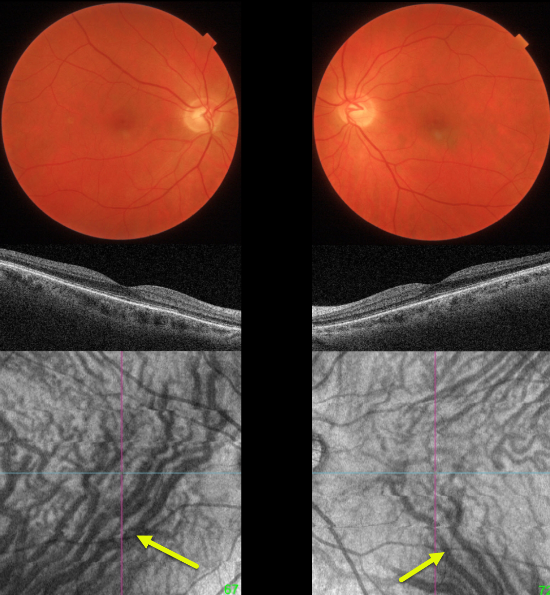
This 51YO minus 5 diopter myope presented without visual complaint for a routine yearly examination. Vision was 20/25 OU.
Both maculas are normal on fundus photo and B-scan OCT. However, en face OCT shows bilateral dilation of the inferior vortex system. The right inferior vortex also drains nearly the entire macula.
Learning Points:
The choroidal venous system, as with the neurosensory retinal vasculature, usually respects the horizontal meridian. With pachychoroid disorders, these anastomoses develop, and in this case, each inferior vortex system drains nearly the entire macula.
For a wonderful review highlighting the concept of venous overload choroidopathy, see Spaide et al, Progress Retinal Eye Research 2022;86:100973.
Pachychoroid, meaning “thick” choroid, is secondary to large dilated Haller vessels. Increased oncotic pressure from these dilated vessels can cause RPE dysfunction, serous detachments, and macular neovascularization.
Our patient has asymptomatic pachychoroid despite having a ‘normal’ choroidal thickness on B-scan OCT. The choroidal thickness depends on the refractive error. A high myope’s choroid should be quite thin, but if it appears as a ‘normal’ thickness, it is thick for that eye.
We also often see pachychoroid disease in elderly patients whose choroid is abnormally thickened for their age but appears ‘normally’ thick when not accounting for this. Although our patient’s retinal findings are completely normal clinically, these subclinical pachychoroid findings put her at risk for the various pachychoroid disorders
For an absolutely fascinating editorial discussing the ambiguity of how best to define what ‘pachychoroid’ actually means, see Spaide Retina 2021;41:231-237.
SUBRETINAL HEMORRHAGE
Originally posted on @retina.rocks January 31, 2023
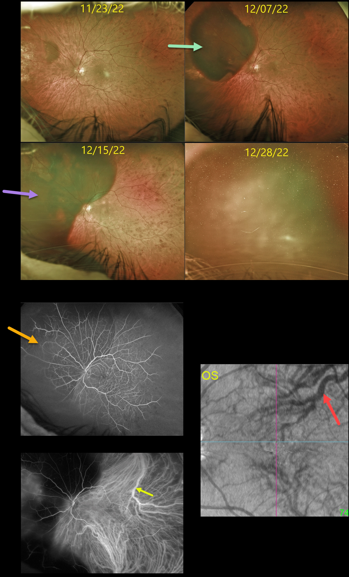
This diabetic male had been followed for over 5 years in our practice, receiving PRN intravitreal injections for diabetic macular edema.
On 8/10/22, he presented with a large superotemporal subretinal hemorrhage in his right eye that progressed into the macula (not shown). Vitrectomy with evacuation of the subretinal blood was performed.
Three months later, an inferior retinal detachment with proliferative vitreoretinopathy was noted (not shown). Staged cataract surgery followed by vitrectomy with silicone oil was performed.
At the 12/7/22 visit, the patient complained of new inferotemporal peripheral field loss, noting that “the same thing is happening in my left eye.”
Unfortunately, a superonasal subretinal hemorrhage was indeed found, and intravitreal Avastin injection was given the following day. However, one week later, the subretinal blood spread posteriorly.
Fluorescein angiography shows nonproliferative diabetic retinopathy with blockage superonasally from the subretinal blood. ICG angiography shows a large dilated superotemporal choroidal vein. En face OCT shows abnormal vortex systems, including a large, dilated superotemporal vortex vein.
On 12/28/22, the subretinal blood spread further with breakthrough bleeding into the vitreous.
Cataract surgery was performed, followed by vitrectomy; however, intraoperatively, once the vitreous hemorrhage was removed, he had a massive subretinal hemorrhage with an inoperable retinal detachment with proliferative vitreoretinopathy.
We believe our patient has an extremely unusual and aggressive variant of pachychoroid disease, as evidenced by the abnormal choroidal vasculature noted on ICG and en face OCT.
We have no explanation for why both eyes developed these massive subretinal hemorrhages, or why they developed almost concurrently. We plan on injecting his right eye with ongoing intravitreal Eylea in hopes of preventing recurrent subretinal hemorrhages.
GIANT RPE TEAR
Anjana Mirajkar, Manish Nagpal, Navneet Mehrotr, and Akansha Sharma
Originally posted on @retina.rocks January 20, 2023
This 62YO male presented with 1 month of vision loss in his left eye. Vision was 20/30 OD and 20/60 OS.
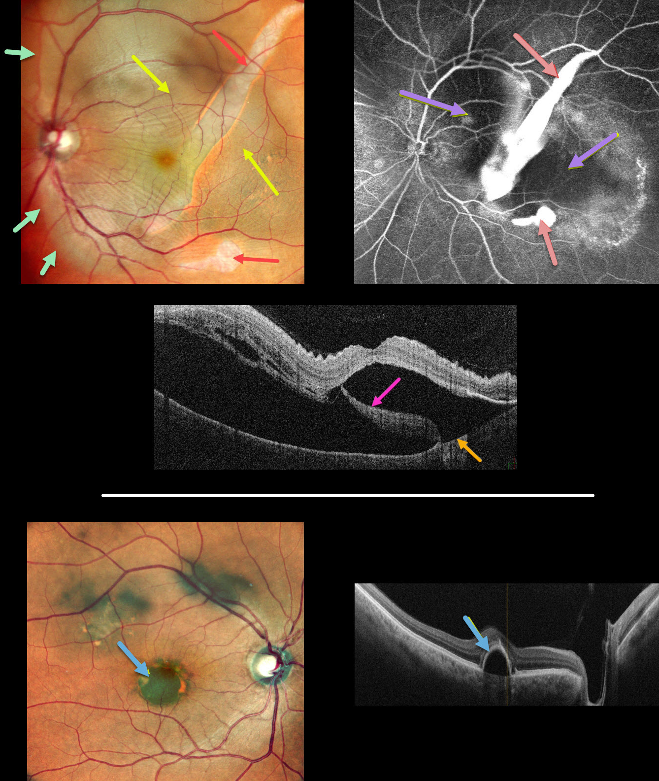
Multicolor imaging of his left eye shows a giant RPE tear extending from the inferior macula into the superotemporal midperiphery, along with a much smaller tear just inferior to the macula. The beds of the RPE tears are depigmented. Increased subretinal pigmentation is noted on either edge of the giant tear. Subretinal fluid variably elevates the macula with inner retinal folds radiating from the optic nerve temporally. This subretinal fluid extends into the superior and temporal midperipheries.
B-scan OCT shows marked subretinal fluid. Bare Bruch’s membrane is noted temporally, and the RPE tear is lifted nasally and floating within the serous detachment.
Fluorescein angiography shows marked hyperfluorescent window defects from the absent RPE with blockage on either side of the giant tear from the redundant retracted RPE. He was placed on a 5-day tapering course of oral steroids but was subsequently lost to follow-up.
An asymptomatic retinal pigment epithelial detachment (PED) is noted in his right macula. The choroid does not appear thickened on OCT B-scan.
Learning Points:
A tear of the RPE is most commonly seen in RPE detachments (PEDs) associated with neovascular AMD, but can also occur in numerous other etiologies, including central serous chorioretinopathy and polypoidal choroidal vasculopathy. The tear leaves a depigmented region where the RPE was and is now absent, along with a pigmented, relatively straight subretinal lesion from the scrolled RPE edge.
Our patient’s presentation is unique in that RPE tears are usually much smaller and tend to rip in just one direction. The edges of our patient’s RPE retracted both nasally and temporally. RPE tears are usually singular, and ours had an additional, much smaller secondary tear more inferiorly.
Although our patient’s findings seem to be in the pachychoroid spectrum, the lack of a thickened choroid on OCT scanning makes the etiology less certain.
See Ersoz et al for a great review on RPE tear classification, pathogenesis, predictors and management (Surv Ophthalmology 2017;62:493-505).
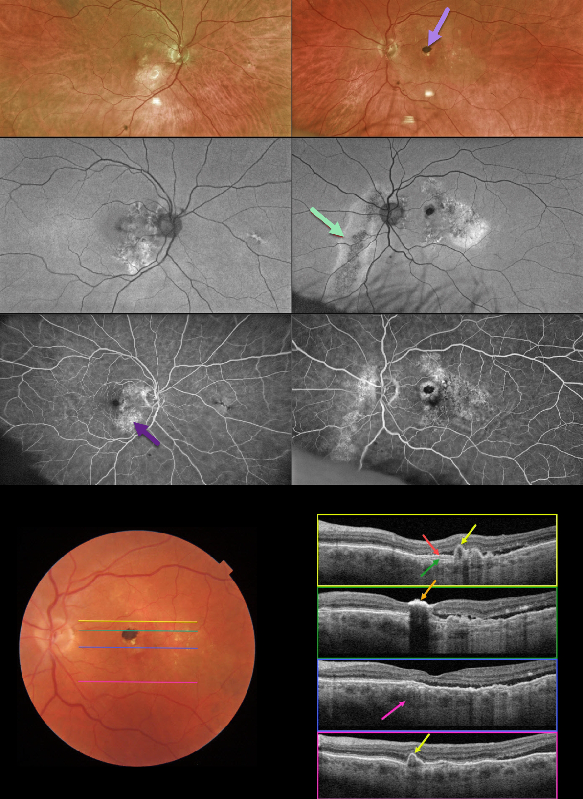
PACHYCHOROID SPECTRUM
Originally posted on @retina.rocks November 22, 2022
This 78YO male was referred for asymptomatic fundoscopic changes. Vision was 20/25 OD and 20/30 OS. He had a history of laser treatment in his left eye decades earlier.
Optos imaging shows bilateral areas of pigmentary loss in each macula, extending inferiorly. A hyperpigmented laser scar is noted in the left superior macula.
Fundus autofluorescence (FAF) shows variable hypo- and hyper-FAF, along with a prominent gutter extending from the left nerve inferonasally.
Fundus fluorescein angiography (FFA) shows window defects within these areas, with blockage from the hyperpigmented laser scar OS.
Swept-source OCT shows numerous findings. Scanning superior to the laser scar shows several choroidal polyps. A shallow elevation of the RPE exposes the underlying Bruch’s membrane. Scanning through the laser scar shows intense hyperreflectivity with posterior shadowing. Scanning through the macular center shows temporal subretinal fluid, along with a thickened choroid. Finally, scanning in the inferior macula shows an additional polyp.
Learning Points:
This case has an overwhelming amount of features from the pachychoroid spectrum, which includes idiopathic central serous retinopathy (ICSC), pachychoroid neovasculopathy, polypoidal choroidal vasculopathy, pachychoroid pigment epitheliopathy, peripapillary pachychoroid syndrome, and choroidal excavation.
The thermal laser treatment many years ago may have been for pachychoroid neovasculopathy, a recently described phenotype (see Pang and Freund, Retina 2015;35:1-9) found in the pachychoroid spectrum. These lesions are particularly common in patients with ICSC, and often remain silent as a flat, irregular RPE elevation on OCT without exudative complications.
Our patient likely had further progression of their pachychoroid disease, resulting in multiple polyps, which are found in polypoidal choroidal vasculopathy (PCV). PCV is a variant of type 1 macular neovascularization, also in the pachychoroid spectrum. PCV is likely distinct from age-related macular degeneration, occurring in a different population (more common in African Americans and Asians), with a relative lack of drusen, and the presence of a thicker choroid.
In addition to the findings above, the patient had a large gutter in his left eye, a common finding in ICSC. Gutters form from pathologies with chronic leakage. Conditions that form gutters other than ICSC include choroidal hemangiomas, choroidal nevi, and uveal melanoma.
For more information on the pachychoroid spectrum, see Cheung et al, Eye 2019;33:14-33 for a great pachychoroid disease review.
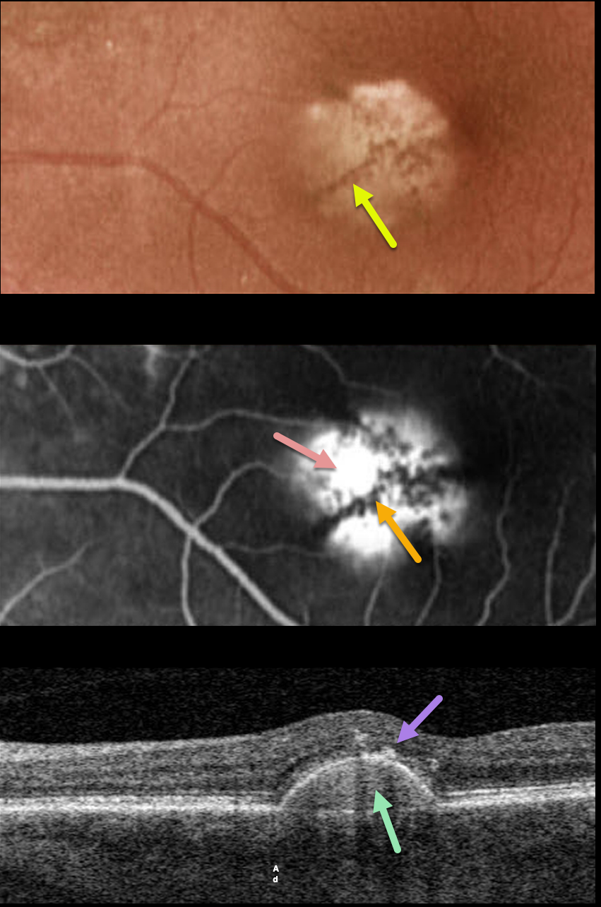
PACHYCHOROID PIGMENT EPITHELIAL DETACHMENT (PED)
Originally posted on @retina.rocks September 13, 2022
This 75YO female has been followed for several years with a stable, asymptomatic chronic retinal pigment epithelial detachment (PED) and 20/40 vision.
Optos color imaging shows a solid, variably depigmented, round PED in the inferonasal region of the left fovea. Radiating linear pigmentation is noted.
Fluorescein angiography shows hyperfluorescent pooling beneath the PED, with blockage from the overlying radiating pigment.
Swept-source OCT shows the PED with a hyperreflective core, overlying hyperreflective foci (representing the clinical radiating pigment), and a relatively thickened choroid for a patient of this age.
Learning Points:
Retinal pigment epithelial detachments are common in patients with AMD and are often associated with macular neovascularization. Non-vascularized PED is also seen as part of the pachychoroid spectrum.
The overlying pigment figure is pathognomonic for a chronic PED, and is seen even when these lesions flatten. Isolated avascular PEDs such as these do not require treatment.
Although our patient was in her mid-70s, there were no drusen and each fundus was otherwise normal. The chronicity of the PED, the lack of other signs of AMD, and the relatively thickened choroid all point to this being an avascular, pachychoroid PED.
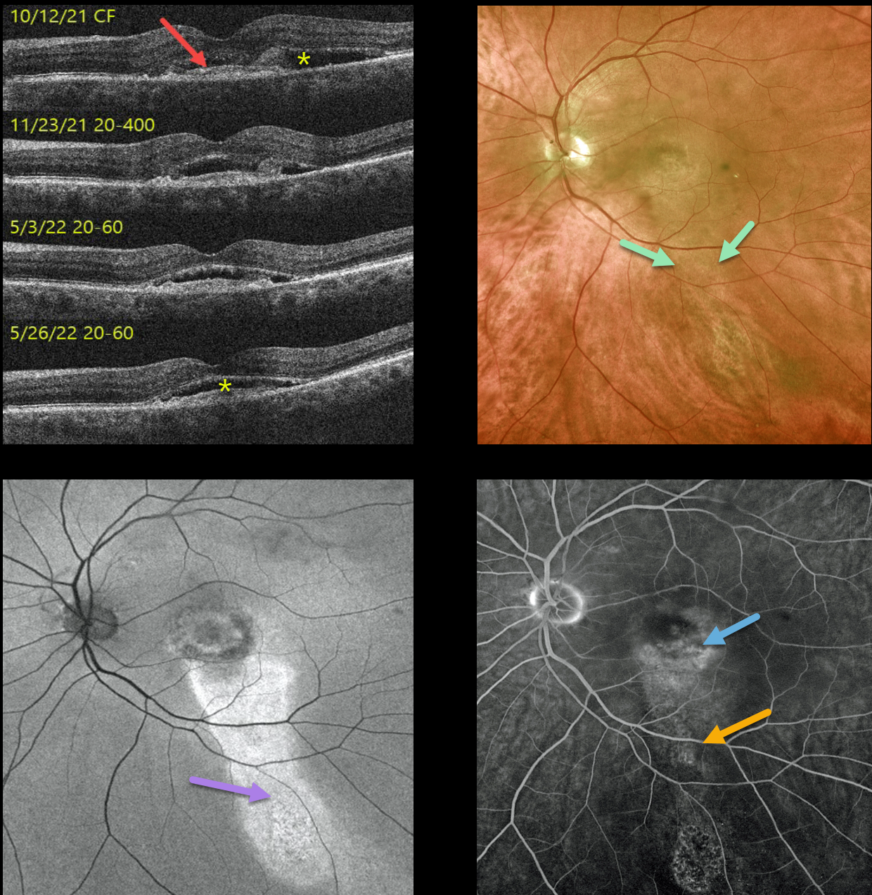
PACHYCHOROID NEOVASCULOPATHY WITH GUTTER
Originally posted on @retina.rocks July 1, 2022
This 69YO male presented with asymptomatic vision loss in his left eye. Vision was 20/30 OD and counting fingers OS.
A small central scar with fluid was present in the left macula (initial photo not shown). OCT scanning shows a shallowly elevated type 1 macular neovascularization with associated subretinal fluid. An intravitreal Avastin injection was given that day, and 1 month later, the fluid had improved. Vision was 20/400. A second intravitreal Avastin was given.
He was then lost to follow-up for about 6 months while he was out of state receiving intravitreal Eylea every 6 weeks. When he returned, his vision was 20/60 with shallow residual subretinal fluid. Optos color imaging shows central macular pigmentary changes with a gutter of inferior pigment loss. Fundus autofluorescence (FAF) more dramatically shows the hyper-FAF inferior gutter. Fluorescein angiography shows mild staining of the foveal MNV with an inferior gutter of window defects.
Due to an upcoming trip to Alaska and Canada, he requested trying photodynamic therapy (PDT) in an attempt to minimize the treatment burden. Several weeks after PDT, vision remained at 20/60, and the subretinal fluid showed slight improvement on OCT. We will see him again when he returns from his travels.
Learning Points:
Pachychoroid neovasculopathy is a recently described phenotype (see Pang and Freund, Retina 2015;35:1-9) found in the pachychoroid spectrum, which includes idiopathic central serous retinopathy (ICSC), pachychoroid neovasculopathy, polypoidal choroidal vasculopathy, pachychoroid pigment epitheliopathy, peripapillary pachychoroid syndrome, and choroidal excavation.
These lesions are particularly common in patients with ICSC and often present as a flat, irregular RPE elevation on OCT without exudative complications. The absence of drusen in our patient’s fellow eye, the thickened choroid, and gutter all point to the diagnosis of pachychoroid neovasculopathy with prior ICSC.
See Cheung et al, Eye 2019;33:14-33 for a great pachychoroid disease review.
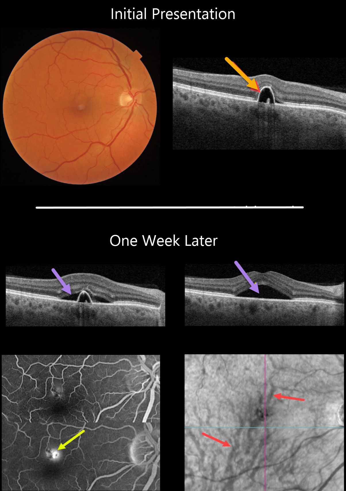
IDIOPATHIC CENTRAL SEROUS CHORIORETINOPATHY
Originally posted on @retina.rocks February 16, 2022
This 55YO male presented on 1/24/22 with a small symptomatic pigment epithelial detachment (PED) in the superior right fovea. Vision was 20/30.
One week later, he was subjectively worse, although vision remained at 20/30. There was a new small blister of foveal subretinal fluid, best captured on swept-source OCT.
Optos fluorescein angiography shows a typical blot leak along the dome of the PED. En face OCT shows a choroidal venous anastomosis.
Learning Points:
The choroidal venous system, as with the neurosensory retinal vasculature, usually respects the horizontal meridian. With pachychoroid disorders, these anastomoses develop, and in this patient’s case, the inferior vortex system drains nearly the entire macula.
For a wonderful review highlighting the concept of venous overload choroidopathy, see Spaide et al, Progress Retinal Eye Research 2022;86;100973.
PERIPHERAL EXUDATIVE HEMORRHAGIC CHORIORETINOPATHY + PACHYCHOROID
Originally posted on @retina.rocks December 22, 2021
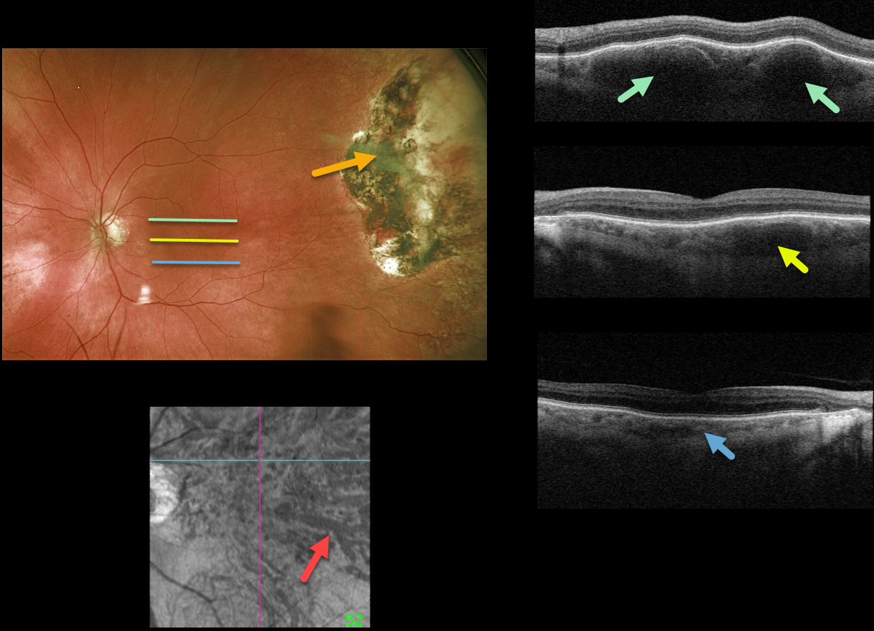
This 85YO male underwent cryotherapy 7 years earlier for a symptomatic hemorrhagic ectopic choroidal neovascularization in his left eye. Optos imaging shows the current appearance of the inactive cryotherapy scarring.
Although the macula has a normal fundoscopic appearance, Triton swept-source OCT scanning is far from normal. B-scans are shown through the top, central, and inferior macula. Although the inferior macular choroid appears relatively normal, a thickened outer choroid (pachyvessels) is noted more centrally, and is huge superiorly. En face imaging through this abnormal choroid is shown.
Learning Points:
The pachychoroid spectrum is a collection of conditions with a thick (“pachy”) choroid. These pachyvessels can cause a range of disorders, including idiopathic central serous chorioretinopathy, pachychoroid pigment epitheliopathy, choroidal excavation, pachychoroid neovasculopathy, polypoidal choroidal vasculopathy, pachydrusen, and peripapillary pachychoroid syndrome.
They are also associated with ectopic neovascularization (aka peripheral exudative hemorrhagic chorioretinopathy), although the choroid in these cases shows increased thickening towards the temporal periphery (see Shroff et al, Retina 2021;41:1518-1525).
FOCAL CHOROIDAL EXCAVATION
Originally posted on @retina.rocks October 6, 2021
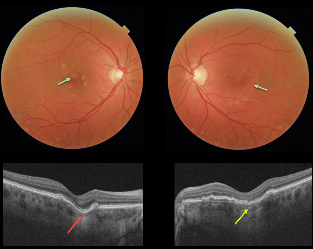
This 60YO female has a history of idiopathic central serous chorioretinopathy that was previously treated successfully with photodynamic therapy in her left eye. Bilateral macular drusen are noted.
OCT shows a definite focal choroidal excavation (FCE) in her right eye, and a possible FCE in her left eye.
Learning Points:
Focal choroidal excavation, originally described by Jampol et al. in 2006, is likely part of the pachychoroid spectrum, as it is usually associated with a thickened choroid and pachyvessels.
The FCE can be observed, although symptomatic manifestations, including active central serous retinopathy or macular neovascularization, often require treatment.
Spaide and Ryan noted this finding in about two-thirds of patients with central serous retinopathy (Am J Ophthalmology 2015;160;1211-1216). An excellent review is by Chung et al (Retina 2017;37;199-221).
PIGMENT EPITHELIAL DETACHMENT (PED)
Originally posted on @retina.rocks April 26, 2021
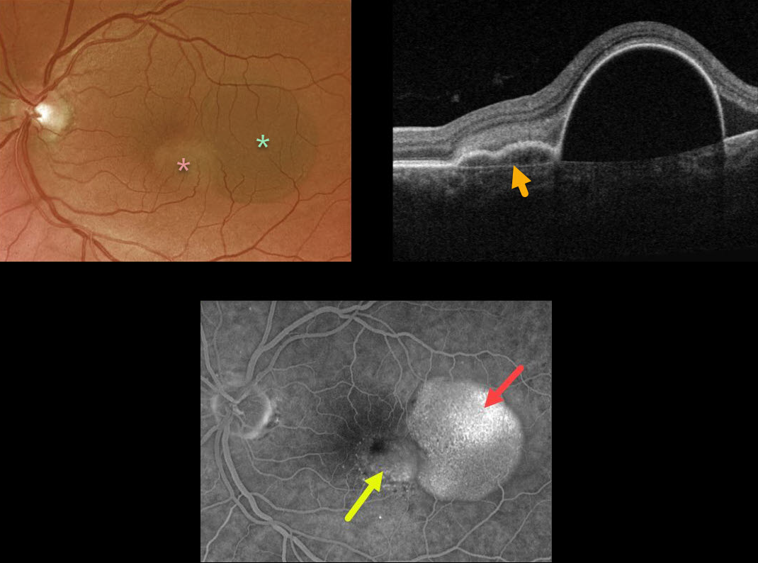
This 57YO female presented with counting-finger vision from a large RPE detachment (PED). The PED was located in the temporal macula, with a smaller notch of irregular RPE elevation more centrally.
Triton swept-source OCT shows a shallow PED at the notch overlying Bruch’s membrane. Optos fluorescein angiography shows late uneven filling of the PED and late speckled staining of the notch.
Anti-VEGF therapy was recommended.
Learning Points:
Originally described by Gass (Retina 1984;4:205-220), a notched PED is a sign of an occult macular neovascularization (MNV). The notch represents a type 1 MNV, which tethers the overlying RPE from the surrounding more highly elevated PED. Hydrostatic leakage from the MNV may contribute to the larger PED.
CHOROIDAL EXCAVATION
Originally posted on @retina.rocks February 17, 2021
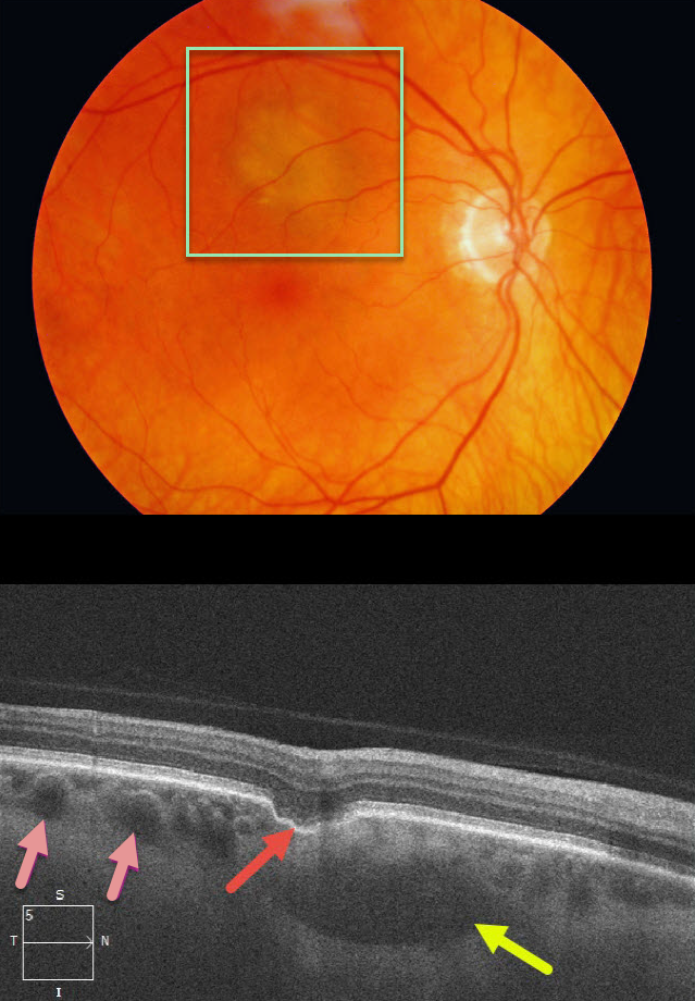
This patient was referred for an asymptomatic, slightly depigmented subretinal lesion in the superior right macula. OCT through this lesion reveals a focal choroidal excavation, choroidal pachyvessels, and probable loculated outer choroidal fluid.
Learning Points:
Focal choroidal excavation (FCE), originally described by Jampol et al in 2006, is likely part of the pachychoroid spectrum since it is usually associated with a thickened choroid and pachyvessels. The FCE can be observed, although symptomatic manifestations, including active central serous retinopathy or macular neovascularization, often require treatment.
Our patient also showed accumulation of fluid in the outer choroid. Spaide and Ryan noted this finding in about two-thirds of patients with central serous retinopathy (Am J Ophthalmology 2015;160;1211-1216). The presence of an FCE, pachyvessels, and loculated posterior choroidal fluid in our patient is intriguing and supports the association between pachychoroid disorders and FCE.


