

Papilledema + Idiopathic Intracranial Hypertension (IIH)
<< Back to Cases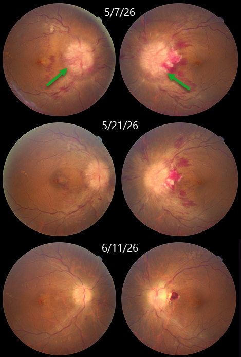
PAPILLEDEMA
Abhishek Karra and Ravindra Karra
Originally posted on @retina.rocks August 5, 2026
This 44YO obese female presented with several days of decreased vision bilaterally. Vision was counting fingers OU. Fundus photography shows severe bilateral disc swelling.
MRI of the brain and orbit was suggestive of partial empty sella with prominent perioptic CSF spaces and no intracranial mass lesions. All these were suggestive of idiopathic intracranial hypertension (IIH). She was referred to neurology, where a spinal tap established the diagnosis of IIH, and she was started on acetazolamide 1000 mg qday. One month later, vision improved to 20/20 OU with marked improvement in the papilledema.
Learning Points:
IIH, formerly known as pseudotumor cerebri, is caused by elevated intracranial pressure primarily due to decreased cerebrospinal fluid (CSF) drainage caused by elevated cerebral venous pressure. The condition is most often found in obese women of childbearing age. Presenting symptoms include headaches, transient visual obscurations, neck/back pain, and tinnitus/whooshing. Treatment includes weight loss, acetazolamide, and topiramate. Severe cases unresponsive to medical therapy, or those with transverse sinus stenosis, require surgery (CSF diversion procedures, venous sinus stenting, or optic nerve sheath fenestration).
PAPILLEDEMA WITH SPONTANEOUS VITREOUS HEMORRHAGE
The European VitreoRetinal Society (EVRS) and Gordon Shen
Originally posted on @retina.rocks June 5, 2026
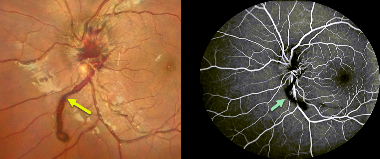
This 20YO female presented with a history of headaches and recent floaters in her right eye. There was a history of idiopathic intracranial hypertension.
Color photography shows a swollen right nerve with overlying preretinal blood extending inferiorly. Fluorescein angiography showed no leakage. Unfortunately, further clinical information and follow-up are not available.
Learning Points:
Vitreous hemorrhage directly from papilledema is exceedingly rare, and the published literature on this specific complication is very limited. Prior reports attribute vitreous hemorrhage to papilledema in eyes with no other pathology. One report suggested that the breakthrough vitreous hemorrhage originated from the optociliary shunt vessels (Fraser et al, J Neuroophthalmology 2012;32:332-334). Our patient offers visual evidence that vitreous blood can arise directly from an edematous nerve.
PAPILLEDEMA
Anand Temkar and Surendra Pal
Originally posted on @retina.rocks September 10, 2025
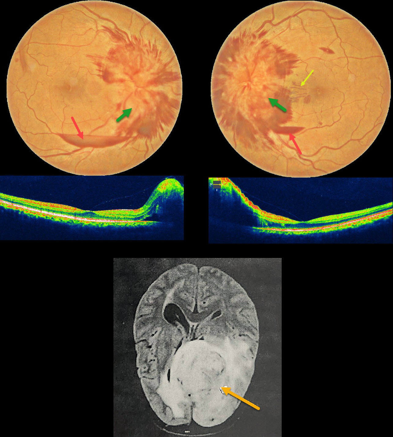
This 50YO female presented with one month of bilateral vision loss. Vision was 20/90 OD and 20/70 OS.
Fundus photography shows severe bilateral optic nerve swelling with surrounding nerve fiber layer hemorrhages. Small inferior subhyaloid hemorrhages are present bilaterally, along with a small outer plexiform layer hemorrhage in the left nasal macula.
MRI scanning showed a high-grade glioma, and she was immediately referred to neurosurgery for further management.
Learning Points:
Glioblastoma multiforme, also known as a grade IV astrocytoma, represents the most common and aggressive primary malignant brain tumor. A variety of neuro-ophthalmic signs and symptoms result from direct involvement of the visual pathway or elevated intracranial pressure, including vision loss or visual field defects, afferent pupillary defect, extraocular muscle motility abnormalities, and papilledema. Although most patients rarely survive beyond 1 year from diagnosis, the prognosis is especially poor with optic nerve involvement.
ANEMIA WITH BILATERAL OPTIC NERVE EDEMA
Fraser McKay
Originally posted on @retina.rocks March 27, 2025
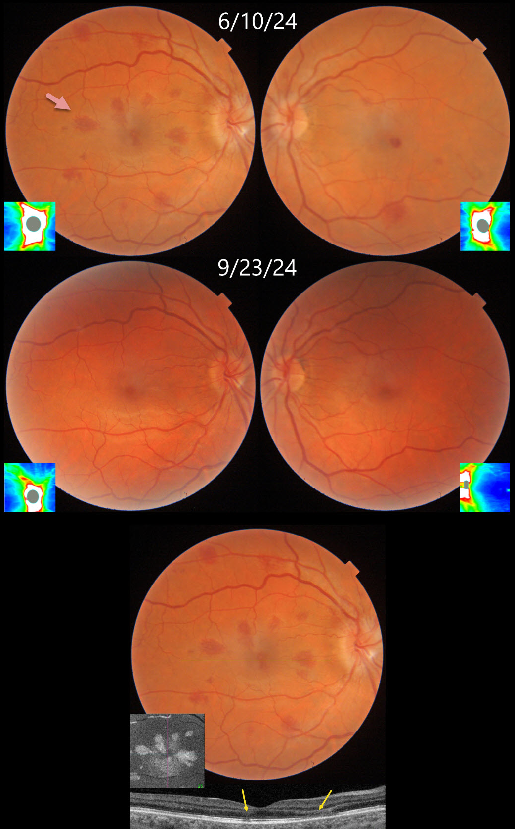
This 34YO female underwent uterine ablation 2 weeks earlier for heavy menstruation and severe secondary iron deficiency anemia. Although we don’t know what her hemoglobin level was prior to the procedure, it had risen to 10.7 postoperatively. Vision was 20/30 OD and 20/50 OS.
Triton color photography shows bilateral teardrop-shaped macular outer plexiform layer (OPL) hemorrhages, with greater involvement in her right eye. Numerous more peripheral deep and white-centered hemorrhages are also noted. Swept-source OCT confirms the location of the macular blood. En face OCT shows a more dramatic view of their petaloid shape. There is also mild optic nerve edema, especially in the right eye. The hemorrhages and nerve swelling mostly resolved with iron supplementation several months later.
Learning Points:
Anemia-related retinal findings most commonly include hemorrhages in the retina and sub-internal limiting membrane. Severe anemia is a rare cause for optic nerve swelling and should always be considered in patients with atypical idiopathic intracranial hypertension (Biousse et al, AJO 2003;135:437-446). The underlying pathogenesis of anemia-induced optic nerve swelling remains unknown.
Radiating deep macular hemorrhages are seen in multiple conditions, including Valsalva and trauma (Baumal et al, Br J Ophthalmol 2021;105:374-380). We have not previously seen these hemorrhages in isolated anemia. Their unique radiating appearance is caused by blood accumulating in Henle’s layer. Kon Graversen et al described hemorrhagic unilateral retinopathy, an idiopathic disorder with a similar appearance (Retina 2014;34:483-489).
RETINITIS PIGMENTOSA WITH PAPILLEDEMA
Tejaswita Verma and Manish Nagpal
Originally posted on @retina.rocks November 20, 2024
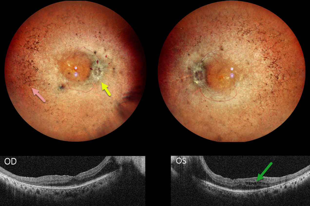
This 30YO male presented with 3 weeks of decreased vision. His father has a history of retinitis pigmentosa (RP). Vision was 20/30 OD and 20/80 OS.
Pseudocolor SLO imaging shows typical bilateral RP scarring with intraretinal pigment migration extending from the peripheral maculas into the retinal peripheries. Bilateral macular puckers and disc swelling with right peripapillary inner retinal fluid are also seen. OCT scanning confirms the epimacular membranes. Bilateral pericentral outer macular atrophy is noted, along with some cystic edema in the left inner and outer nuclear layers.
Learning Points:
RP is a clinically and genetically heterogeneous group of inherited retinal disorders. The disease can present sporadically or with any inheritance pattern (autosomal dominant, autosomal recessive, sex-linked, or mitochondrial). It is characterized by diffuse, progressive dysfunction of predominantly rod photoreceptors, with subsequent RPE degeneration and intraretinal pigment migration. Visual impairment usually presents as night blindness and progressive visual field loss.
Bilateral optic disc edema is not typical for RP. Unfortunately, we were unable to work up our patient for a secondary cause because he was lost to follow-up immediately.
ANEMIA
Anand Temkar and Manish Nagpal
Originally posted on @retina.rocks October 24, 2024
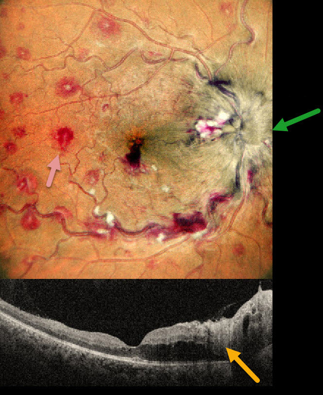
This 35YO male presented with 1 month of vision loss in his right eye. There was no past medical history. Vision was 20/40 OD and 20/20 in his normal OS.
MultiColor imaging shows a markedly swollen optic nerve with extensive multifocal deep, white-centered retinal hemorrhages. OCT shows marked mostly outer retinal edema extending from the nerve towards the macular center.
Extensive laboratory evaluation was negative except for a microcytic anemia with a hemoglobin of 2.9 g/dl. This was felt to be caused by an iron deficiency due to poor diet. Intravitreal anti-VEGF injections were recommended, along with oral prednisone for the optic nerve swelling. Unfortunately, he was immediately lost to follow-up.
Learning Points:
Anemia-related retinal findings most commonly include hemorrhages in the retina and sub-internal limiting membrane. Severe anemia is a rare cause for papilledema and should always be considered in patients with atypical idiopathic intracranial hypertension (Biousse et al, AJO 2003;135:437-446). The underlying pathogenesis of anemia-induced optic nerve swelling remains unknown. It is unclear why our patient had unilateral findings from a systemic disorder.
PAPILLEDEMA
Originally posted on @retina.rocks September 9, 2024
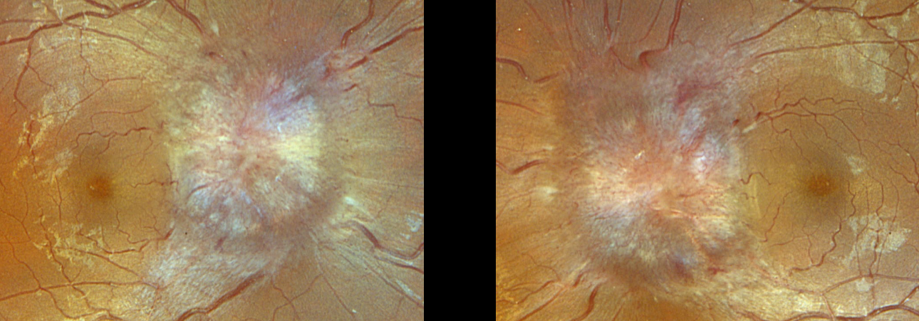
This 19YO obese female presented with 2 weeks of headaches, dizziness, and blurred vision. Vision was counting fingers OD and 20/400 OS. Blood pressure was 129/91.
Optos color RGB imaging shows severe bilateral optic disc swelling. The lumbar puncture opening pressure was extremely elevated at 43cm H2O, establishing the diagnosis of idiopathic intracranial hypertension (pseudotumor cerebri), and she was started on Diamox 500mg BID. Unfortunately, she was immediately lost to follow up.
LEUKEMIC OPTIC NERVE INFILTRATION WITH PSEUDOPAPILLEDEMA
Anjana Mirajkar and Manish Nagpal
Originally posted on @retina.rocks July 8, 2024
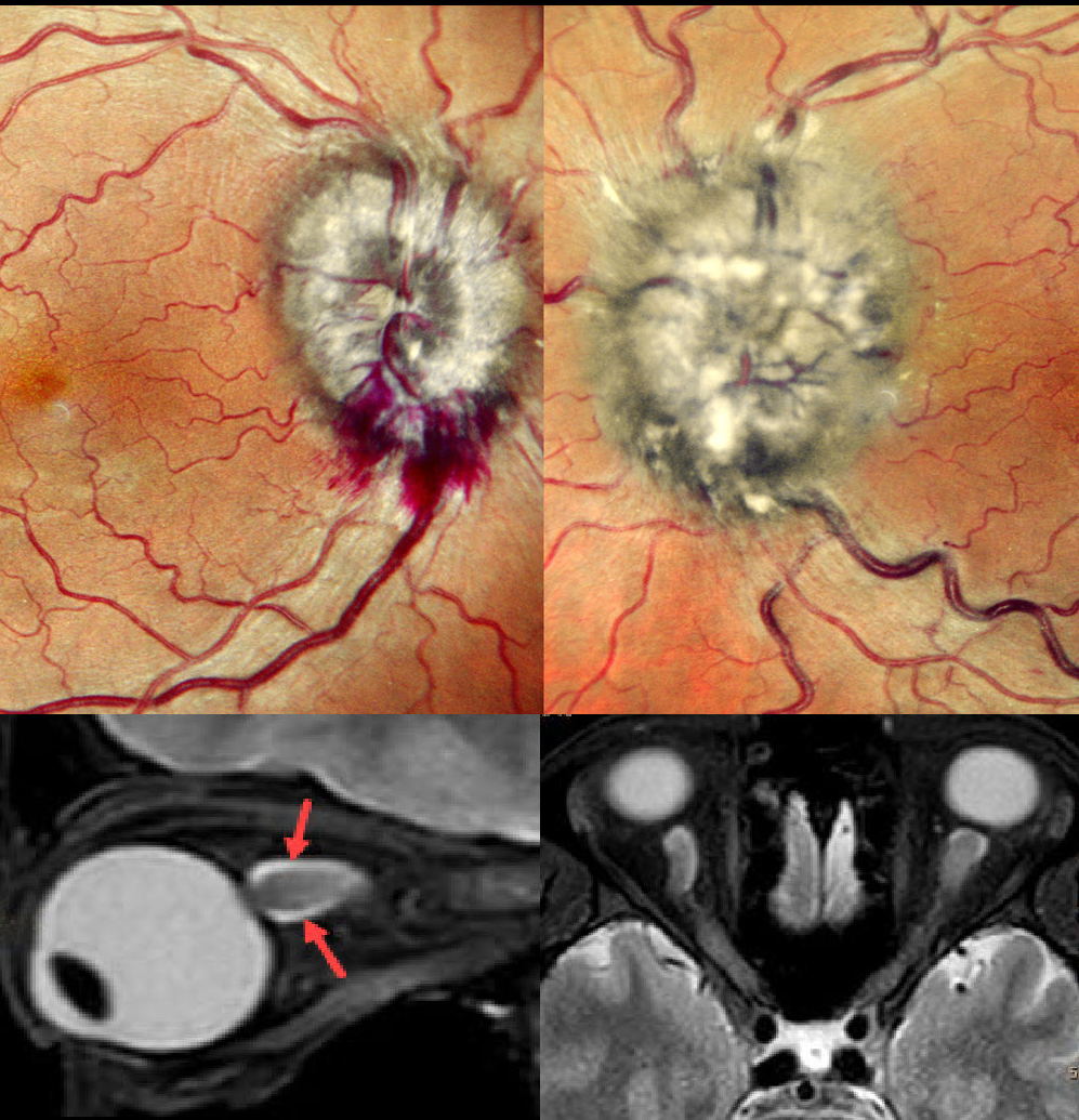
This 40YO male with a known 10-year history of chronic myeloid leukemia complained of vision loss in his left eye for 3 weeks. Vision was 20/20 OD and hand motion OS.
Fundus examination revealed marked bilateral optic nerve swelling, with the left eye greater than the right eye, and some nerve fiber layer hemorrhages along the right inferior disc margin. MRI scanning was consistent with leukemic infiltration of the optic chiasm and optic nerves. She was referred back to her oncologist.
Learning Points:
Leukemia with central nervous system involvement can rarely present with leukemic optic nerve infiltration. This can masquerade as papilledema (bilateral swollen nerves from elevated intracranial pressure) or nerve edema from inflammatory or infectious causes (Miller et al, Graefe’s 2021;259;1315-1322).
PAPILLEDEMA
Originally posted on @retina.rocks September 2, 2022
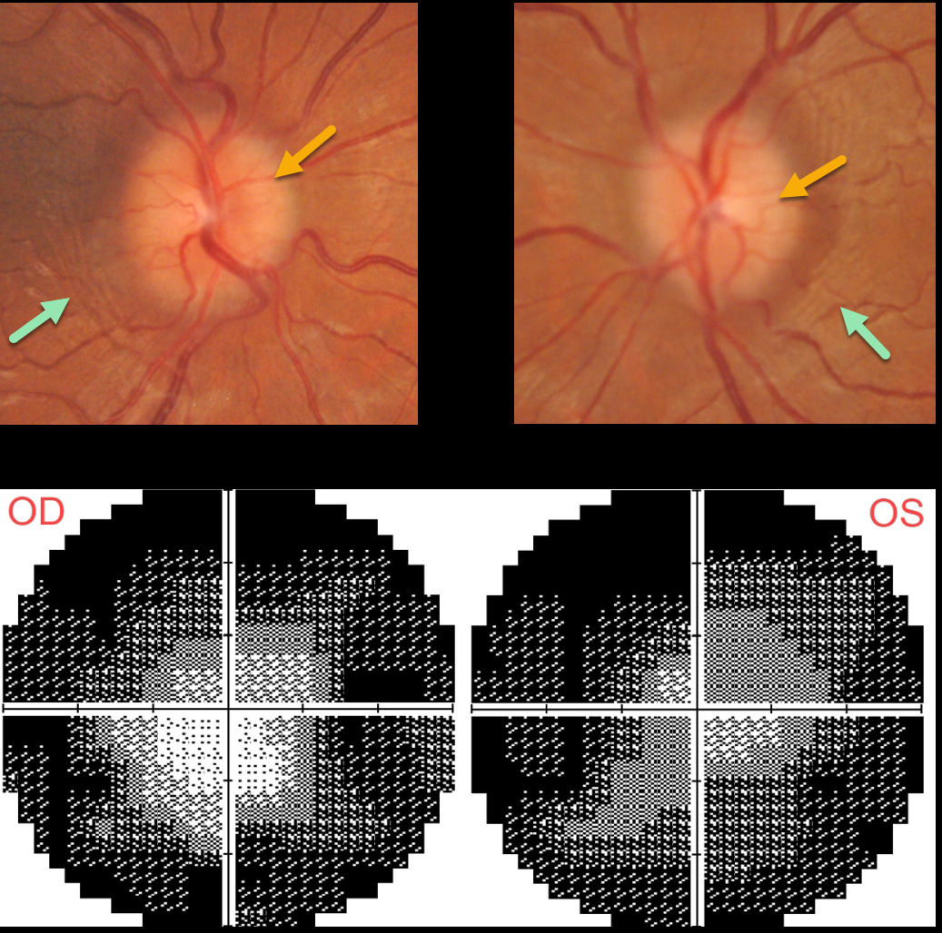
This 30YO female was diagnosed with idiopathic intracranial hypertension (pseudotumor cerebri) in 2020. Her initial lumbar puncture opening pressure was elevated at 25cm H2O.
She was lost to follow-up for about 2 years, when she presented with recurrent headaches, a ‘whooshing’ sound in her ears, and blurred vision. She was off of Topamax for about a year. Vision was 20/30 OD and 20/40 OS.
Both optic nerves are mildly edematous. The retinal nerve fiber layer (not shown) is somewhat thickened bilaterally. Despite the modest nerve edema, prominent temporal circumferential retinal folds (Paton’s folds) surround both nerves. Visual fields are severely constricted.
Learning Points:
In 1911 Paton and Holmes established that papilledema was due to elevated intracranial pressure and not inflammation as was previously thought (Brain 1911;33:389-432).
They also noted that nerve edema was associated with “a series of folds which run concentric with the edge of the disc” which they felt represented retinal folds.
Sibony and Kupersmith, correlating OCT with fundus photography, found that these folds represented a variety of structural stresses on the nerve and peripapillary tissues, including peripapillary wrinkles, inner retinal folds, choroidal folds and outer retinal folds/creases (Ophthalmology 2016;123:1397-1399).
PAPILLEDEMA
Originally posted on @retina.rocks March 18, 2022
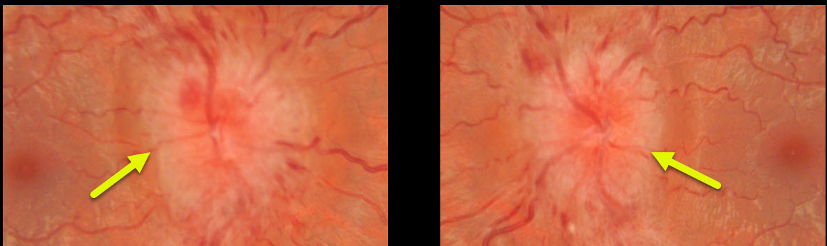
This 22YO female presented with severe bilateral optic nerve edema and enlarged blind spots. She reported recent weight gain following the placement of an IUD.
MRI and MRV scanning were normal. Lumbar puncture opening pressure was extremely elevated at 55cm H2O, establishing the diagnosis of idiopathic intracranial hypertension (pseudotumor cerebri).
She was evaluated by neurosurgery, and a lumboperitoneal shunt was placed, improving the patient’s symptoms and nerve appearance (not shown).
PAPILLEDEMA
Originally posted on @retina.rocks July 28, 2021
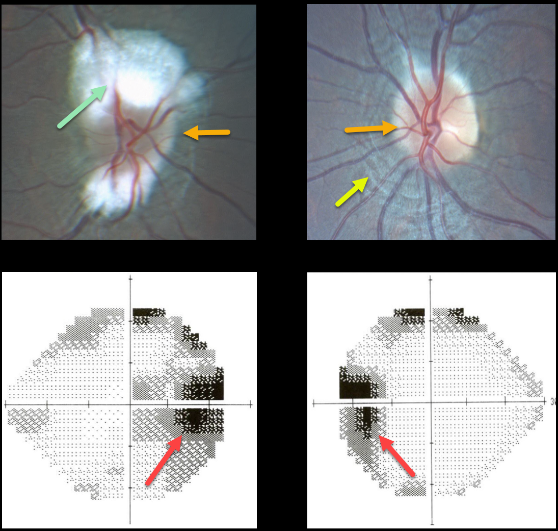
This 14YO presented with mild bilateral optic nerve edema and enlarged blind spots. Evaluation of the right nerve was difficult due to the myelinated nerve fiber layer.
There are prominent circumferential retinal lines (Paton’s folds) surrounding the left nerve.
MRI and MRV scanning were normal, and lumbar puncture opening pressure was elevated at 26mm H2O. The patient was diagnosed with idiopathic intracranial hypertension (pseudotumor cerebri).
Learning Points:
In 1911, Paton and Holmes established that papilledema was due to elevated intracranial pressure rather than inflammation, as previously thought (Brain 1911;33:389-432).
They also noted that nerve edema was associated with “a series of folds which run concentric with the edge of the disc,” which they felt represented retinal folds.
Sibony and Kupersmith, correlating OCT with fundus photography, found that these folds represented a variety of structural stresses on the nerve and peripapillary tissues, including peripapillary wrinkles, inner retinal folds, choroidal folds, and outer retinal folds/creases (Ophthalmology 2016;123:1397-1399).
PAPILLEDEMA
Originally posted on @retina.rocks June 29, 2021
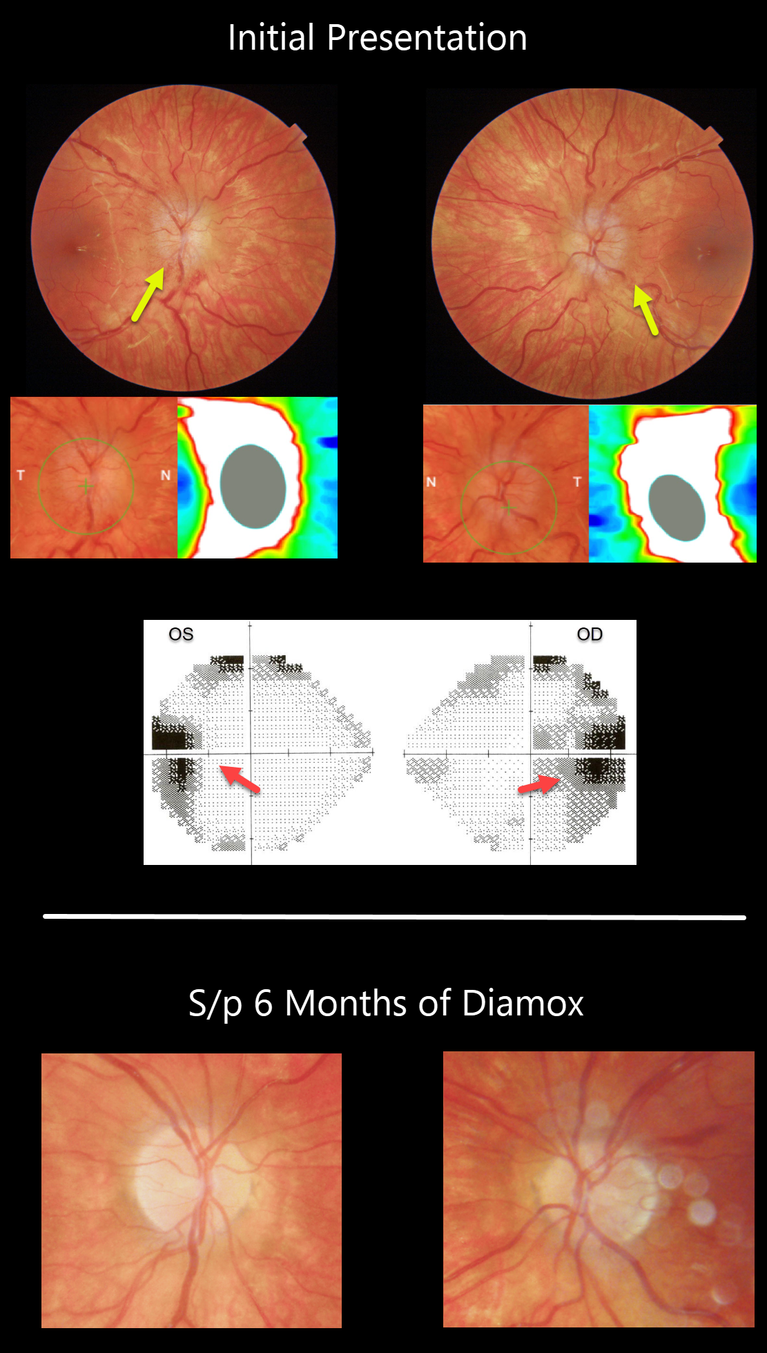
This 13YO girl presented with headaches and tinnitus with 20/25 vision OU. There was moderate bilateral optic nerve swelling, beautifully imaged with Triton swept-source OCT. Visual fields showed enlarged blind spots.
MRI scanning was normal, and her lumbar puncture had a severely elevated opening pressure of 49mm H2O, establishing the diagnosis of idiopathic intracranial hypertension (IIH).
After six weeks of oral Diamox, her headaches and tinnitus had improved significantly, and the optic nerve edema had virtually resolved bilaterally.
Learning Points:
Remember that papilledema is not synonymous with optic nerve swelling. Papilledema is defined as bilateral optic nerve swelling due to elevated intracranial pressure.
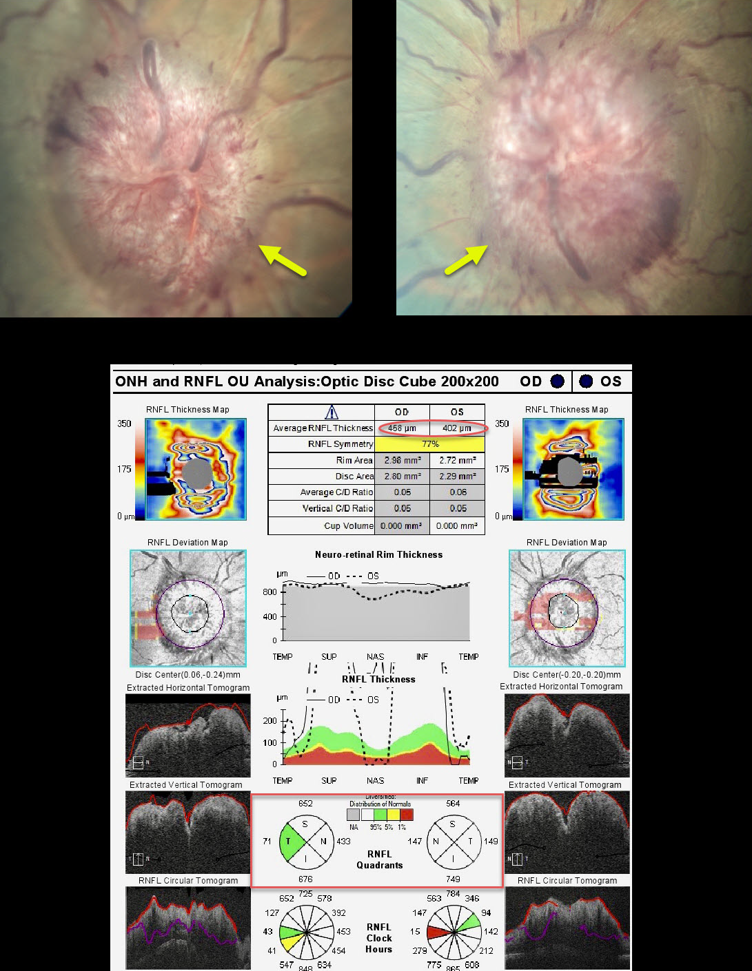
PAPILLEDEMA
Originally posted on @retina.rocks February 23, 2021
This patient presented with diplopia from bilateral esotropia and reduced abduction in each eye due to sixth nerve palsies. There were no headaches or visual disturbances.
Her nerves showed severe papilledema with severe retinal nerve fiber layer thickening on OCT.
An MRI showed no mass lesions, but her lumbar puncture opening pressure was 41 cm H2O.
Despite an aggressive weight loss regimen and oral acetazolamide, her intracranial pressure remained elevated and required a ventriculoperitoneal shunt.
Learning Points:
Idiopathic intracranial hypertension (pseudotumor cerebri) can occasionally cause a downward displacement of the brainstem, stretching the sixth nerve as it exits the pons towards Dorello’s canal at the apex of the petrous bone.
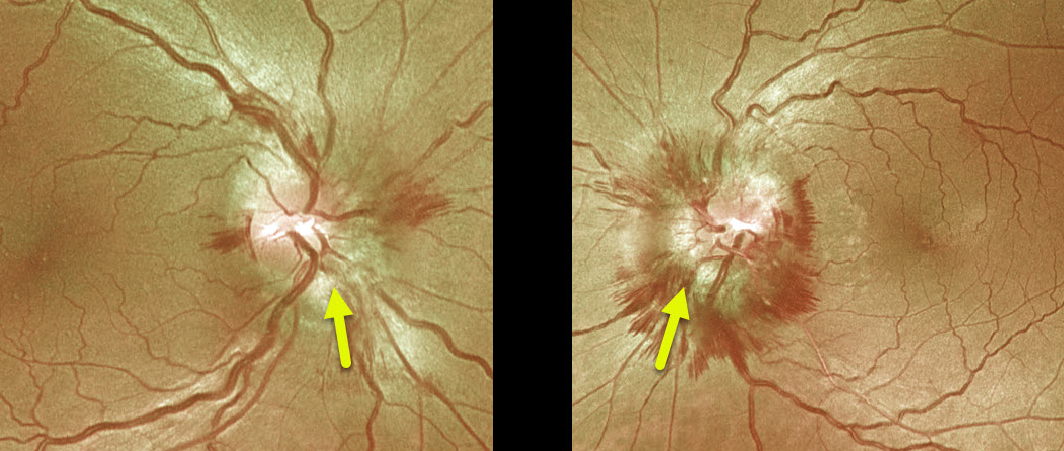
PAPILLEDEMA
Originally posted on @retina.rocks September 17, 2020
This is a beautiful example of papilledema in a patient with idiopathic intracranial hypertension (pseudotumor cerebri).
This patient had an opening lumbar puncture pressure of 28 cm H2O.
Note the absence of lipid, which, for some reason, is much more common in patients with malignant hypertension or neuroretinitis.
Learning Points:
Remember that papilledema is not synonymous with optic nerve swelling. Papilledema is defined as bilateral optic nerve swelling due to elevated intracranial pressure.
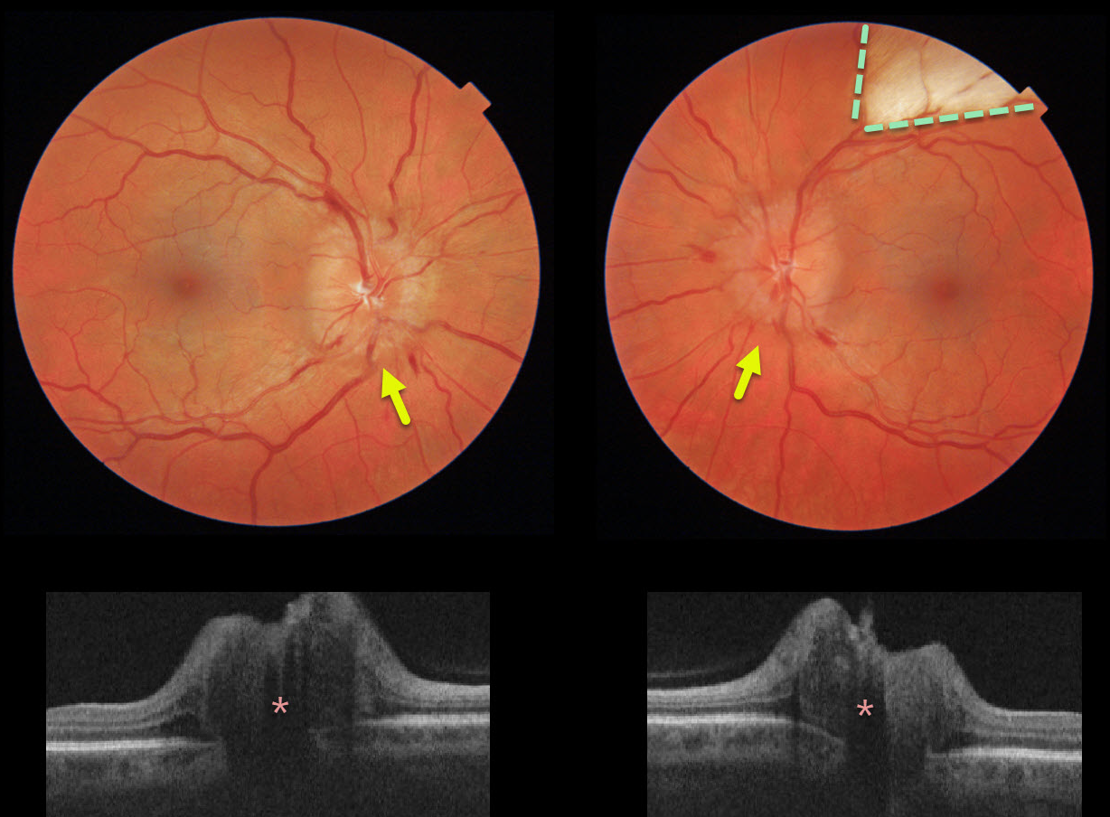
PAPILLEDEMA
Originally posted on @retina.rocks June 26, 2020
This healthy 40yo female presented with chronic headaches. She was on no systemic medications, and her blood pressure was normal. Her nerves were swollen bilaterally, and a myelinated nerve fiber layer was also noted in her left eye.
The retinal nerve fiber layer on OCT was severely thickened bilaterally.
Learning Points:
The diagnosis of idiopathic intracranial hypertension (pseudotumor cerebri) was made after an MRI scan was normal and a lumbar puncture revealed a markedly elevated opening pressure.
She is currently being successfully managed with oral acetazolamide.
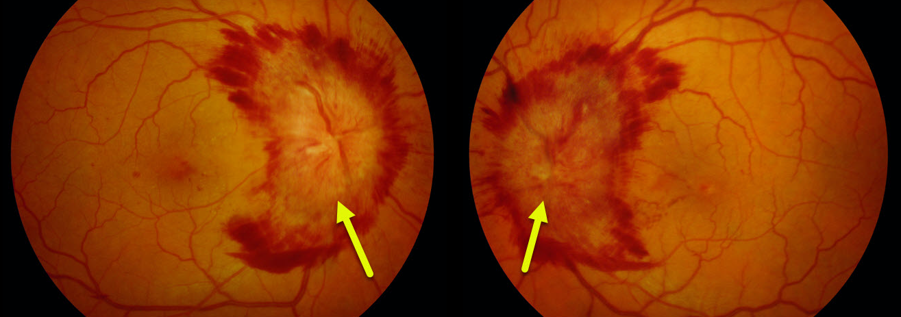
PAPILLEDEMA
Originally posted on @retina.rocks February 3, 2020
This patient had papilledema with severe disc hemorhages. MRI detected a compressive meningioma as the cause for the elevated intracranial pressure.
Learning Points:
Optic nerve swelling has numerous causes, including neurologic (optic neuritis and elevated intracranial pressure) and retinal (retinal vein occlusion, neuroretinitis, sarcoidosis, Harada’s) causes.
Bilateral optic nerve swelling due to elevated intracranial pressure is known as papilledema.
Neurologic imaging is important to establish the correct diagnosis.


