

Papillitis
<< Back to Cases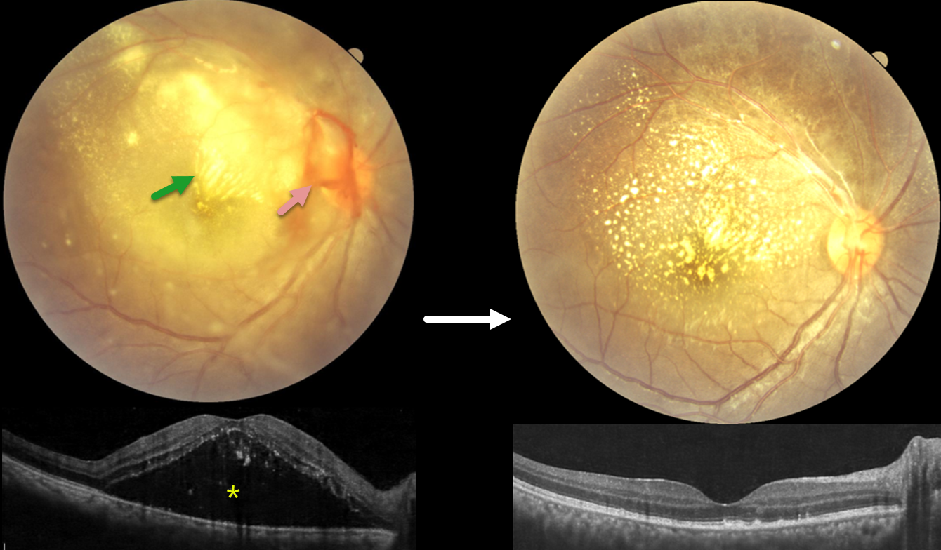
SCRUB TYPHUS POST-FEVER RETINITIS
Anand Temkar, Surendra Pal, Vasumathi Vijay, and Manorama Baheti
Originally posted on @retina.rocks June 3, 2026
This 28YO female presented with 4 days of blurred vision in her right eye. For the past 2 weeks, she has complained of generalized weakness, malaise, and body pain. Vision was counting fingers OD and 20/20 in her normal OS.
Fundus photography shows variable, dense subretinal lipid, particularly confluent in the superonasal macula. There is some preretinal blood around the nerve, and the retina is somewhat yellow and opaque temporally and inferiorly. Patches of inner retinal opacification are noted outside the macula superiorly. OCT shows hyperreflective dots from lipid exudate in the outer retina, with a large serous detachment. Laboratory testing, including CBC, ESR, syphilis, TB, HIV, TORCH profile, and chest X-ray, was negative. We started a 1-week tapering course of oral prednisone 40mg and doxycycline 100mg BID. Three days after our initial examination, she mentioned that she had 2 weeks of mild intermittent fever. Weil-Felix testing was positive, and she was subsequently diagnosed with scrub typhus and asked to continue the doxycycline for a full 2-week course.
The clinical picture gradually improved, and about 6 weeks following presentation, vision improved to 20/40 despite significant residual lipid. OCT scanning through the macular center was mostly normalized. We continue to follow her closely.
Learning Points:
Rickettsial infections cause posterior segment involvement in a substantial proportion of patients, often asymptomatic despite significant fundoscopic findings. The most common rickettsial organisms causing retinal disease include Rickettsia rickettsii (Rocky Mountain spotted fever), Rickettsia conorii (Mediterranean spotted fever), and Rickettsia typhi (murine typhus).
Scrub typhus, caused by Orientia tsutsugamushi and transmitted by a larval mite (chigger) bite, has ocular involvement in about 20% of patients. Retinal findings include retinal hemorrhages, branch retinal vein occlusion, retinal vasculitis/retinitis, and papillitis (Ganekal et al, Ind J Ophthalmol 2021;69:1167-1171).
The ocular and systemic findings of vasculitis and perivasculitis result from both direct endothelial cell destruction by the organism and exaggerated immune responses. Doxycycline is the drug of choice; treatment should last at least 7 days and continue for at least 3 days after fever has subsided and clinical improvement is evident. Visual prognosis is generally favorable with appropriate treatment.
CMV PAPILLITIS AND RETINITIS
Akansha Sharma and Manish Nagpal
Originally posted on @retina.rocks September 18, 2025
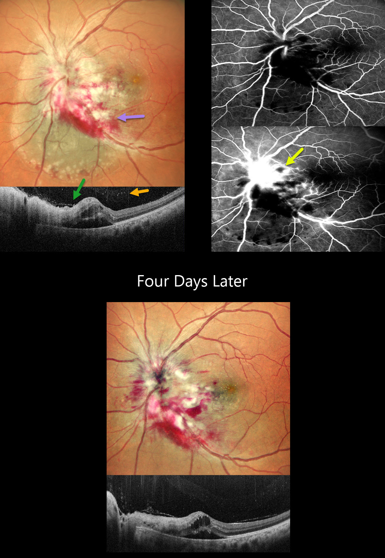
This 42YO female with a known history of HIV infection presented with 1 month of vision loss in her left eye. Her last CD4 count was 38 despite being compliant with HAART therapy. Her husband was HIV positive. Vision was 20/20 in her normal OD and 20/400 in OS.
Multicolor SLO imaging shows active cytomegalovirus (CMV) retinitis involving the nerve, which extends nasally and inferotemporally. Subretinal fluid is noted clinically and on OCT. OCT also shows full-thickness disorganization and hyperreflectivity, along with overlying vitreous cells. Fluorescein angiography shows early blockage with late disc and retinal leakage.
Oral valganciclovir and biweekly intravitreal injections were started. Four days later, vision improved to 20/90 with improvement in the papilledema and retinitis.
Learning Points:
CMV retinitis develops as a reactivation of latent CMV in immunosuppressed individuals. Before effective antiretroviral treatment emerged in the mid to late 1990’s, CMV retinitis developed in up to 40% of HIV/AIDS patients, often within the last 6 months of life. Treatment includes a combination of systemic and intravitreal medications, including ganciclovir, foscarnet, and cidofovir. Rhegmatogenous retinal detachment is a common late sequela and requires vitrectomy with silicone oil.
RADIATION RETINOPATHY AND PAPILLOPATHY
Mattie Adams
Originally posted on @retina.rocks May 27, 2025
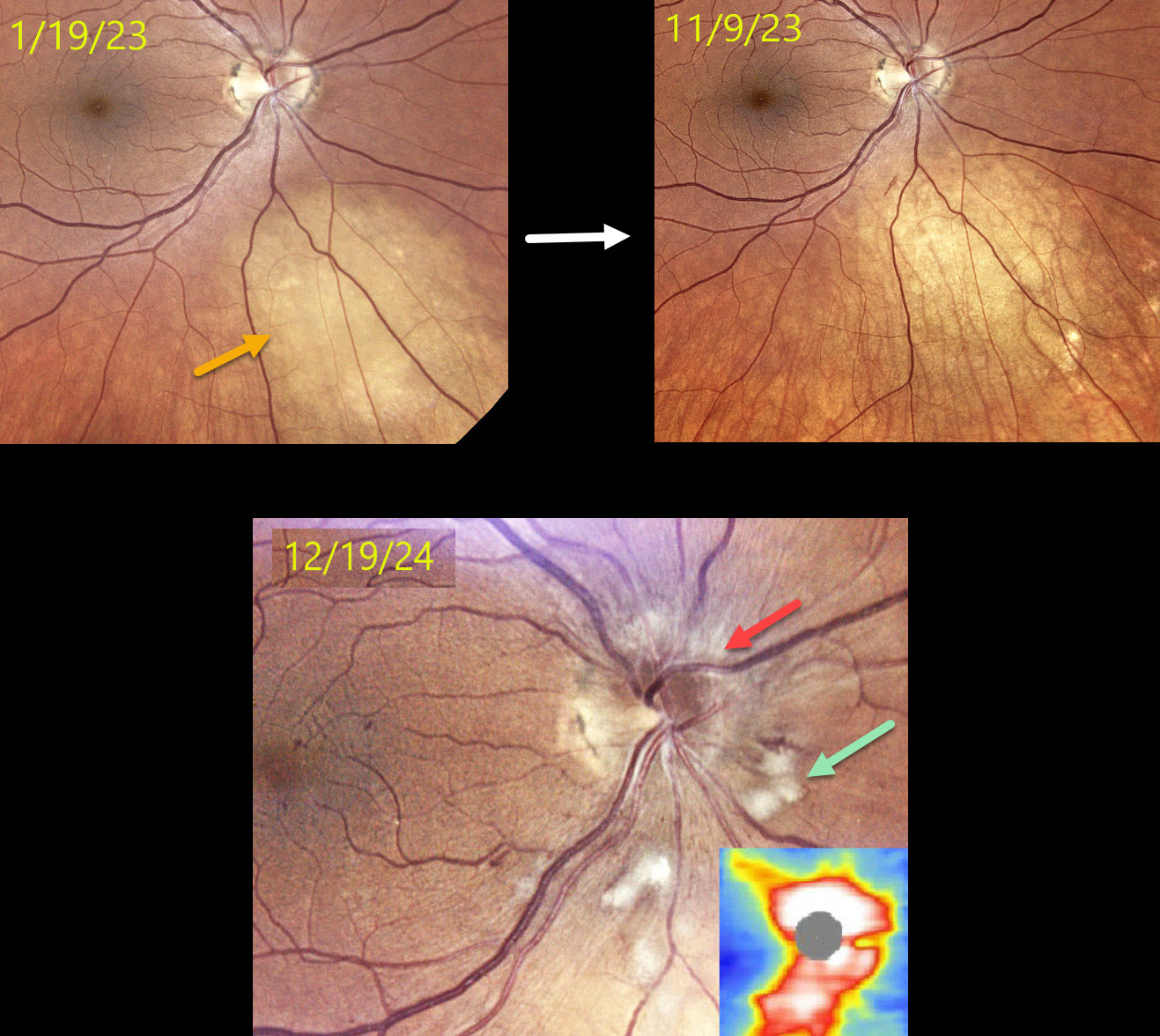
This 52YO female presented on 1/19/23 with a history of metastatic breast cancer and an asymptomatic uveal metastasis in her right eye. The lesion completely regressed following external beam radiation.
When examined on 12/19/24, she remained visually asymptomatic with 20/60 vision. Color photography shows a new, mildly swollen nerve, which is confirmed on OCT. Scattered nerve fiber layer infarcts (cotton-wool spots, CWS) and some nasal macular telangiectasia are also noted.
Learning Points:
Radiation retinopathy usually occurs 3 months to 3 years after external-beam radiation or plaque radiotherapy. The ocular findings closely resemble those seen with diabetic retinopathy. Radiation optic neuropathy (Carey et al, Br J Ophthalmol 2023;107:743-749) can also develop.
Since our patient was completely asymptomatic without macular edema, we recommended close observation. We will begin intravitreal anti-VEGF therapy if she develops macular edema or worsening nerve findings.
SYPHILITIC PAPILLITIS
Navneet Mehrotra and Tamanna Patel
Originally posted on @retina.rocks March 12, 2025
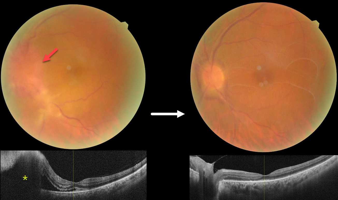
This 52YO male presented with 3 weeks of vision loss in his left eye. He has a history of diabetes, hypertension, and HIV. He was diagnosed elsewhere with optic neuritis and started on intravenous methylprednisolone with no improvement. Vision was 20/30 in his normal OD and counting fingers OS. There was a mild nongranulomatous anterior uveitis OS on slit lamp examination.
Color photography shows a markedly swollen nerve with possible retinitis extending inferotemporally. OCT scanning confirms the disc elevation with subretinal fluid extending beneath the nasal macula.
On further questioning, there was a recent history of a genital chancre, and the treponema pallidum hemagglutination assay was positive for syphilis. Following a 2-week course of parenteral penicillin, vision improved to 20/120 with marked improvement in the posterior segment findings. Unusual premacular vitreous membranes are noted.
Learning Points:
Ocular manifestations of syphilis vary widely and include virtually all variations of anterior and posterior segment inflammation. In our experience, placoid syphilis is the most common posterior segment presentation, although a recent systematic review and meta-analysis found papillitis to be the most reported finding in syphilitic uveitis (Zhang et al, J of Ophthalmology 2017;1; 6594849). Papillitis can be isolated or associated with other findings.
Any patient who tests positive for syphilis should always be checked for HIV since coinfection, as in our patient, is common.
TUBERCULAR SERPIGINOUS-LIKE CHOROIDITIS
Originally posted on @retina.rocks March 11, 2025
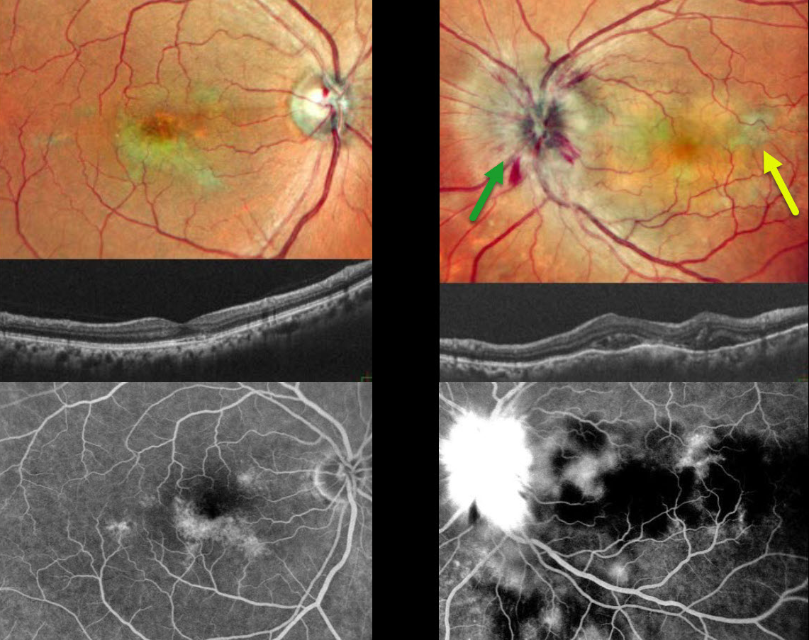
This 54YO male presented with 2 weeks of vision loss in his left eye. He was diagnosed elsewhere with papilledema, and brain MRI scanning was normal. Vision was 20/20 in his asymptomatic OD and counting fingers OS. Pseudocolor SLO imaging of the OS shows marked disc swelling with radiating hemorrhages in the nerve fiber layer. Some deep, hypopigmented retinal lesions are also seen.
OCT scanning shows irregular choroidal thickening vs choroidal folds with overlying pockets of variably reflective subretinal fluid. The right eye shows multifocal serpiginous-like areas of chorioretinal scarring with variable outer retinal thinning on OCT.
On fluorescein angiography, the macular lesions stain OD. The left nerve shows profound leakage, and the choroidal lesions show variable blockage and staining.
We suspected tubercular serpiginous-like choroiditis OD with acute tubercular papillitis and choroiditis OS. Chest X-ray showed a mass pressing on the trachea. Chest CT confirmed the mass, and subsequent biopsy revealed granulomatous thyroiditis. His Mantoux skin test was positive. We advised emergent Infectious disease consultation regarding starting anti-tubercular therapy. Unfortunately, he was immediately lost to follow-up.
Learning Points:
Although tuberculosis most commonly presents with pulmonary involvement, extrapulmonary sites can include the gastrointestinal, skin, cardiovascular, genitourinary, and central nervous systems, as well as the eyes. Ocular involvement, like syphilis, can mimic virtually any type of uveitis, including anterior, intermediate, posterior, and panuveitis, retinitis, and retinal vasculitis, neuroretinitis, optic neuropathy, choroidal granuloma, choroiditis, and scleritis.
Tubercular serpiginous-like choroiditis mimics serpiginous but affects younger patients with more multifocal and peripheral recurrences and progression if untreated (Agarwal et al, AJO 2020;220:160-169). Although the exact mechanism is not yet clear, a direct or indirect infectious trigger by the Mycobacterium tuberculosis is thought to cause the choroiditis. Its origin is probably immunogenic since it seems to respond to corticosteroids and other immunosuppressants.
OPTIC NERVE PAPILLITIS
Originally posted on @retina.rocks June 18, 2021
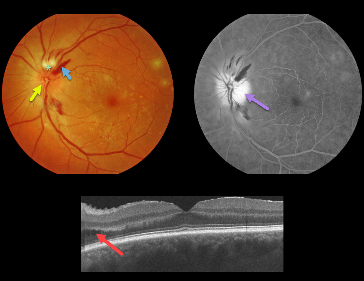
This 66YO diabetic male presented with an asymptomatic unilateral swollen nerve, peripapillary hemorrhages, and nerve fiber layer ischemia. Vision was 20/25, and there was no afferent pupillary defect (APD).
OCT scanning shows some mild cystic fluid in the peripapillary inner and outer nuclear layer. Fluorescein angiography shows diffuse optic nerve leakage.
Blood pressure was normal. He denied exposure to deer ticks or cat scratches, and the MRI was normal. The funduscopic findings spontaneously normalized within a few months.
Learning Points:
Diabetic papillopathy, often a diagnosis of exclusion, is most commonly seen in type 2 diabetics. It has a good visual prognosis with observation alone. A small physiologic cup may predispose to this condition (see Regillo et al, Arch Ophthlamol 1995;113:889-895).
Unlike ischemic optic neuropathy, vision is usually quite good, there is usually no APD, the nerve swelling is more diffuse than sectorial, and there is no altitudinal field defect.
The differential for unilateral acute optic nerve swelling is broad, including diabetes, ischemic (anterior ischemic optic neuropathy), and inflammatory causes (optic neuritis, Bartonella, syphilis, sarcoidosis).
Papillitis must be differentiated from papilledema, which is bilateral optic nerve swelling caused by elevated intracranial pressure.
We have been burned more than once by unilateral nerve edema being the only presenting sign of severe hypertension. Although nerve swelling is usually bilateral in hypertensive retinopathy, we always check blood pressure in all patients presenting with unilateral or bilateral optic nerve swelling.


