

Paracentral Acute Middle Maculopathy (PAMM)
<< Back to Cases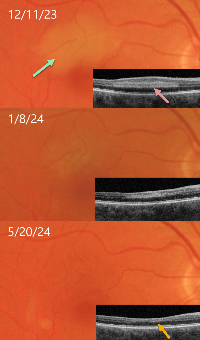
PARACENTRAL ACUTE MIDDLE MACULOPATHY (PAMM)
Originally posted on @retina.rocks September 11, 2024
This 68YO male presented on 12/11/23 with a several-day history of a paracentral scotoma in his right eye. Vision was 20/30 OD and 20/25 in his normal OS.
Triton color imaging shows an area of retinal opacification in the superior macula. This area is hyperreflective on swept-source OCT. The lesion gradually resolved over several months. When last examined on 5/20/24, the funduscopic appearance normalized. The area of prior mid-retinal hyperreflectivity was replaced by retinal thinning, with the inner plexiform/inner nuclear/outer plexiform layers compressed into a single hyperreflective line.
Learning Points:
Paracentral acute middle maculopathy (PAMM) is a phenotype caused by acute mid-retinal ischemia due to obstruction of the deep and intermediate capillary plexi. PAMM is associated with an increasing constellation of conditions, including retinal vein and artery occlusions. It is unclear if patients with a pure PAMM phenotype require an immediate stroke protocol referral (Bousquet et al, Retina 2023;43:1827-1832).
In our practice, we have seen several elderly patients who developed a complete CRAO within days of presenting with PAMM, so we do send these patients for emergent evaluation. We do not believe that typical PAMM in younger patients or found as a secondary finding in other ocular conditions requires a stroke workup. Our patient’s medical evaluation was fortunately negative.
PARACENTRAL ACUTE MIDDLE MACULOPATHY (PAMM)
Originally posted on @retina.rocks April 25, 2023
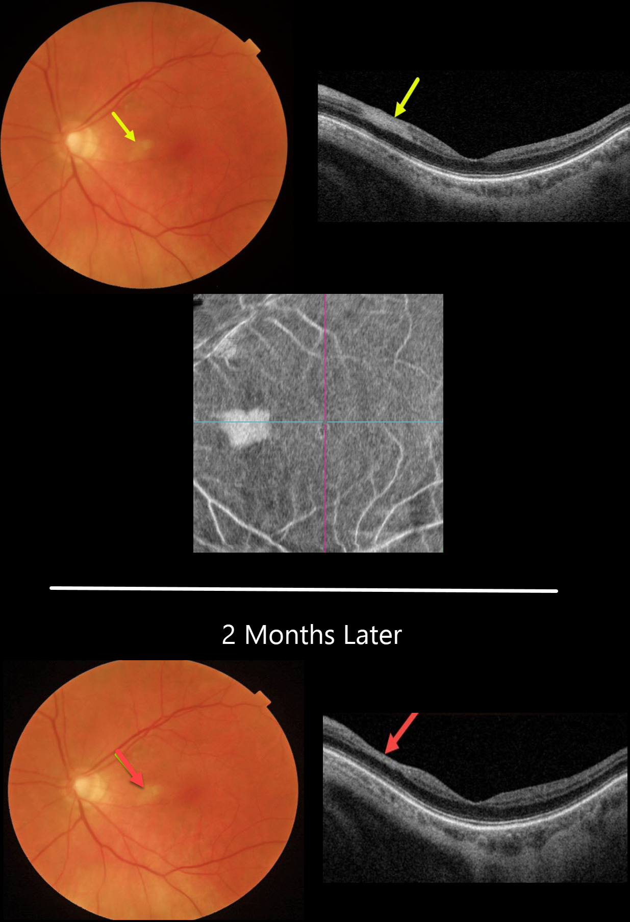
This 81YO female presented with 1 week of a temporal paracentral scotoma in her left eye. Vision was 20/50 OS.
Color imaging shows an area of inner retinal opacification in the nasal macula. This area is hyperreflective on swept-source OCT. En face imaging shows another perspective for this lesion.
Two months later, the retinal opacification was smaller with a small area of secondary inner retinal thinning.
Due to our patient’s age and somewhat atypical appearance of the PAMM lesion, which involved both the inner and middle retinal layers, she was immediately referred to the nearest stroke center.
This revealed a critical ipsilateral internal carotid artery stenosis, which was successfully repaired with emergent surgery.
Learning Points:
Paracentral acute macular maculopathy (PAMM) is a phenotype caused by acute mid-retinal ischemia due to obstruction of the deep and intermediate capillary plexi. PAMM is associated with an increasing constellation of conditions, including retinal vein and artery occlusions. It is unclear if patients with a pure PAMM phenotype require an immediate stroke protocol referral.
In our practice, we have seen several elderly patients who developed a complete CRAO within days of presenting with PAMM, so we do send these patients for emergent evaluation. We do not believe that typical PAMM in younger patients or found as a secondary finding in other ocular conditions requires a stroke workup.
CRAO
Ryan Kern
Originally posted on @retina.rocks January 10, 2023
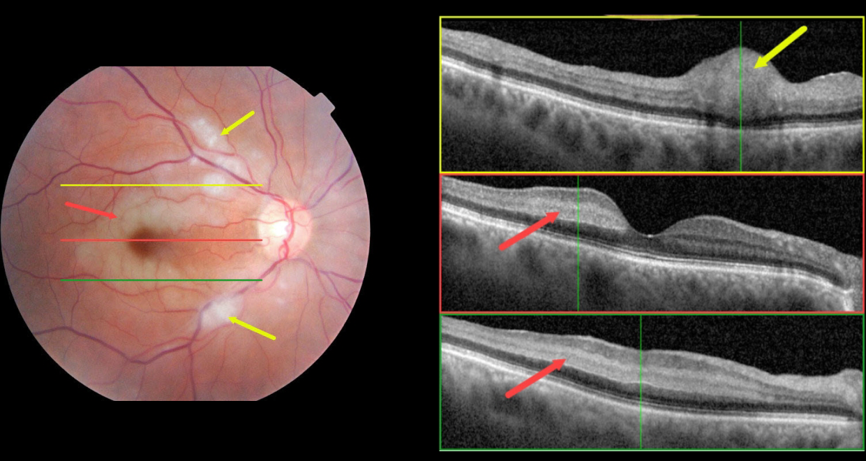
This 70 YO female presented with acute vision loss in her right eye starting 1 day earlier. Vision was 20/70 OD and 20/25 in her normal left eye.
Color fundus photography shows an incomplete central retinal artery occlusion (CRAO) with focal areas of acute inner retinal infarcts along with paracentral areas of deeper retinal opacification.
OCT scanning through the superior macula shows a focal area of retinal opacification and thickening that extends from the outer plexiform layer to the internal limiting membrane. Scans of the central and inferior macula show hyperreflectivity involving the middle retinal layers (outer plexiform, inner nuclear, and inner plexiform).
Blood pressure was 183/110. Emergent stroke evaluation was fortunately negative. Her blood pressure was stabilized, and baby aspirin was started. Visual acuity improved to 20/30 one month later.
This case was submitted by Ryan Kern.
Learning Points:
Paracentral acute macular maculopathy (PAMM) is a phenotype caused by acute mid-retinal ischemia due to obstruction of the deep (DCP) and intermediate (ICP) capillary plexi. PAMM is associated with an increasing constellation of conditions, including retinal vein and artery occlusions.
It is unclear if patients with a pure PAMM phenotype require an immediate stroke protocol referral. In our practice, we have seen several elderly patients who developed a complete CRAO within days of presenting with PAMM, so we do send these patients for emergent evaluation. We do not believe that typical PAMM in younger patients or found as a secondary finding in other ocular conditions requires a stroke workup.
Our patient’s fundus appearance has more of a Purtscher ’s-like appearance with focal areas of cotton wool spot-like inner retinal ischemia (although on OCT these involved the middle retinal layers as well) and mid-retinal ischemia.
However, given her age and history, acute onset, and unilateral presentation, her diagnosis is most consistent with an incomplete CRAO.
For a great recent PAMM review, see Scharf et al, Progress in Retinal and Eye Research 2021;81;100884.
CRVO + RAO + PAMM
Originally posted on @retina.rocks March 4, 2022
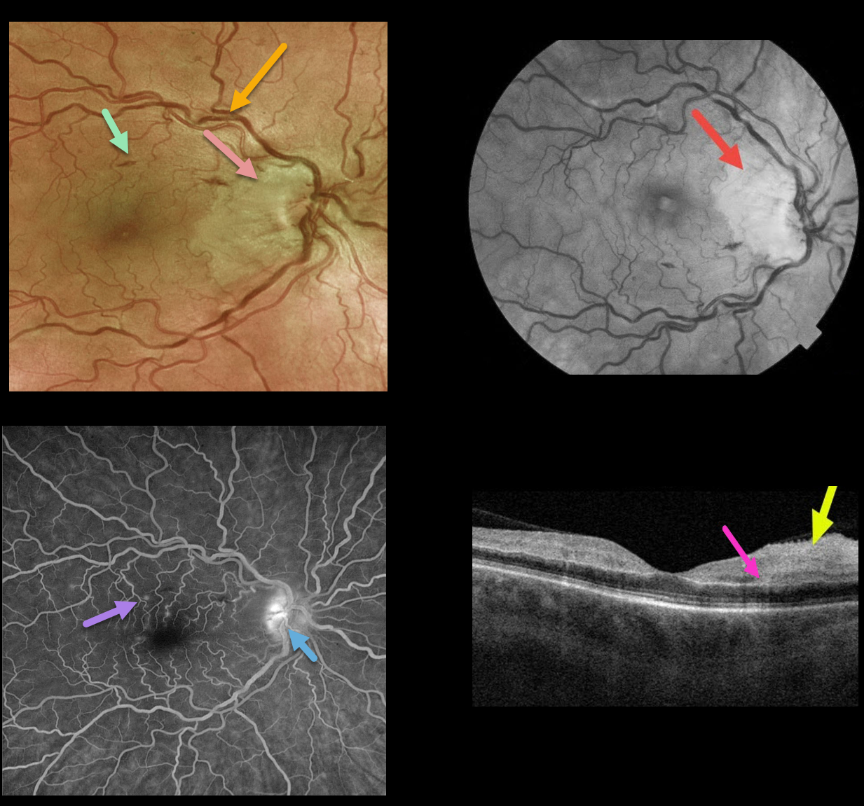
This 50YO female presented with counting finger vision in her right eye due to an acute central retinal vein occlusion (CRVO) with a nasal macular retinal artery occlusion (RAO).
Optos fundus photography shows retinal vascular tortuosity, mild retinal hemorrhages, a mildly swollen nerve, and white ischemic nasal macular retina. The ischemic retinal changes are most apparent on the red-free photo.
Fluorescein angiography (FA) shows some mild focal areas of macular leakage with late staining of the temporal disc.
OCT shows areas of inner retinal edema and hyperreflectivity. There is also a small area of mid-retinal hyperreflectivity consistent with paracentral acute middle maculopathy (PAMM).
Learning Points:
Cilioretinal artery occlusion is usually non-embolic and seen in association with giant cell arteritis, secondary to a CRVO, or as an isolated event. The occlusion may be due in part to arterial compression from a swollen nerve.
Pichi et al felt that CRVO PAMM-type lesions, as seen in our patient, are due to hypoperfusion from increased downstream intraluminal pressure (Br J Ophthalmol 2019;103:1137-1145).
We couldn’t find a definite cilioretinal artery in our patient, although the ischemia was within the distribution of one if one were present. Interestingly, the nasal macular capillaries appeared perfused on FA. However, FA captures just the inner retinal capillary plexus.
Although the OCT shows evidence of acute inner capillary plexus ischemia, the angiographically perfused inner retina supports a more PAMM-type mechanism in our case.
PARACENTRAL ACUTE MIDDLE MACULOPATHY (PAMM)
Gehad Hassan Youssef
Originally posted on @retina.rocks February 10, 2022
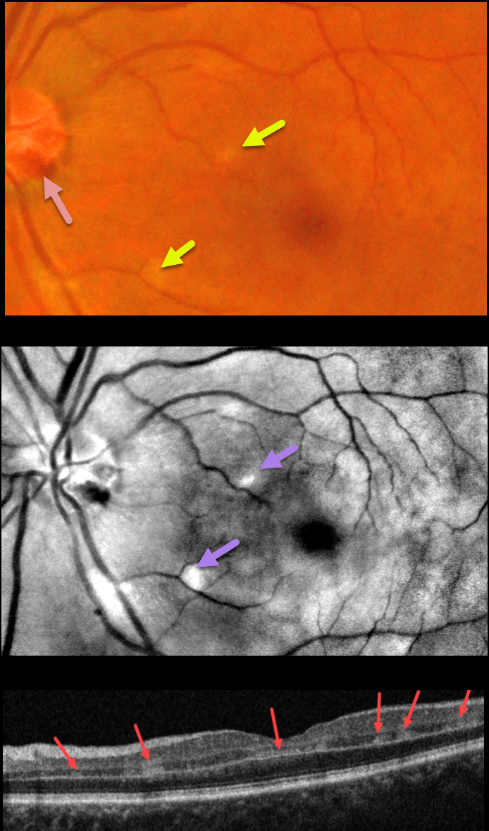
This 57YO female presented with sudden painless vision loss in her left eye. There was no significant past ocular or medical history. Vision was 20/20 in her normal right eye and 20/130 in her left eye.
Fundus photography shows several small hemorrhages along the temporal disc margin and faint patches of scattered whitish retinal opacification, which are much more pronounced in a perivenular distribution in the red-free image.
OCT B-scan shows multiple patches of focal hyperreflectivity involving the inner plexiform, inner nuclear, and outer plexiform layers.
Learning Points:
Paracentral acute middle maculopathy (PAMM) is a recently described phenotype that consists of acute mid-retinal ischemia, occurring as an idiopathic entity or in other ocular disorders, including Purtscher’s, diabetic retinopathy, retinal vascular occlusions, and acute macular neuroretinopathy.
For a great recent review, see Scharf et al, Progress in Retinal and Eye Research 2021;81;100884.
Perivenular PAMM may be the only presenting sign for an incomplete central retinal artery occlusion or mild central retinal vein occlusion (see Zhao et al, AJO 2022;234:15-19). Our patient may indeed have a very mild CRVO, given the optic nerve hemorrhages.
PARACENTRAL ACUTE MIDDLE MACULOPATHY (PAMM)
Lovro Soco
Originally posted on @retina.rocks October 1, 2021
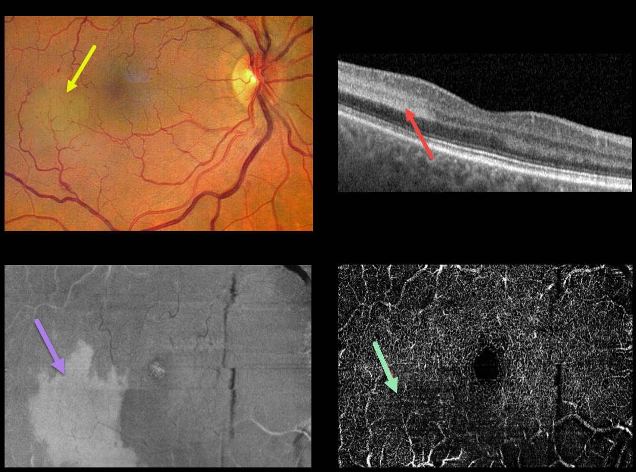
This 63YO patient complained of a recent scotoma in the mid-periphery of her right eye. Vision was 20/20 OU. She gave a history of hypertension with a recent elevated BP of 200/110. She denied a history of diabetes.
Fundus photography shows a patch of deep retinal whitening in the temporal macula. There were a few scattered dot and blot retinal hemorrhages in the right eye, which may have been related to her recently elevated blood pressure.
OCT shows a well-demarcated hyperreflective region spanning the inner plexiform, inner nuclear, and outer plexiform layers. Wide-field en face OCT shows a placoid area of hyperreflectivity within this region. Wide-field OCT angiography of the deep capillary plexus shows a relative flow defect within this area.
Learning Points:
Paracentral acute middle maculopathy (PAMM) is a recently-described phenotype. It consists of acute mid-retinal ischemia found as an idiopathic entity or in other ocular disorders, including Purtscher’s, diabetic retinopathy, retinal artery and retinal vein occlusions, and acute macular neuroretinopathy.
For a great recent review, see Scharf et al, Progress in Retinal and Eye Research 2021;81;100884.
PARACENTRAL ACUTE MIDDLE MACULOPATHY (PAMM)
Originally posted on @retina.rocks February 3, 2021
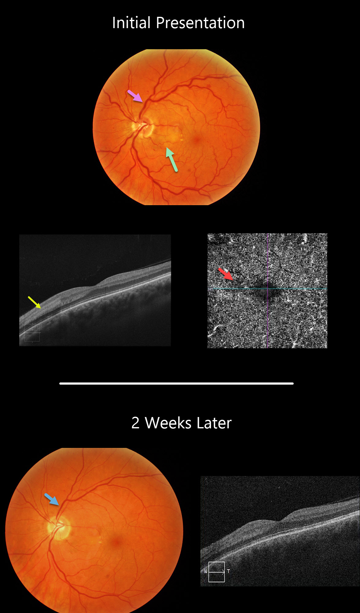
This 45YO female presented with 20/40 vision in her left eye with an acute negative scotoma temporal to her visual axis. This corresponded with a focal area of retinal whitening in the nasal macula.
This appeared as hyperreflectivity of the inner plexiform, inner nuclear, and outer plexiform layers on OCT and a relative flow void on OCT angiography.
Two weeks later, the retina and OCT findings improved, and vision was 20/30. This may have represented a mild, spontaneously resolving central retinal vein occlusion, as the initial fundus photo shows very mild, scattered retinal hemorrhages with dilated veins, and the venous caliber normalized at her exam 2 weeks later.
Learning Points:
Paracentral acute maculopathy (PAMM) is a recently described phenotype.
It consists of acute mid-retinal ischemia found as an idiopathic entity or in other ocular disorders, including Purtscher’s, diabetic retinopathy, retinal artery and retinal vein occlusions, and acute macular neuroretinopathy.
CRAO
Originally posted on @retina.rocks December 24, 2020
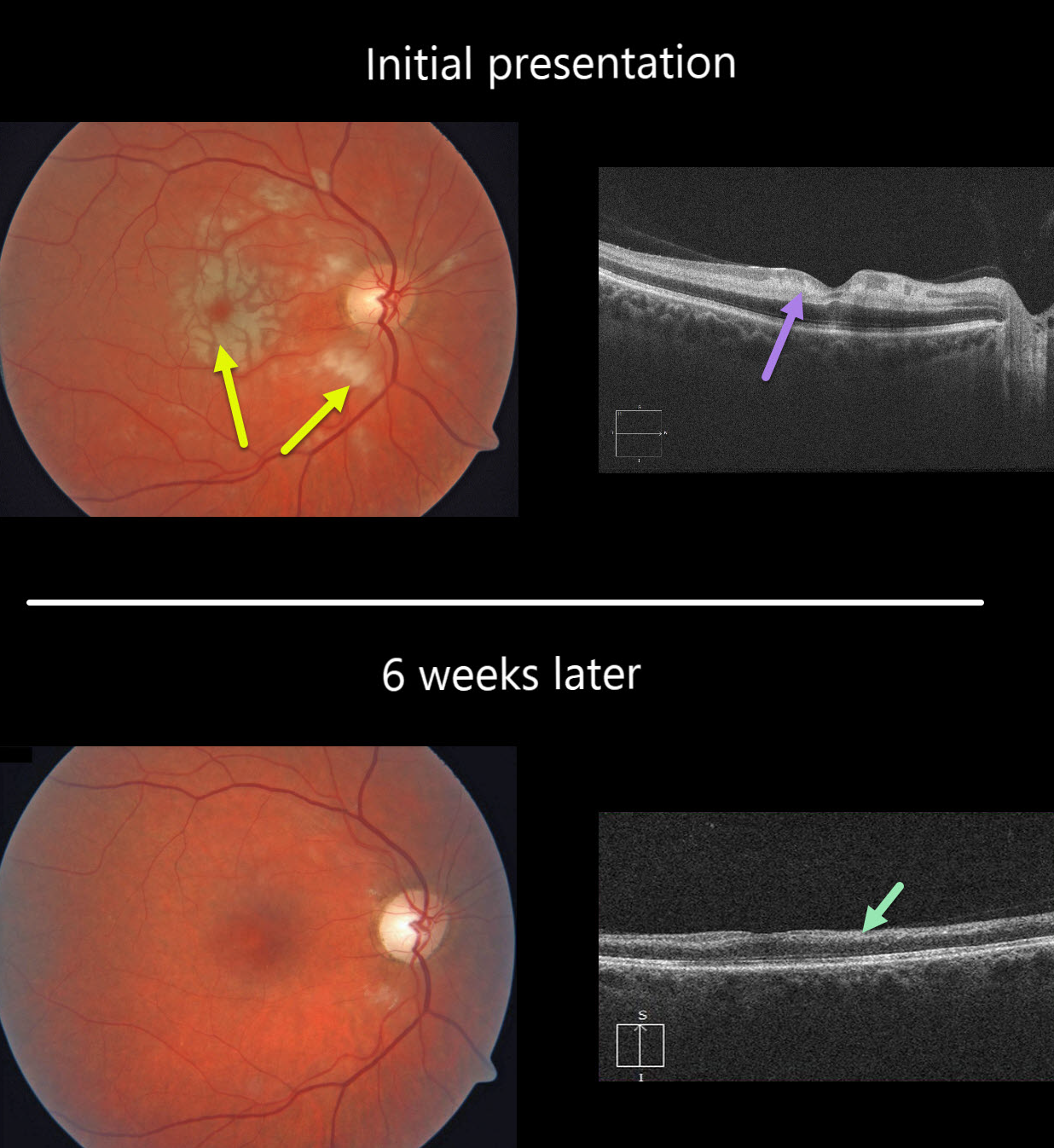
This healthy 49YO female presented with severe, sudden vision loss in her right eye.
There were multiple white macular patches similar to Purtscher’s retinopathy, but without retinal hemorrhages.
Optical coherence tomography (OCT) showed multiple mid-retinal hyperreflective paracentral acute macular maculopathy (PAMM) lesions. Since we felt this could represent an incomplete central retinal artery occlusion (CRAO), we sent her immediately to the nearest stroke center for further assessment.
At her six-week follow-up, the retinal appearance virtually normalized. However, OCT showed severe diffuse thinning of the inner non-photoreceptor retinal layers consistent with a resolved CRAO. Her stroke workup was unremarkable.
Learning Points:
Purtscher’s retinopathy was first described by Otmar Purtscher in 1910. Although originally described in a man who fell from a tree with cranial trauma, these white patches of retinal ischemia and hemorrhages are found in numerous other etiologies, including pancreatitis, chest trauma, and collagen vascular diseases.
The findings are called Purtscher’s retinopathy when due to trauma, and Purtscher ‘s-like retinopathy when seen from other causes.
The underlying cause of Purtscher’s is unknown, but is thought to involve leukoembolization due to complement activation. The areas of ischemia include more superficial nerve fiber layer infarcts (cotton wool spots) and deeper ischemia involving the middle retinal layers (PAMM).


