

Pars Plana Cyst
<< Back to Cases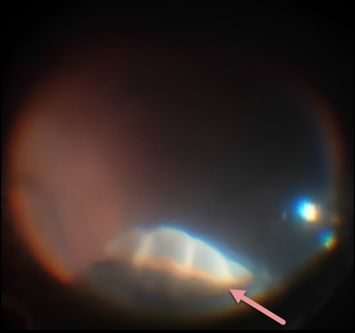
PARS PLANA CYST
Niki Gupta
Originally posted on @retina.rocks May 9, 2023
During vitrectomy for a macular pucker, this intraoperative photo shows an incidental finding of 3-4 pars plana cysts in the superior quadrant found on scleral depression. The cysts were oval, and the inner cyst walls appeared smooth and transparent. Anteriorly, they extended variably over about one-half of the pars plana towards the ciliary processes.
Learning Points:
Pars plana cysts are found in up to 18% of eyes and are more prevalent in the older population. They typically appear as ovoid elevations with a smooth, taut surface.
Pars plana cysts are usually acquired and represent a normal ocular aging change. While the cysts themselves are benign, they may occur secondary to ocular or systemic pathology, including retinal detachment, posterior uveitis, and multiple myeloma.
PARS PLANA CYST
Originally posted on @retina.rocks March 15, 2022
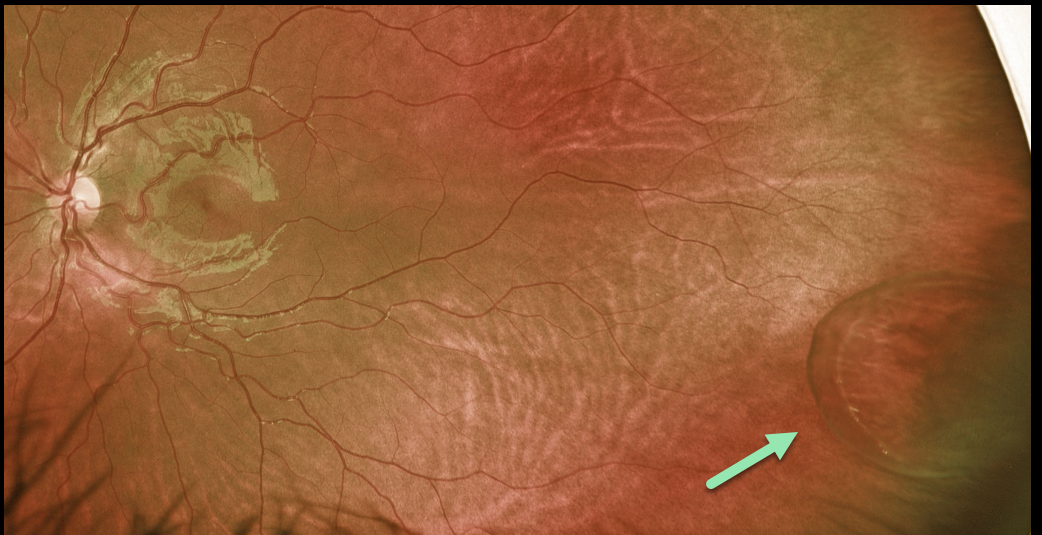
This 9YO female was referred for an asymptomatic lesion in her left eye. She was found to have a large pars plana cyst in the temporal periphery.
Learning Points:
Pars plana cysts are localized to the pars plana region, commonly along a single ora bay. They form when the epithelial cell layers separate between the non-pigmented ciliary epithelium and pigmented epithelium, or solely within the pigmented epithelium.
Although they are benign findings, they need to be differentiated from retinal detachment or retinoschisis.


