

Pars Planitis
<< Back to Cases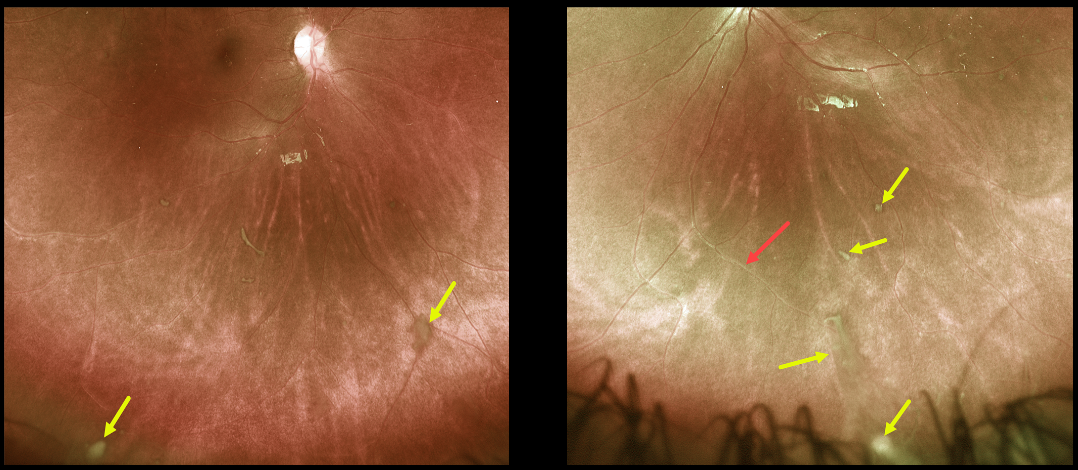
PARS PLANITIS
Originally posted on @retina.rocks June 15, 2021
This healthy 11YO boy was referred for these asymptomatic posterior segment findings.
Optos imaging shows bilateral inferior clumps of vitreous debris (“puff balls”) and vasculitis in the left eye. Not imaged was the bilateral vitreous base inflammation with white pars plana exudate.
Since our patient was completely asymptomatic, we are observing him several times a year.
Learning Points:
One of the more common classification systems for uveitis is based on its primary location within the eye (i.e., anterior vs posterior).
Intermediate uveitis is centered within the retinal periphery and vitreous base, and has a relatively short differential, including sarcoidosis, syphilis, inflammatory bowel disease, multiple sclerosis, and idiopathic (pars planitis).
Pars planitis is unique in the presence of a white vitreous base and pars plana inflammatory material. It is most common in young females, has a waxing-and-waning clinical course over many years, and usually becomes less active with age.
Patients can develop visual symptoms from floaters or cystoid macular edema (CME), and can also develop spill-over anterior uveitis.
Symptomatic vitritis and CME are usually treated with periocular and rarely systemic steroids. Cryotherapy to the inflamed vitreous base is sometimes recommended for more severe and chronic disease (see Sohn et al, AJO 2016;162:35-42).


