

Pentosan Polysulfate Sodium (PPS) Maculopathy
<< Back to Cases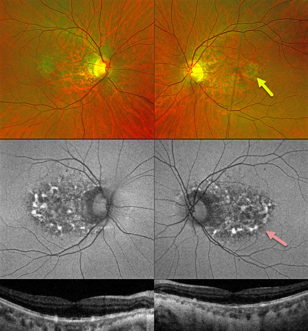
PENTOSAN POLYSUFATE SODIUM TOXICITY
Originally posted on @retina.rocks May 12, 2026
This 48YO was on pentosan polysulfate sodium (PPS, Elmiron) for interstitial cystitis for 2-3 years, but discontinued this drug 2 years earlier. Vision was 20/30 bilaterally.
Fundus color imaging shows bilateral ill-defined macular pigmentary changes. Swept-source OCT shows variable loss of the ellipsoid zone, outer segment bands, and RPE with clumps of hyperreflective material involving or located above the RPE. Optos fundus autofluorescence (FAF) shows patchy hyper- and hypo- FAF extending from the central macula to the nerve.
Learning Points:
Elmiron was approved by the FDA in 1996 for the treatment of interstitial cystitis. A unique PPS retinopathy was recently described, and our patient shows classic findings. Toxicity seems to develop over many years and can mimic more common disorders, including age-related macular degeneration and macular dystrophies.
The risk of toxicity is dose-dependent (Tao et al, Ophthalmology Retina 2025;9:870-882), although toxicity can rarely occur with relatively minimal exposure, as occurred in our patient (Yousif and Johnson, JAMA Ophthalmology 2025;143:362-363). The drug should be discontinued once retinal findings are discovered, although progressive functional and structural changes can develop even after drug cessation (Jung et al, JAMA Ophthalmol 2023;141:260-266).
A recent report found severe asymptomatic adenomatous polyposis in 75% of their PPS maculopathy patients (Feo et al, AJO 2026;287:1-13). Most of their patients required partial or total colectomy or endoscopic resection. They therefore recommended colonoscopy screening in all exposed patients.
PENTOSAN POLYSULFATE SODIUM TOXICITY VS AMD MACULAR ATROPHY
Will Gibson
Originally posted on @retina.rocks September 4, 2023
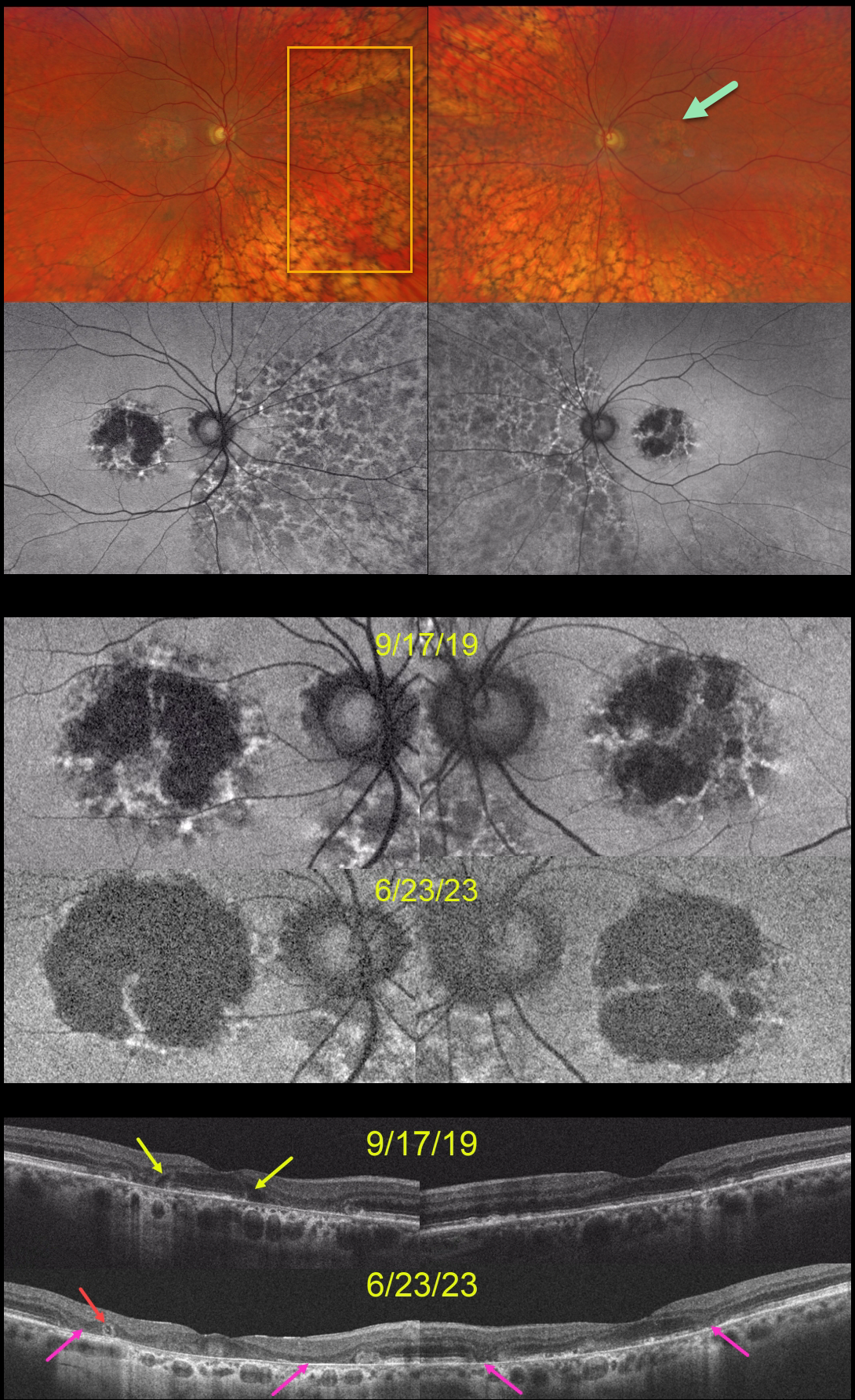
This 59YO was examined on 9/17/19. She was taking Elmiron (pentosan polysulfate sodium, PPS) for 17 years for interstitial cystitis. Vision was 20/40 OD and 20/30 OS.
Optos color RG imaging shows bilateral central-sparing macular atrophy (MA) and extensive, posterior peripheral reticular degeneration of the RPE (PRDRPE).
Fundus autofluorescence shows hypo-FAF from the MA with surrounding punctate and linear areas of hyper-FAF. Based on these findings, she decided to stop taking Elmiron.
When last examined on 6/23/23, vision dropped to 20/800 OD and was relatively stable at 20/40 OS. The MA progressed bilaterally clinically and on FAF and OCT. Hyporeflective OPL degeneration (wedge defects) is noted on the initial 9/17/19 OCT. On the 6/23/23 OCTs, an area of outer retinal tubulation (ORT) is seen temporally OD, and progressive outer retinal and RPE atrophy are noted bilaterally.
Learning Points:
Elmiron was approved by the FDA in 1996 for the treatment of interstitial cystitis. Recently, a unique PPS retinopathy has been described. Toxicity seems to develop over many years and can mimic more common disorders, including age-related macular degeneration and macular dystrophies.
Peripapillary hypoautofluorescence, more densely packed macular autofluorescent changes, and earlier central macular involvement suggest PPS toxicity over other causes (see Barnes et al Ophthalmology Retina 2020;4:1196-1201), including maternally inherited diabetes and deafness (MIDD).
ORT is often noted overlying inactive macular neovascularization with ongoing anti-VEGF therapy and should not be confused with exudative fluid or cysts, which lack a hyperreflective border. The outer hyperreflective band likely represents inner segment mitochondria undergoing fission and translocation towards the nucleus (Litts et al, Retina 2018;38:445-461).
ORT, initially described by Zweifel et al (Arch Ophthalmol 2009;127:1596-1602), is a neurodegenerative condition of the photoreceptors and Muller cells associated with outer retinal and retinal pigment epithelium atrophy, including advanced AMD and inherited retinal diseases.
The hyper-FAF surrounding the initial MA is more typical for PPS toxicity, and progressive atrophy is not uncommon even with stopping therapy (Jung et al, JAMA Ophthalmol 2023;141:260-266). However, in our experience, PPS usually produces more widespread macular findings. Given the PRDRPE, the MA could also be age-related. Or maybe our patient had baseline age-related MA superimposed on Elmiron toxicity?
PENTOSAN POLYSULFATE SODIUM TOXICITY
Originally posted on @retina.rocks March 28, 2023
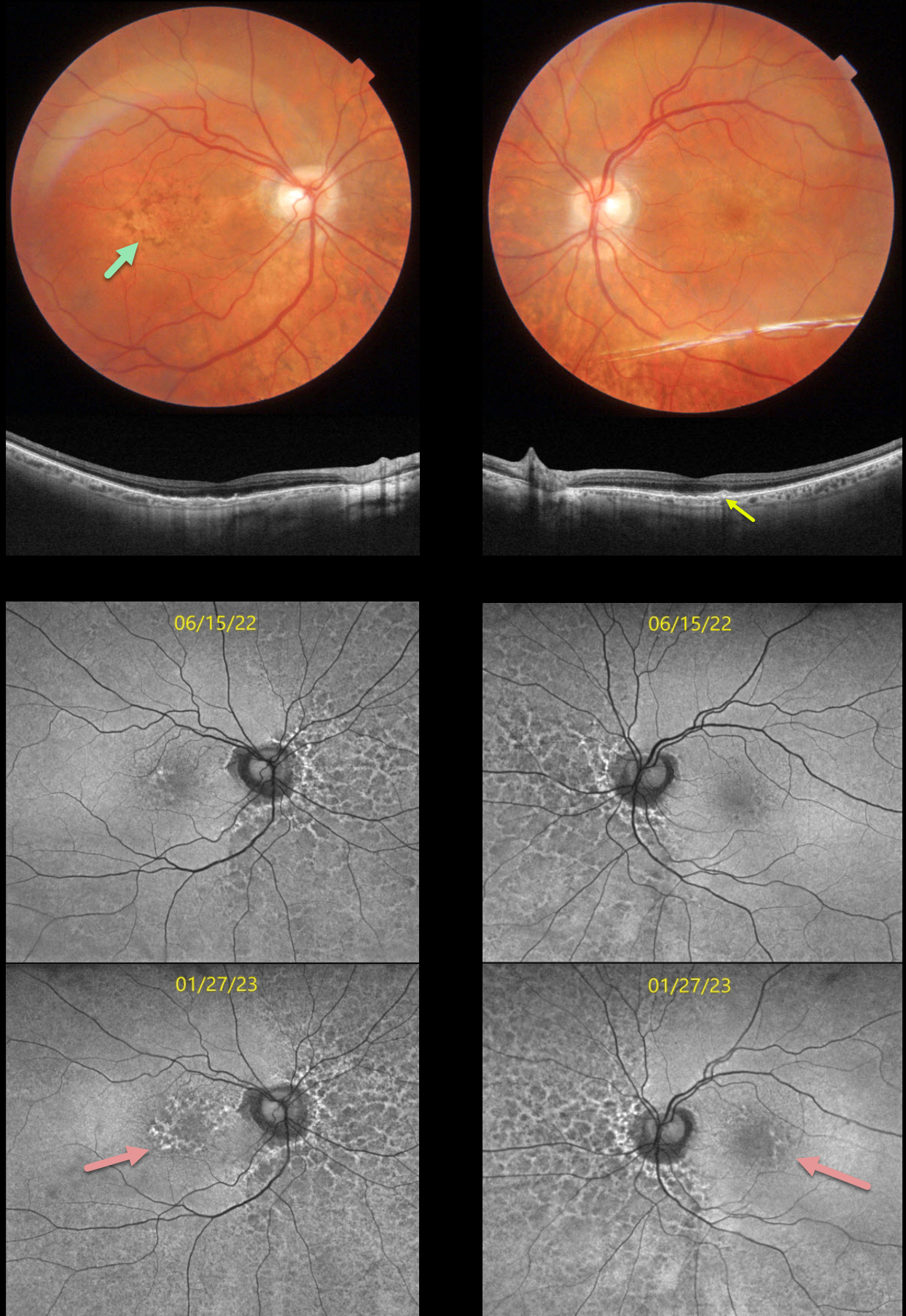
This 74YO female has been followed by us for a few years with known pentosan polysulfate (PPS, Elmiron) retinal toxicity. She has been taking Elmiron for 25 years.
When we initially diagnosed her with these retinal findings, she was hesitant to stop treatment since her interstitial cystitis symptoms were intolerable and dramatically improved with this medicine. She returned for her 6-month examination on 1/27/23 with increased central visual and contrast complaints.
Optos color imaging looked fairly stable. However, fundus autofluorescence (FAF) shows definite progressive macular changes. Macular color photography with swept-source OCT shows variable loss of the outer retinal layers with some focal outer retinal hyperreflective deposits.
Learning Points:
Elmiron was approved by the FDA in 1996 for treating interstitial cystitis. A unique PPS retinopathy was described in 2018, and our patient shows classic findings. Toxicity seems to develop over many years and can mimic more common disorders, including age-related macular degeneration and macular dystrophies.
The drug should be discontinued once retinal findings are discovered, although progressive functional and structural changes can develop even after drug cessation (Jung et al, JAMA Ophthalmol 2023;141:260-266).
We strongly recommend that she discontinue her Elmiron therapy. However, she felt that she would rather completely lose her central vision than face life with the pain from untreated interstitial cystitis and will remain on treatment.
She will discuss other potential therapeutic options with her nephrologist in the meantime.
PENTOSAN POLYSULFATE SODIUM MACULOPATHY
Originally posted on @retina.rocks August 18, 2022
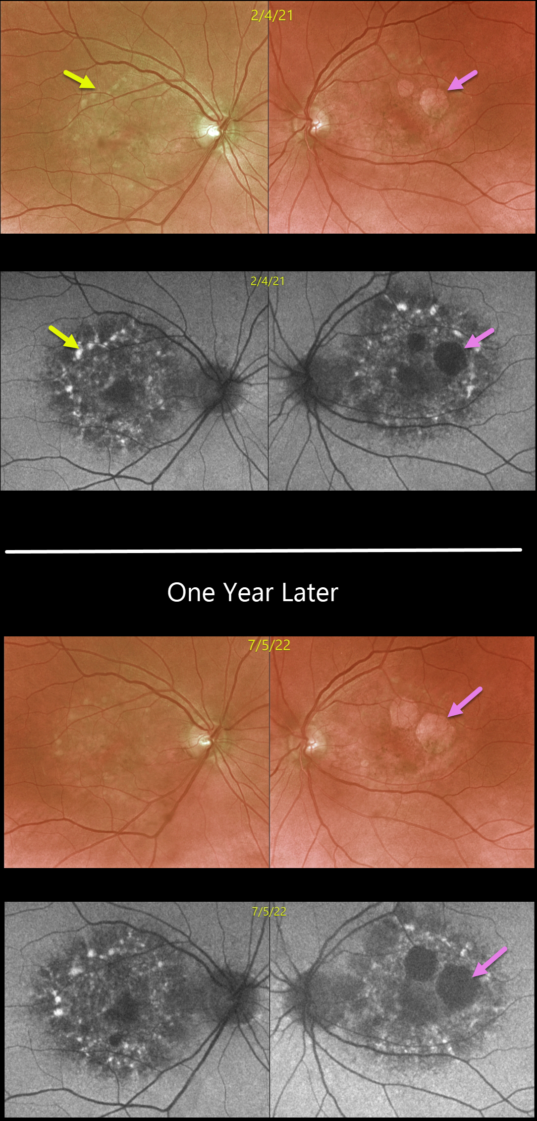
This 64YOF presented in February 2020 with findings of Pentosan polysulfate sodium (PPS, Elmiron) retinal toxicity. She used PPS for 4 years, but discontinued the drug 9 years earlier. We have since followed her yearly.
Optos color imaging of the prior (17 months earlier) and current examinations shows variable yellow-orange atrophic macular pigmentary changes. These findings are best visualized with fundus autofluorescence.
Close inspection of these images shows definite enlargement of the RPE atrophy, especially in her left eye. She remains completely free of visual symptoms with vision of 20/30 OD and 20/25 OS.
Learning Points:
Elmiron was approved by the FDA in 1996 for treating interstitial cystitis. A unique PPS retinopathy was recently described, and our patient shows classic findings.
Toxicity seems to develop over many years and can mimic more common disorders, including age-related macular degeneration and macular dystrophies.
Progressive changes, as in our patient, usually continue despite discontinuation of the causative drug (Shah et al., JAMA Ophthalmology 2020;138:894-900).
PENTOSAN POLYSULFATE SODIUM MACULOPATHY + OUTER RETINAL TUBULATION
Originally posted on @retina.rocks March 1, 2022
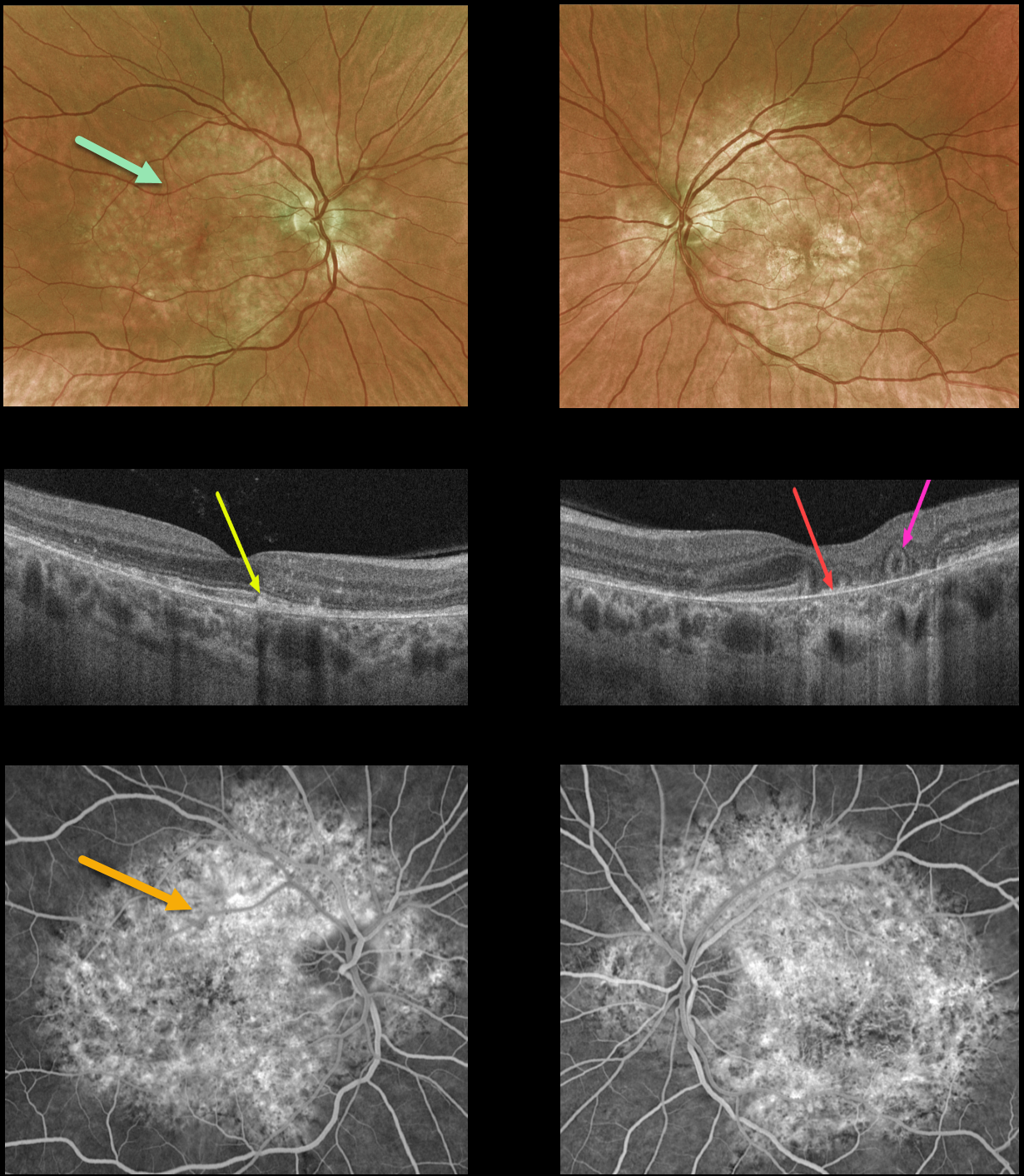
This 54YO female presented with a 9-month history of blurred vision in her left eye. She was on Elmiron (pentosan polysulfate sodium, PPS) for about 20 years due to interstitial cystitis, but stopped the medication before seeing us due to the widespread publicity about its potential retinal toxicity.
Optos color imaging shows variable yellow-orange atrophic pigmentary changes throughout the macular and peripapillary posterior poles.
OCT of the right macula shows scattered hyperreflective lesions mostly within the outer segment layers. The left macular OCT shows variable outer retinal and RPE loss, especially centrally, where bare Bruch’s membrane is visible. There is also an area of outer retinal tubulation (ORT) more temporally.
Fluorescein angiography shows window defects within these regions.
Learning Points:
Elmiron was approved by the FDA in 1996 for treating interstitial cystitis. Recently, a unique PPS retinopathy has been described, and our patient shows classic findings.
Toxicity seems to develop over many years and can mimic more common disorders, including age-related macular degeneration and macular dystrophies.
Peripapillary hypoautofluorescence, more densely packed macular autofluorescent changes, and earlier central macular involvement suggest PPS toxicity over other causes (see Barnes et al Ophthalmology Retina 2020;4:1196-1201), including maternally inherited diabetes and deafness (MIDD).
ORT is often noted overlying inactive macular neovascularization with ongoing anti-VEGF therapy and should not be confused with exudative fluid or cysts, which lack a hyperreflective border. The outer hyperreflective band likely represents inner segment mitochondria undergoing fission and translocation towards the nucleus (Litts et al, Retina 2018;38:445-461).
ORT, initially described by Zweifel et al (Arch Ophthalmol 2009;127:1596-1602), is a neurodegenerative condition of the photoreceptors and Muller cells associated with atrophy affecting the outer retina and retinal pigment epithelium, including advanced AMD and inherited retinal diseases.
PENTOSAN POLYSULFATE SODIUM MACULOPATHY
Originally posted on @retina.rocks October 21, 2021
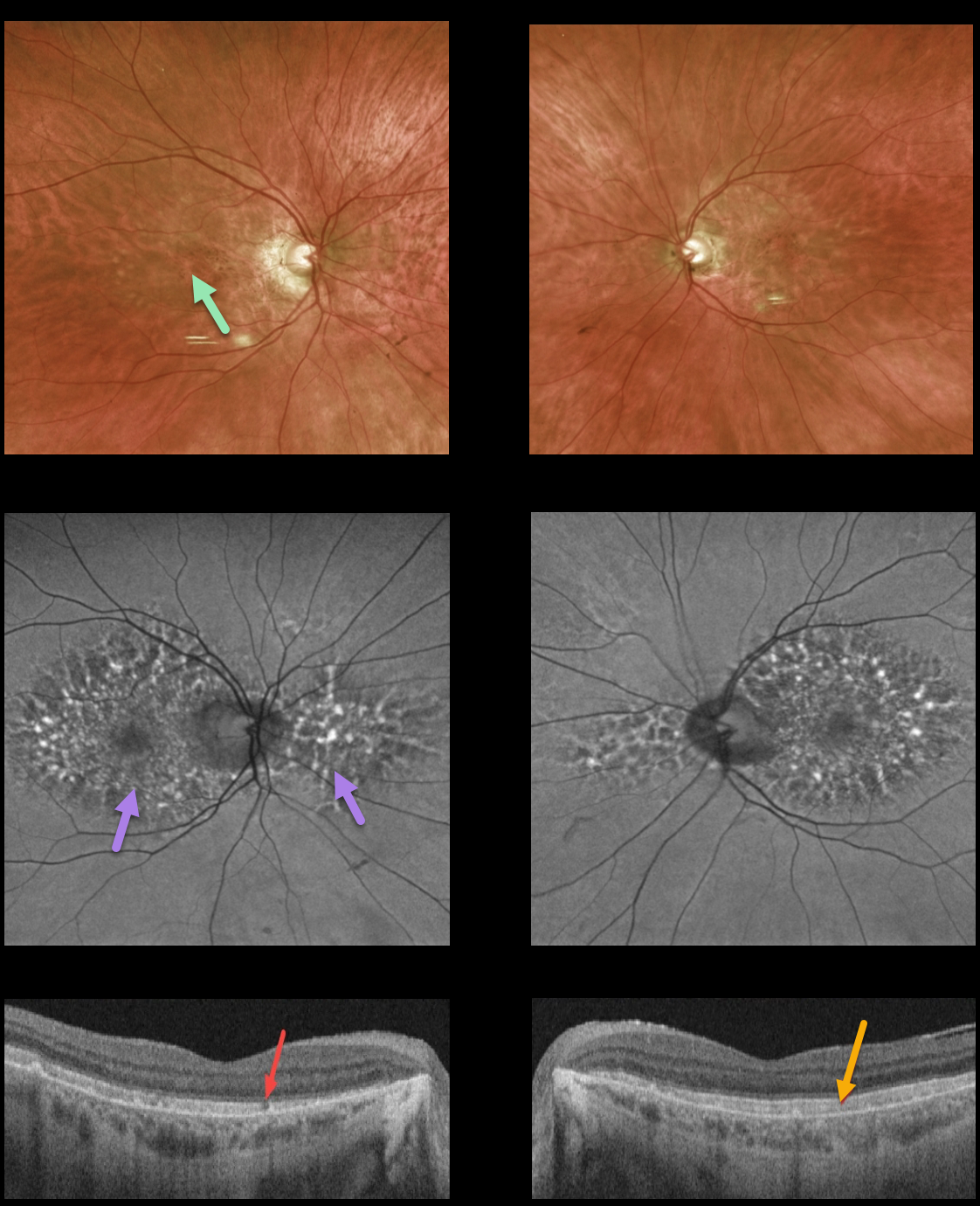
This 74YO female had a long history of Elmiron (pentosan polysulfate, PPS) use. Vision was 20/30 OU.
Variable yellow-orange atrophic pigmentary changes were noted throughout each macula, with more dramatic patches of macular and peripapillary hyper- and hypoautofluorescence.
Triton swept-source OCT showed some patchy ellipsoid loss OD and diffuse hyperreflectivity of the outer segments.
Learning Points:
Elmiron was approved by the FDA in 1996 for treating interstitial cystitis. Recently, a unique PPS retinopathy has been described, and our patient shows classic findings. Toxicity seems to develop over many years and can mimic more common disorders, including age-related macular degeneration and macular dystrophies.
Peripapillary hypoautofluorescence, more densely packed macular autofluorescent changes, and earlier central macular involvement suggest PPS toxicity over other causes (see Barnes et al, Ophthalmology Retina 2020;4:1196-1201), including MIDD. Mitochondrial disease, pattern macular dystrophies, and PPS maculopathy have similar yet distinct findings. We hope the last 3 days of posts will help to clarify these clinically overlapping phenotypes.
PENTOSAN POLYSULFATE SODIUM MACULOPATHY
Originally posted on @retina.rocks March 24, 2021
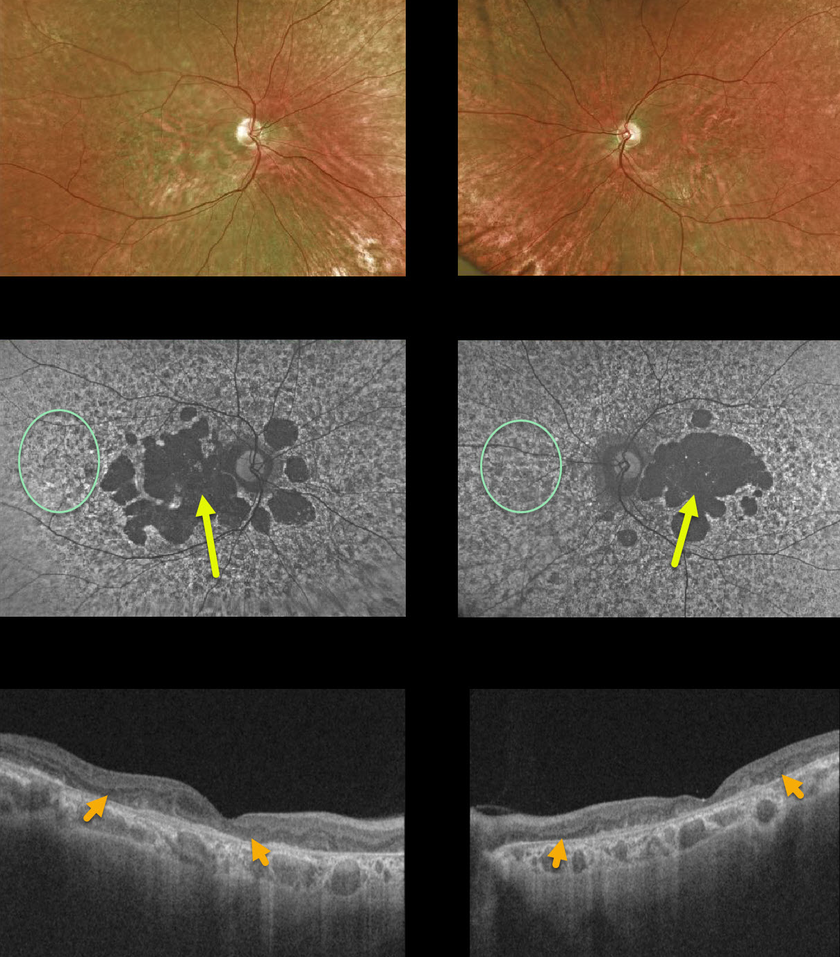
This 65YO female was taking Elmiron (pentosan polysulphate, PPS) for many years beginning in the mid-1990s. Vision was 20/400 OD and counting fingers OS.
Variable yellow-orange atrophic pigmentary changes were noted funduscopically throughout each macula, with more dramatic patches of macular hypoautofluorescence with a peripheral granular hyper- and hypoautofluorescent appearance.
OCT scanning showed variable outer retinal loss.
Learning Points:
Elmiron was approved by the FDA in 1996 for treating interstitial cystitis. Recently, a unique PPS retinopathy has been described, and our patient shows classic findings. Toxicity seems to develop over many years and can mimic more common disorders, including age-related macular degeneration and macular dystrophies.
Peripapillary hypoautofluorescence, more densely packed macular autofluorescent changes, and earlier central macular involvement suggest PPS toxicity over other causes (see Barnes et al, Ophthalmology Retina 2020;4:1196-1201).
PENTOSAN POLYSULFATE SODIUM MACULOPATHY
Originally posted on @retina.rocks June 2, 2020
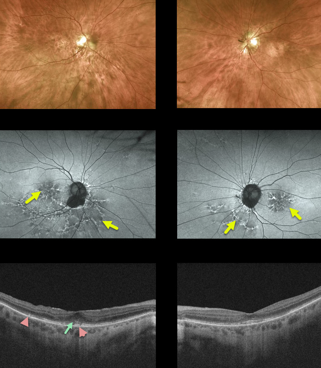
This 72yo female was on pentosan polysulfate sodium (PPS, Elmiron) for 5-10 years in the 1990s and was previously misdiagnosed with dry age-related macular degeneration (AMD).
While her fundus exam may resemble pigmentary changes associated with dry AMD, there are no drusen.
Fundus autofluorescence (FAF) shows a mixture of hyper- and hypo-autofluorescent spots in the macula extending outwards into the mid-peripheries. Drusen and pigmentary changes in AMD typically exhibit variable hypoautofluorescence.
OCT shows some variable attenuation of the EZ band along with hyperreflective deposits above the RPE.
Learning Points:
PPS, sold under the brand name Elmiron, is used to treat interstitial cystitis. PPS maculopathy seems to develop over many years and can mimic more common disorders, including AMD and macular dystrophies. As with hydroxychloroquine, macular changes can progress despite discontinuation of the drug. In our experience, wide-field FAF is critical in establishing the diagnosis.
Although there is no treatment for the underlying maculopathy, patients need to be followed and treated if macular neovascularization develops.


