

Peripapillary Pachychoroid Syndrome
<< Back to Cases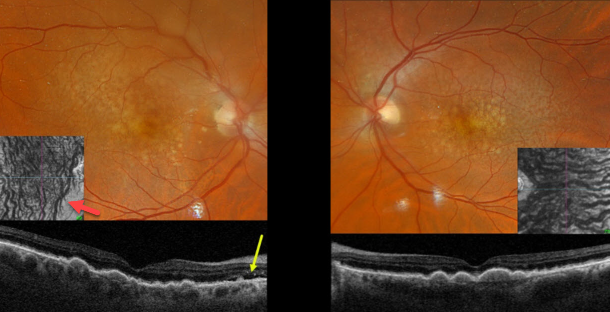
PERIPAPILLARY PACHYCHOROID SYNDROME
Originally posted on @retina.rocks March 20, 2025
This 77YO male was referred for asymptomatic neovascular AMD in his right eye. Vision was 20/50 bilaterally.
Optos color RGB imaging shows extensive bilateral larger drusen and subretinal drusenoid deposits, which are evident on Triton swept-source OCT. Localized subretinal fluid is noted just temporal to the right nerve.
Although the choroid is not particularly thickened, it is relatively thick for a 77YO, and large vessels span the full-thickness choroid. En face choroidal OCT shows markedly abnormal dilated vessels.
Learning Points:
Originally described by Phasukkijwatana et al in 2018 (Retina 2018;38:1652-1667), peripapillary pachychoroid syndrome presents with nasal macular intraretinal and subretinal fluid. These eyes can look very similar to those of patients with optic pit maculopathy, but without the pit. Additional findings include a thicker nasal than temporal macular choroid, choroidal folds (75% of cases), shorter axial lengths (<23mm), and hyperopia. There is no angiographic leakage despite the presence of OCT fluid, which often spontaneously waxes and wanes. Symptomatic fluid can successfully be treated with photodynamic therapy.
We assume our patient’s fluid is related to his pachychoroid disease rather than nAMD. Observation was recommended.
PERIPAPILLARY PACHYCHOROID SYNDROME
Originally posted on @retina.rocks October 16, 2023
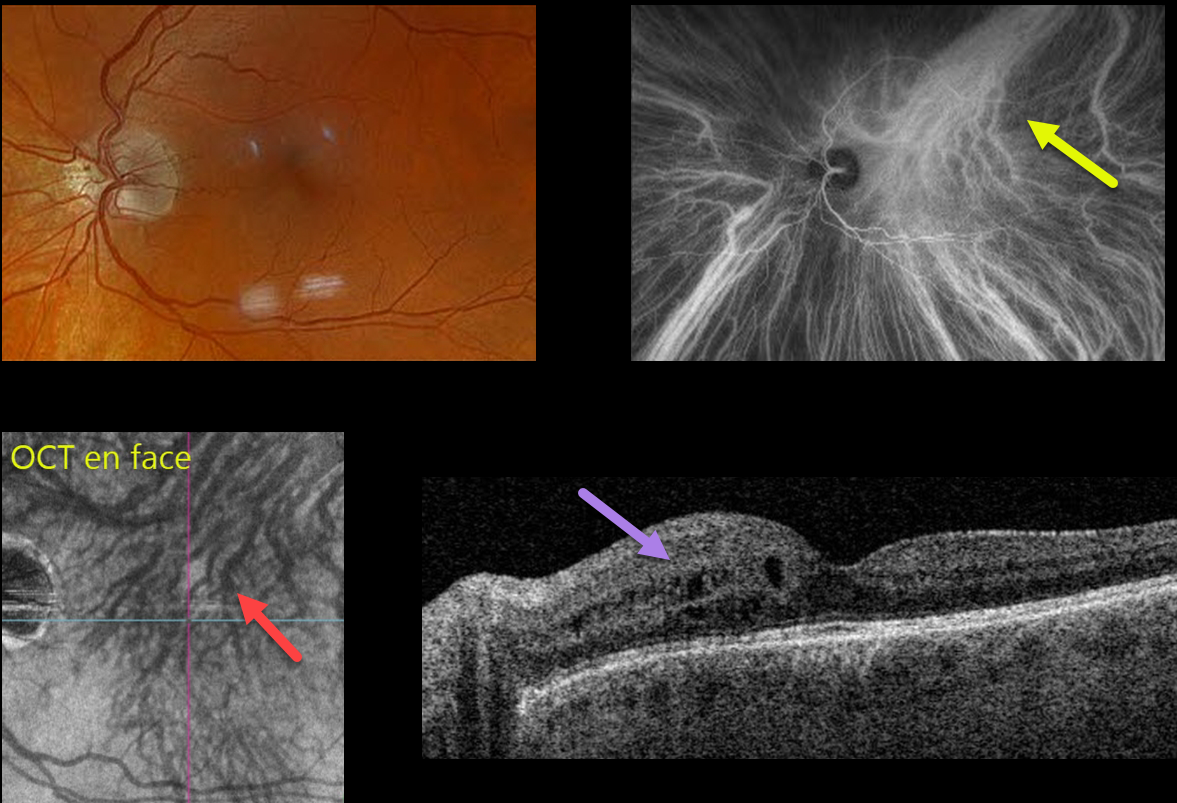
This 59YO male has been followed for the past year with asymptomatic peripapillary pachychoroid syndrome (PPS) in his left eye. Vision is stable at 20/25 OU.
Optos color RGB imaging shows a normal left macula. Indocyanine green (ICG) angiography is normal OD (not shown), but reveals a dilated vortex system superotemporally that drains nearly the entire macula, along with a strikingly similar appearance from the OCT choroidal en face.
Triton swept-source OCT was normal OD (not shown), but shows mild nasal thickening with cysts in the inner nuclear layer OS.
Learning Points:
Originally described by Phasukkijwatana et al in 2018 (Retina 2018;38:1652-1667), PPS presents with nasal macular intraretinal and subretinal fluid. These eyes can look very similar to those of patients with optic pit maculopathy, but without the pit.
Additional findings include a thicker nasal than temporal macular choroid, choroidal folds (75% of cases), shorter axial lengths (<23mm), and hyperopia. There is no angiographic leakage despite the OCT fluid. This fluid will often spontaneously wax and wane. Symptomatic fluid can successfully be treated with photodynamic therapy.
Intervortex venous anastomoses are common in pachychoroid-related disorders (Spaide et al Retina 2021;41:997-1004). In this case, the choroidal anastomosis and dilated superior vortex system were beautifully imaged on both ICG and en face OCT.
In our practice, we continue to find that en face imaging often provides a readily accessible, unique, and compelling perspective for a variety of pathologies, and we hope other clinicians will add this to their daily clinic routine.
PACHYCHOROID SYNDROME
Will Gibson
Originally posted on @retina.rocks March 22, 2023
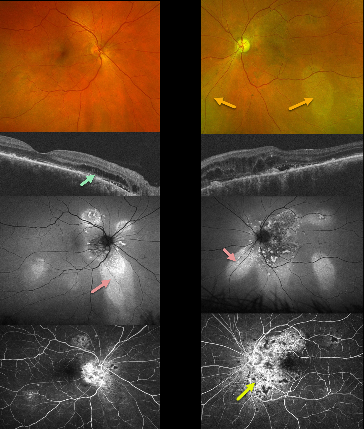
This 51YO male has been followed for years with self-limited episodes of idiopathic central serous retinopathy (ICSC).
Optos color imaging of both eyes shows regions of inferior pigmentary loss, most pronounced in the OS. These findings are much more dramatic on fundus autofluorescence. Swept-source OCT shows variable bilateral loss of the outer retinal bands. Outer retinal fluid with trace subretinal fluid is seen centrally and nasally OS. Fluorescein angiography shows variable staining.
Learning Points:
This patient has numerous features of the pachychoroid spectrum, including idiopathic central serous retinopathy (ICSC), pachychoroid neovasculopathy, polypoidal choroidal vasculopathy, pachychoroid pigment epitheliopathy, peripapillary pachychoroid syndrome, and choroidal excavation.
Our patient’s current findings are dominated by the peripapillary pachychoroid syndrome. Originally described by Phashukkuwatana et al (Retina 2018;1652-1667), findings include peripapillary choroidal thickening associated with nasal macular subretinal and/or intraretinal fluid. Most patients also have chorioretinal folds.
The fluid waxes and wanes, and is responsive to photodynamic therapy if treatment is needed for persistent symptomatic foveal fluid (Iovino et al, Eye 2022;36:716-723). Our patient is being observed for now.
PERIPAPILLARY PACHYCHOROID SYNDROME WITH UNILATERAL BDUMP
Originally posted on @retina.rocks October 15, 2020
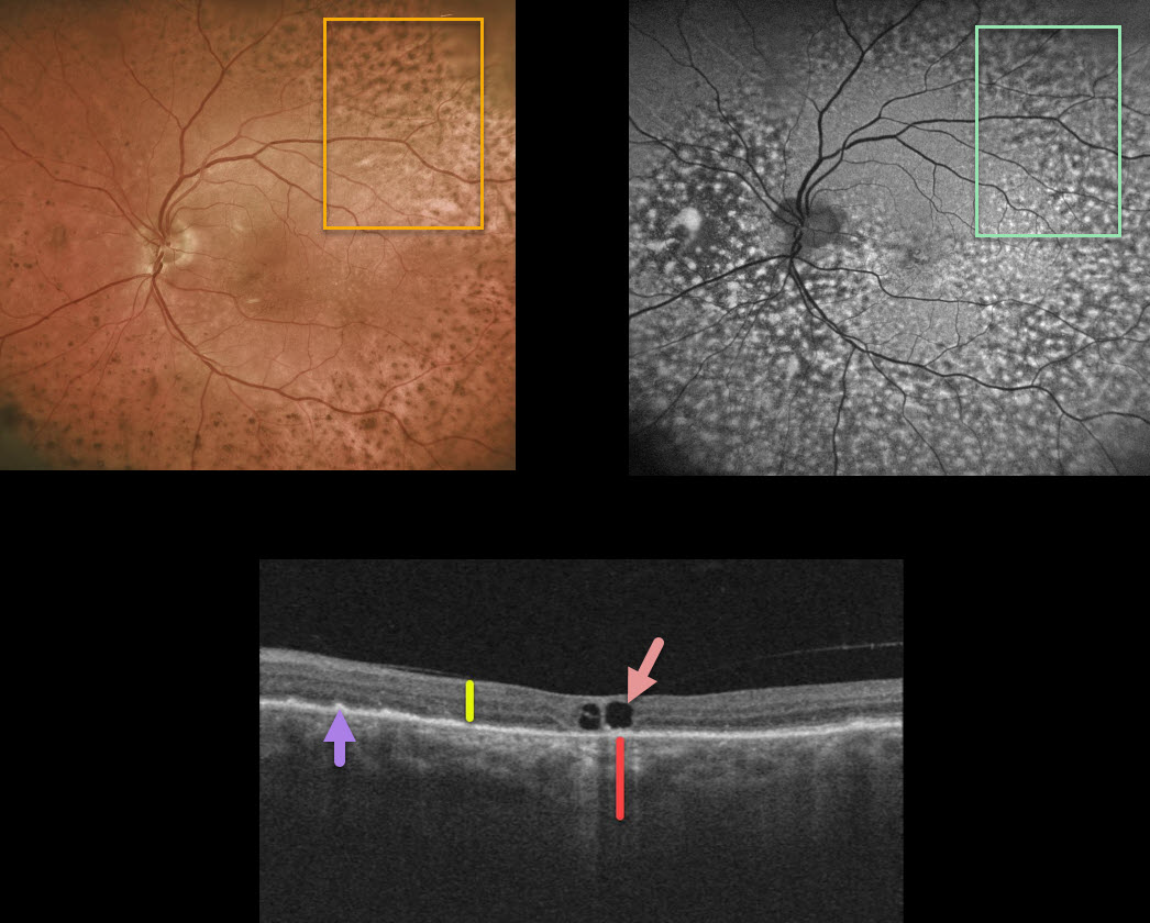
While being followed for peripapillary pachychroid syndrome (PPS) with periodic photodynamic therapy for symptomatic foveal fluid OS, this patient presented with new diffuse pigmentary deposits in the left eye only.
Fundus autofluorescence shows a dramatic pattern of hyperautofluorescent and hypofluorescent spots scattered throughout the fundus. These findings, although unilateral, look very similar to those in the paraneoplastic condition, bilateral diffuse uveal melanocytic proliferation (BDUMP).
Macular OCT scanning shows a diffusely thickened choroid when compared to the neurosensory retina. Other OCT findings here include a few large central foveal cysts, outer retinal atrophy, and RPE changes.
Our patient has no known prior cancer diagnoses, and recently had a negative CT abdominal scan and chest x-ray. He continues to be followed closely by his internist since BDUMP often precedes the diagnosis of a systemic malignancy by months to years.
Learning Points:
Typical PPS findings include thickened choroid, chorioretinal folds, shorter axial lengths, and hyperopia.
In BDUMP, the thickened choroid and pigmentary changes are secondary to infiltration by benign uveal melanocytes, possibly in response to a primary visceral tumor that produces melanocytic growth factors.
PERIPAPILLARY PACHYCHOROID SYNDROME
Originally posted on @retina.rocks August 18, 2020
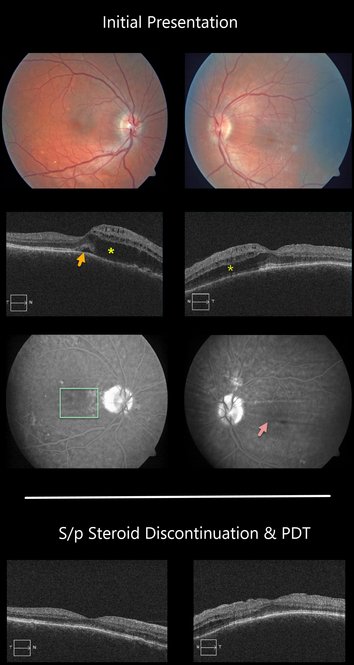
This patient has peripapillary pachychoroid syndrome (PPS), a recently described bilateral condition characterized by nasal macular intraretinal and subretinal fluid.
These eyes can look very similar to those of patients with optic pit maculopathy, but without the pit. Additional findings include a thicker nasal macular choroid than temporal macular choroid, choroidal folds (75% of cases), shorter axial lengths (<23mm), and hyperopia.
There is no angiographic evidence of leakage despite fluid noted on OCT.
The patient worsened when initially placed on steroids, but made a tremendous improvement after steroids were discontinued, followed by photodynamic therapy.
Learning Points:
PPS is part of the pachychoroid spectrum, where patients have a thick choroid. Probably the most common and well-known pachychoroid disease is idiopathic central serous chorioretinopathy. Just like ICSC, this condition worsens with steroid use.
See Phasukkijwatana et al Retina, 2018;38:1652-1657 for more on PPS.


