

Peripheral Exudative Hemorrhagic Chorioretinopathy (PEHCR)
<< Back to Cases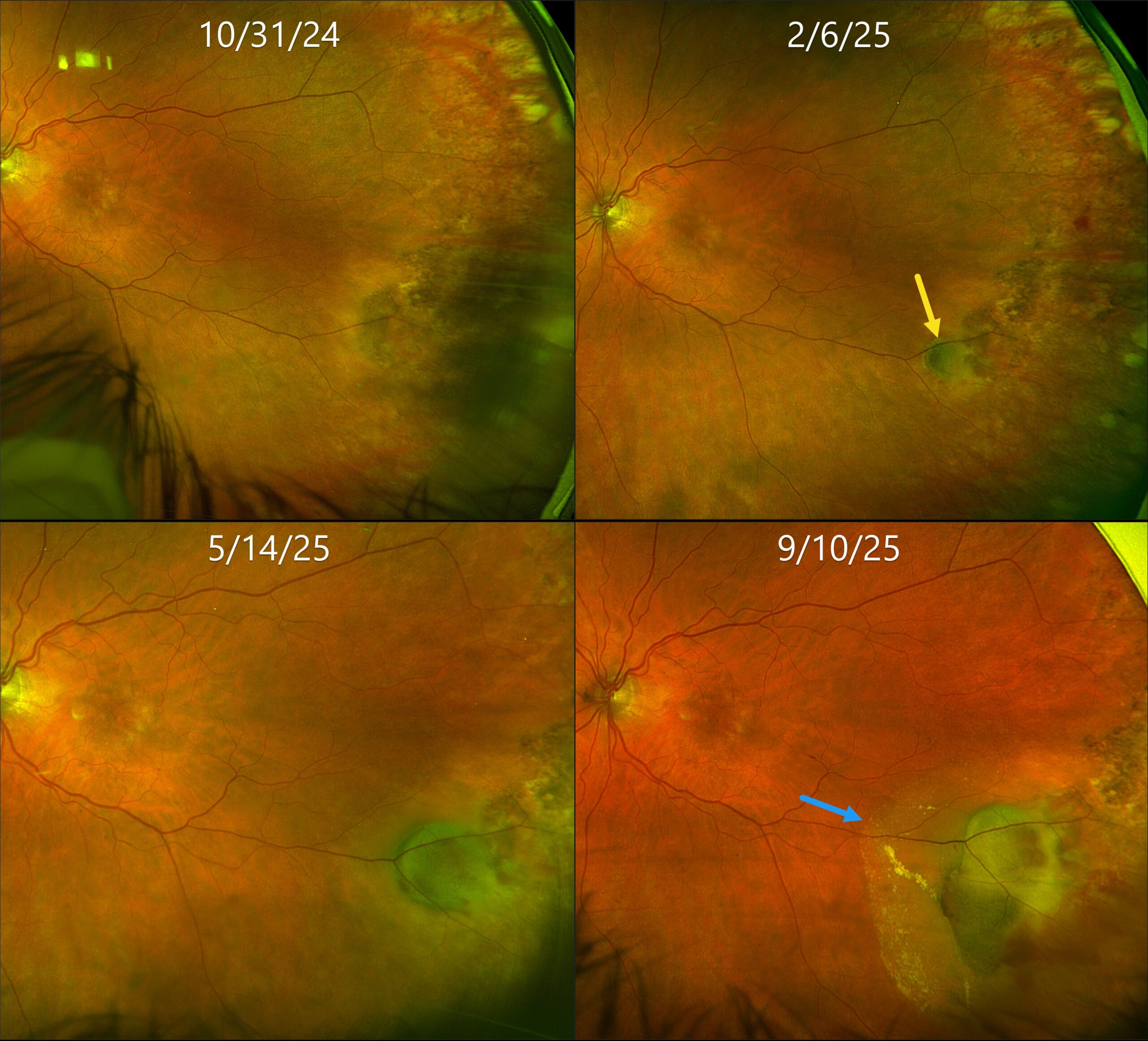
PERIPHERAL EXUDATIVE HEMORRHAGIC CHORIORETINOPATHY
Originally posted on @retina.rocks June 19, 2026
This 80YO female was initially referred for asymptomatic retinal findings in her left eye. Vision was 20/25.
Optos color RG imaging shows flat pigmented subretinal scarring in the left inferotemporal periphery. Three months later, a subretinal pigmented nodular lesion is noted along with a small dot of temporal blood. Over the following 7 months, this subretinal lesion enlarged with increasing blood and subretinal fluid approaching the macula. Although she remained asymptomatic, intravitreal anti-VEGF therapy was started.
Learning Points:
Ectopic choroidal neovascularization (CNV), commonly referred to as peripheral exudative hemorrhagic chorioretinopathy (PEHCR), is an exudative process similar to wet AMD but located in the retinal periphery. Ectopic CNV can often masquerade as a choroidal mass or uveal melanoma. It can also cause extensive choroidal/suprachoroidal, subretinal, and vitreous hemorrhage, more commonly in patients who are on blood thinners. The subretinal blood often spontaneously regresses without treatment, although anti-VEGF injections can be used for symptomatic or increasing exudation.
SHOULD THIS LESION BE EMERGENTLY LASERED???
Originally posted on @retina.rocks March 11, 2026
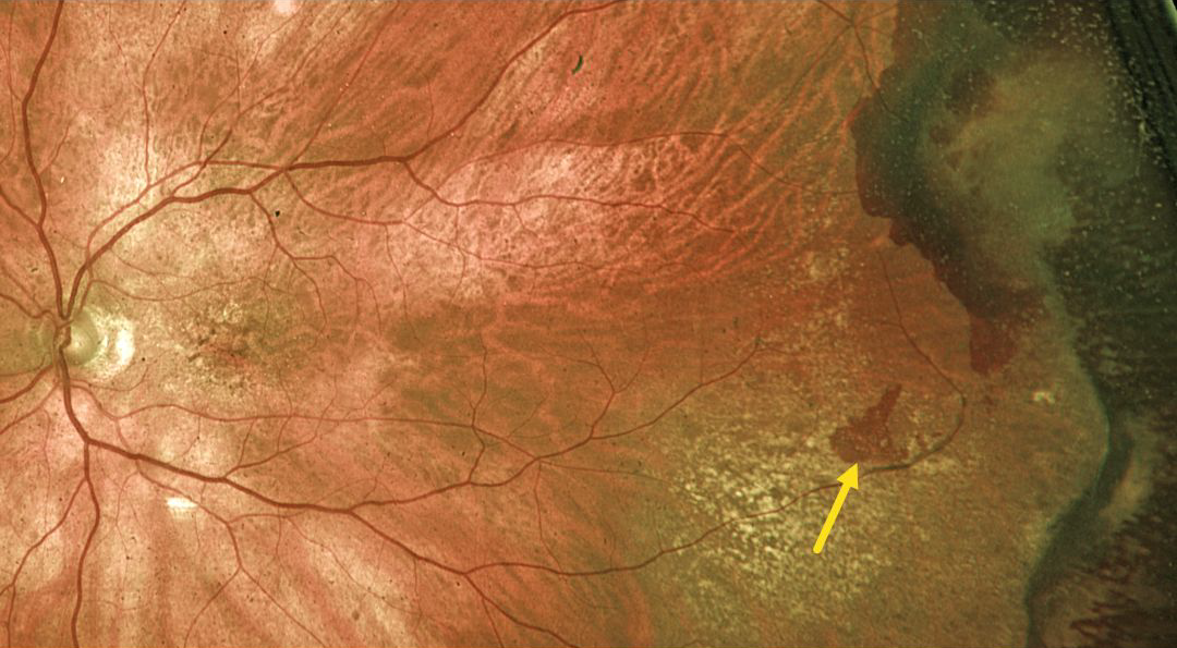
This 89YO male was referred for an asymptomatic retinal tear with possible retinal detachment in his left eye. Vision was 20/60.
Optos color RG imaging shows a red horseshoe-shaped lesion in the temporal midperiphery. Would you recommend immediate thermal laser demarcation of this retinal tear?
We hope not! This patient has asymptomatic and chronic ectopic choroidal neovascularization (CNV), which is evidenced by the more peripheral subretinal scarring and superotemporal dark subretinal blood. Ectopic CNV can often masquerade as a choroidal mass or uveal melanoma. It can also cause extensive choroidal/suprachoroidal, subretinal and vitreous hemorrhage, more commonly in patients who are on blood thinners. The subretinal blood often spontaneously regresses without treatment, although anti-VEGF injections can be used for symptomatic or increasing exudation.
We recommended observation and expect this lesion to remain relatively stable.
PERIPHERAL EXUDATIVE HEMORRHAGIC CHORIORETINOPATHY
Originally posted on @retina.rocks April 17, 2024
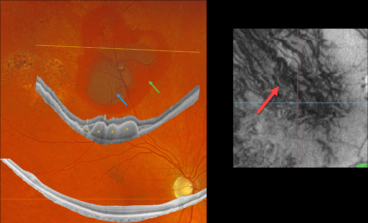
This 64YO diabetic male presented with asymptomatic retinal findings in his left eye. Vision was 20/40 OU.
Optos color RGB imaging shows superior subretinal and sub-RPE blood. Optos Silverstone widefield swept-source OCT through this lesion shows prominent multiple choroidal polyps. Subretinal pigmentary clumping temporal to the blood likely represents findings from absorbed subretinal blood. Moderate non-proliferative retinopathy is noted.
En face imaging is quite abnormal with vortex anastomoses and dilated choroidal vessels.
Observation was recommended.
Learning Points:
Pachychoroid, meaning “thick” choroid, is secondary to large, dilated Haller vessels. Increased oncotic/mechanical pressure from these dilated vessels can cause RPE dysfunction, serous detachments, and macular neovascularization.
What defines a ‘pachy’ choroid remains quite unclear (Spaide, Retina 2021;41:231-237), since pachychoroid-related findings can be present despite a ‘normal’ macular choroidal thickness as in our patient.
Spaide recently described venous overload choroidopathy, a unifying concept that better explains the underlying pathogenesis (Progress Retinal Eye Research 2022;86:100973).
The pachychoroid spectrum also includes ectopic neovascularization (aka peripheral exudative hemorrhagic chorioretinopathy), with the choroid showing progressive thickening towards the temporal periphery (Shroff et al, Retina 2021;41:1518-1525).
Our patient’s widefield OCT beautifully shows the choroidal polyps responsible for the peripheral subretinal bleeding.
SUBRETINAL HEMORRHAGE
Originally posted on @retina.rocks January 31, 2023
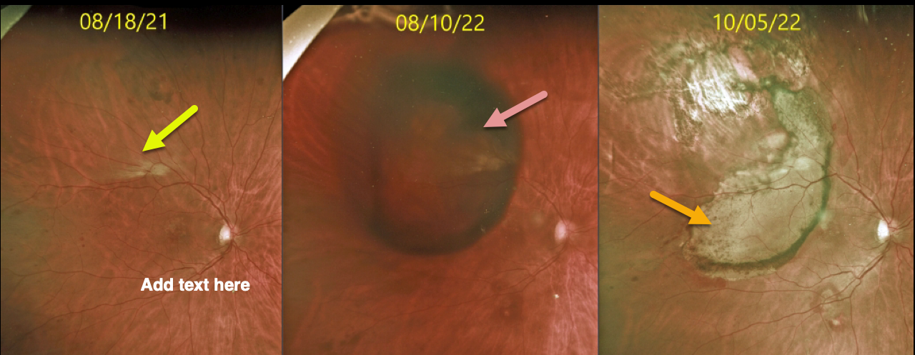
This diabetic male had been followed for over 5 years in our practice, receiving PRN intravitreal injections for diabetic macular edema.
On 8/10/22, he presented with a large superotemporal subretinal hemorrhage in his right eye that progressed into the macula (not shown). Vitrectomy with evacuation of the subretinal blood was performed.
Three months later, an inferior retinal detachment with proliferative vitreoretinopathy was noted (not shown). Staged cataract surgery followed by vitrectomy with silicone oil was performed.
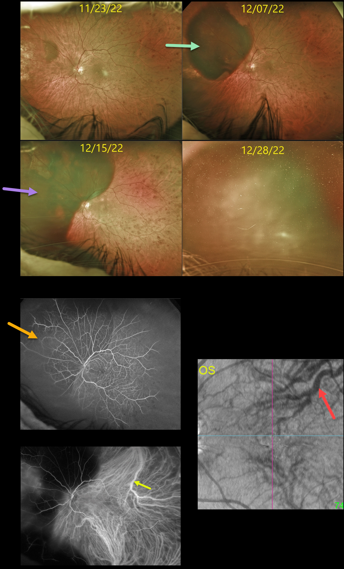
At the 12/7/22 visit, the patient complained of new inferotemporal peripheral field loss, noting that “the same thing is happening in my left eye.”
Unfortunately, a superonasal subretinal hemorrhage was indeed found, and intravitreal Avastin injection was given the following day. However, one week later, the subretinal blood spread posteriorly.
Fluorescein angiography shows nonproliferative diabetic retinopathy with blockage superonasally from the subretinal blood. ICG angiography shows a large dilated superotemporal choroidal vein. En face OCT shows abnormal vortex systems, including a large, dilated superotemporal vortex vein.
On 12/28/22, the subretinal blood spread further with breakthrough bleeding into the vitreous.
Cataract surgery was performed, followed by vitrectomy; however, intraoperatively, once the vitreous hemorrhage was removed, he had a massive subretinal hemorrhage with an inoperable retinal detachment with proliferative vitreoretinopathy.
We believe our patient has an extremely unusual and aggressive variant of pachychoroid disease, as evidenced by the abnormal choroidal vasculature noted on ICG and en face OCT.
We have no explanation for why both eyes developed these massive subretinal hemorrhages, or why they developed almost concurrently. We plan on injecting his right eye with ongoing intravitreal Eylea in hopes of preventing recurrent subretinal hemorrhages.
PERIPHERAL EXUDATIVE HEMORRHAGIC CHORIORETINOPATHY
Originally posted on @retina.rocks October 24, 2022
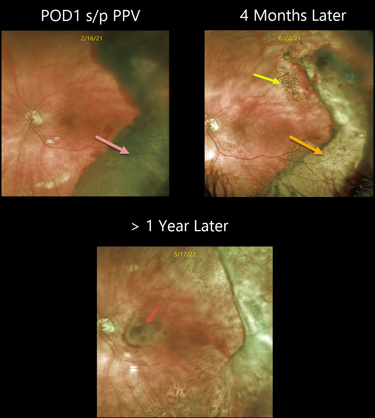
This 58YO diabetic male was seen originally with mild scattered retinal hemorrhages from nonproliferative retinopathy, along with a faint patch of myelinated nerve fiber layer.
He presented one year later after waking with new nasal field loss. Vision was 20/70, and he had a new, large, superotemporal subretinal hemorrhage just outside the macula.
Following 2 monthly intravitreal Avastin injections, the subretinal blood resolved, with secondary subretinal scarring and yellowing devitalized blood. Vision is 20/60.
Learning Points:
Ectopic choroidal neovascularization (CNV) can often masquerade as a choroidal mass or uveal melanoma. It can also cause extensive choroidal/suprachoroidal, subretinal, and vitreous hemorrhage, more commonly in patients who are on blood thinners.
The subretinal blood often spontaneously regresses without treatment, although anti-VEGF injections can be used for symptomatic or increasing exudation.
PERIPHERAL EXUDATIVE HEMORRHAGIC CHORIORETINOPATHY
Originally posted on @retina.rocks May 26, 2022
This 80YO female with a known history of dry AMD presented with a 3-week history of poor vision in her left eye. Vision was hand motion from a dense vitreous hemorrhage. B-scan ultrasonography showed a thickened temporal subretinal lesion without retinal detachment (not shown).
One day following vitrectomy, vision improved to 20/100. The temporal subretinal and RPE blood somewhat resembled a uveal malignant melanoma. However, the well-demarcated location and shape of the lesion, along with the posterior rim of subretinal blood, helped establish the diagnosis of a breakthrough vitreous hemorrhage from ectopic choroidal neovascularization (CNV), also known as peripheral exudative hemorrhagic chorioretinopathy (PEHCR).
Four months later, the resolved devitalized blood was turning yellow. The coarse subretinal pigmentation, most prominent superotemporally, is pathognomonic for prior subretinal blood.
She was subsequently lost to follow-up until she presented with a 6-month history of decreased vision. Although the PEHCR ridge continued to contract, vision was counting fingers due to a new macular neovascularization. Anti-VEGF therapy was started.
Learning Points:
PEHCR is an exudative process similar to wet AMD but located in the retinal periphery. Ectopic CNV can often masquerade as a choroidal mass or uveal melanoma.
It can also cause extensive choroidal/suprachoroidal, subretinal, and vitreous hemorrhage, more commonly in patients who are on blood thinners.
The subretinal blood often spontaneously regresses without treatment, although anti-VEGF injections can be used for symptomatic or increasing exudation.
PERIPHERAL EXUDATIVE HEMORRHAGIC CHORIORETINOPATHY
Originally posted on @retina.rocks March 21, 2022
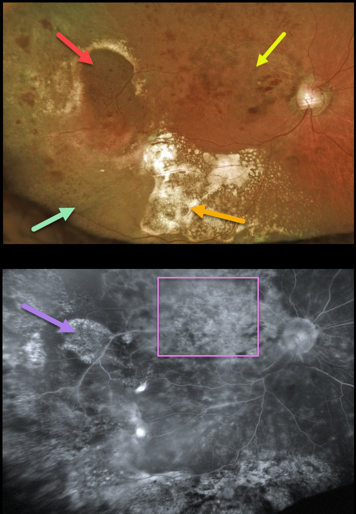
This 94YO female presented with ectopic choroidal neovascularization (CNV), also known as peripheral exudative hemorrhagic chorioretinopathy (PEHCR) in her right temporal periphery. Inferior to this lesion are clumps of subretinal pigment. Confluent subretinal lipid is also noted more inferiorly.
Vision was 20/400 due to macular edema from an unrelated major superotemporal branch retinal vein occlusion (BRVO). The inner retinal hemorrhages from the BRVO extend into the region of the ectopic subretinal lesion, somewhat confusing the clinical picture.
Fluorescein angiography shows variable diffuse staining of the ectopic CNV and secondary pigmentary changes, along with diffuse leakage from the BRVO.
Learning Points:
PEHCR is an exudative process similar to wet AMD but located in the retinal periphery. Ectopic CNV can often masquerade as a choroidal mass or uveal melanoma. It can also cause extensive choroidal/suprachoroidal, subretinal, and vitreous hemorrhage, more commonly in patients who are on blood thinners.
The subretinal blood often spontaneously regresses without treatment, although anti-VEGF injections can be used for symptomatic or increasing exudation. The inferior subretinal pigment in our patient is a classic finding for resolved subretinal blood.
PERIPHERAL EXUDATIVE HEMORRHAGIC CHORIORETINOPATHY
Originally posted on @retina.rocks January 28, 2022
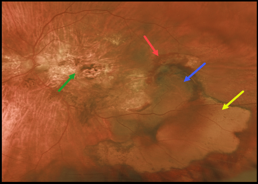
This 77YO female has been followed for years with vision of 20/200 OD and counting fingers OS from inactive age-related disciform scars. She then came in with sudden symptomatic vision loss and floaters in her left eye.
Vision was hand motion OS from a breakthrough vitreous hemorrhage from an inferotemporal ectopic choroidal neovascularization (CNV) that grew from the edge of the original macular scar. She was not taking anticoagulants or aspirin.
Following two monthly intravitreal Avastin injections, vision remained at hand motion. The vitreous hemorrhage had cleared, and the subretinal blood was resolving.
Optos imaging shows the original macular scarring, along with varying stages of resolving rub-RPE blood, subretinal blood, and devitalized, yellow subretinal blood.
Learning Points:
Ectopic CNV, also known as peripheral exudative hemorrhagic chorioretinopathy, is an exudative process similar to wet AMD but located in the retinal periphery.
Ectopic CNV can often masquerade as a choroidal mass or uveal melanoma. It can also cause extensive choroidal/suprachoroidal, subretinal, and vitreous hemorrhage, more commonly in patients who are on blood thinners.
The subretinal blood often spontaneously regresses without treatment, although anti-VEGF injections can be used for symptomatic or increasing exudation.
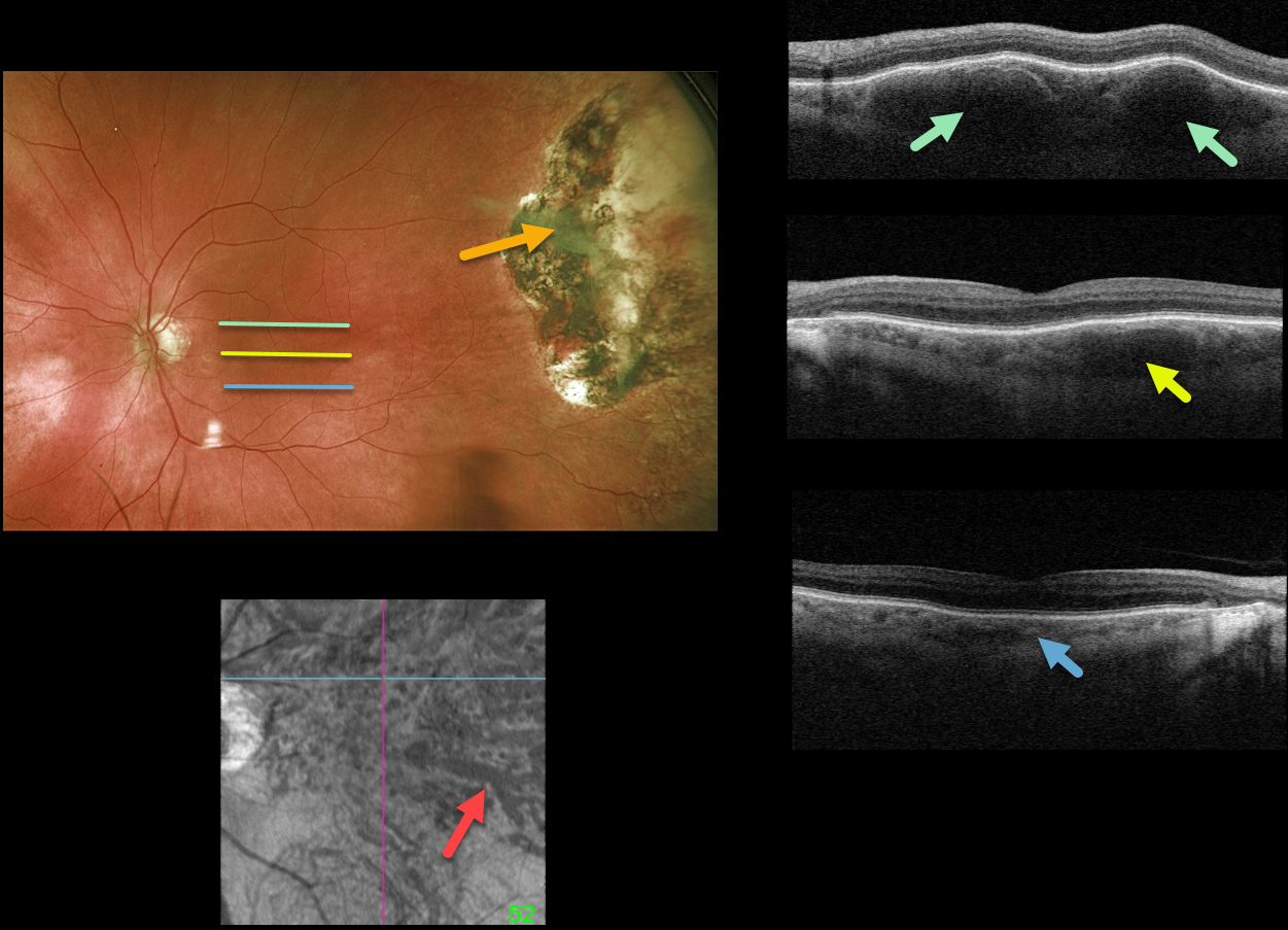
PERIPHERAL EXUDATIVE HEMORRHAGIC CHORIORETINOPATHY + PACHYCHOROID
Originally posted on @retina.rocks December 22, 2021
This 85YO male underwent cryotherapy 7 years earlier for a symptomatic hemorrhagic ectopic choroidal neovascularization in his left eye. Optos imaging shows the current appearance of the inactive cryotherapy scarring.
Although the macula has a normal fundoscopic appearance, Triton swept-source OCT scanning is far from normal. B-scans are shown through the top, central, and inferior macula. Although the inferior macular choroid appears relatively normal, a thickened outer choroid (pachyvessels) is noted more centrally, and is huge superiorly. En face imaging through this abnormal choroid is shown.
Learning Points:
The pachychoroid spectrum is a collection of conditions with a thick (“pachy”) choroid. These pachyvessels can cause a range of disorders, including idiopathic central serous chorioretinopathy, pachychoroid pigment epitheliopathy, choroidal excavation, pachychoroid neovasculopathy, polypoidal choroidal vasculopathy, pachydrusen, and peripapillary pachychoroid syndrome.
They are also associated with ectopic neovascularization (aka peripheral exudative hemorrhagic chorioretinopathy), although the choroid in these cases shows increased thickening towards the temporal periphery (see Shroff et al, Retina 2021;41:1518-1525).
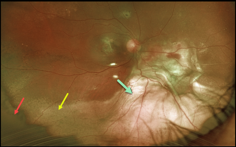
PERIPHERAL EXUDATIVE HEMORRHAGIC CHORIORETINOPATHY
Originally posted on @retina.rocks May 31, 2021
This is a beautiful Optos image of a 67YO female with counting finger vision 1-week following pars plana vitrectomy for a breakthrough vitreous hemorrhage.
There is a rim of dark yellow vitreous skirt, resolving devitalized subretinal blood, and elevated subretinal fibrotic scarring.
Learning Points:
Ectopic choroidal neovascularization (CNV), also known as peripheral exudative hemorrhagic chorioretinopathy, is an exudative process similar to wet AMD but located in the retinal periphery.
Ectopic CNV can often masquerade as a choroidal mass or uveal melanoma. It can also cause extensive choroidal/suprachoroidal, subretinal, and vitreous hemorrhage, more commonly in patients who are on blood thinners.
The subretinal blood often spontaneously regresses without treatment, although anti-VEGF injections can be used for symptomatic or increasing exudation.
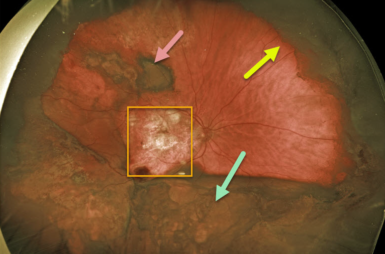
PERIPHERAL EXUDATIVE HEMORRHAGIC CHORIORETINOPATHY
Originally posted on @retina.rocks March 30, 2021
This 80YO male had 20/80 vision in his left eye with ongoing Eylea injections for wet age-related macular degeneration (AMD). Vision was hand motion in his right eye due to an inactive disciform scar.
Several months ago, he complained of decreased vision OD and was found to have a dense vitreous hemorrhage. About 6 weeks later, he underwent vitrectomy in this eye to try to restore his peripheral vision.
One day postoperatively, Optos photography showed the residual vitreous skirt, subretinal blood, and elevated choroidal/suprachoroidal yellow blood.
Learning Points:
Ectopic choroidal neovascularization (CNV), also known as peripheral exudative hemorrhagic chorioretinopathy, is an exudative process similar to wet AMD but located in the retinal periphery.
Ectopic CNV can often masquerade as a choroidal mass or uveal melanoma. It can also cause extensive choroidal/suprachoroidal, subretinal, and vitreous hemorrhage, more commonly in patients who are on blood thinners (our patient was on Pradaxa).
The subretinal blood often spontaneously regresses without treatment, although anti-VEGF injections can be used for symptomatic or increasing exudation.
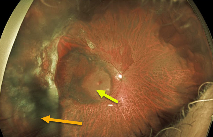
PERIPHERAL EXUDATIVE HEMORRHAGIC CHORIORETINOPATHY
Originally posted on @retina.rocks January 15, 2021
This 71YO male had a vitrectomy in 2018 for a breakthrough vitreous hemorrhage from ectopic choroidal neovascularization (CNV). He did well with 20/40 vision until a recurrent vitreous hemorrhage developed two years later.
The vitreous cleared with several monthly intravitreal Avastin injections, but vision remains at hand motion due to submacular blood, some of which is yellow and devitalized. We discussed repeat vitrectomy with subretinal TPA, but he elected for in-office anti-VEGF injections. He is not on any systemic blood thinners.
Learning Points:
Ectopic CNV, also known as peripheral exudative hemorrhagic chorioretinopathy, is a degenerative and exudative process similar to wet AMD but located in the retinal periphery.
Ectopic CNV can often masquerade as a choroidal mass or uveal melanoma. It can also cause extensive subretinal and vitreous hemorrhage, more commonly in patients who are on blood thinners.
The subretinal blood often spontaneously regresses without treatment, although anti-VEGF injections can be used for symptomatic or increasing exudation.
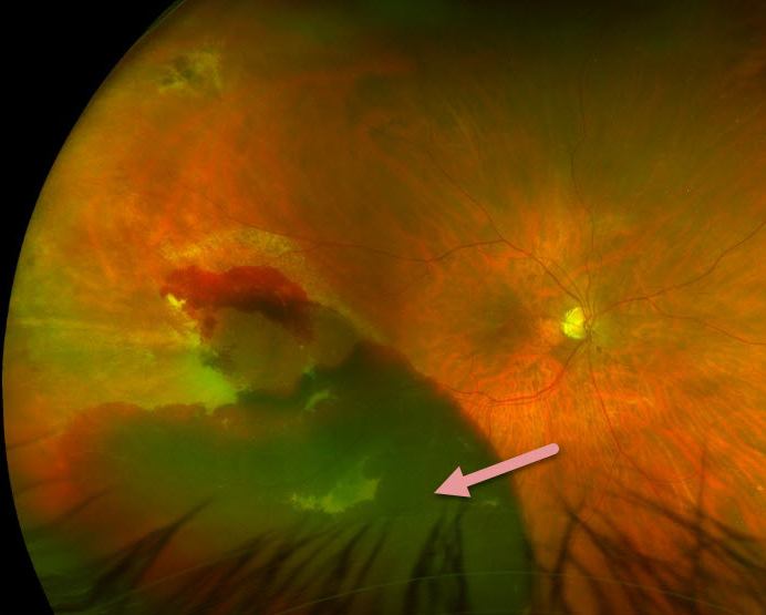
PERIPHERAL EXUDATIVE HEMORRHAGIC CHORIORETINOPATHY
Originally posted on @retina.rocks June 12, 2020
This 65YO female has a fresh peripheral subretinal hemorrhage.
The subretinal blood often regresses spontaneously, as occurred in this patient (later images not shown).
Learning Points:
Peripheral exudative hemorrhagic chorioretinopathy (PEHCR) is essentially choroidal neovascularization occurring in the retinal periphery.
PEHCR can be mistaken for uveal melanoma because the dark subretinal blood can simulate a pigmented choroidal tumor.
PEHCR seems to share features with polypoidal choroidal vasculopathy.
Anti-VEGF therapy is helpful when needed. Patients can also develop a breakthrough vitreous hemorrhage, which also sometimes requires vitrectomy.
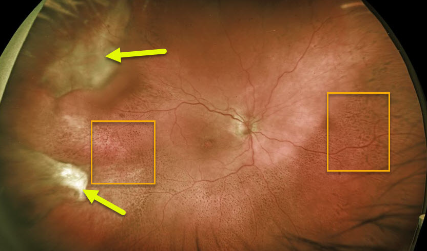
PERIPHERAL EXUDATIVE HEMORRHAGIC CHORIORETINOPATHY
Originally posted on @retina.rocks January 30, 2020
This patient has involuted neovascularization with subretinal fibrosis in the temporal and superotemporal periphery.
The coarse pigment clumping seen throughout the temporal, inferior and nasal retina is pathognomonic for prior resolved subretinal blood.
Fortunately the entire process spares the macular center, leaving the patient with normal central vision.
Learning Points:
Ectopic choroidal neovascularization, also known as peripheral exudative hemorrhagic chorioretinopathy, is a degenerative and exudative process like wet AMD but found in the retinal periphery.
Ectopic neovascularization can appear like a choroidal mass or uveal melanoma and is often misdiagnosed as such. It can also cause subretinal and vitreous hemorrhage.


