

Peripheral Microvascular Abnormalities
<< Back to Cases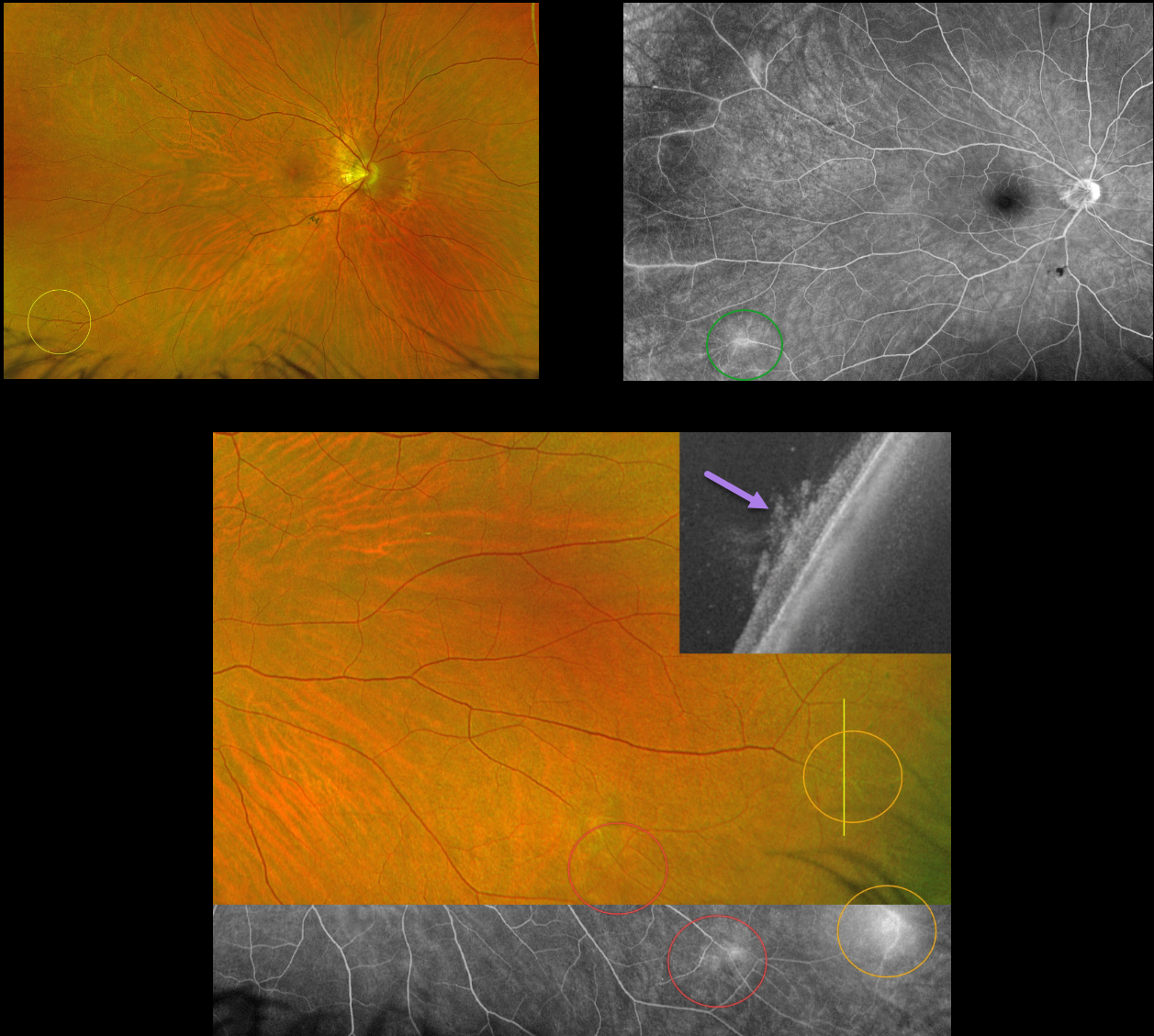
POSSIBLE CELIAC RETINAL NEOVASCULARIZATION
Paulo Eduardo Stanga, Javier Valentin Bravo, and Sebastian Stangam
Originally posted on @retina.rocks June 6, 2023
This 55YO female presented with asymptomatic peripheral retinal vascular findings. She has a history of high myopia and celiac disease. Vision was 20/20 OU.
Optos imaging of the OD shows telangiectatic vessels in the inferotemporal periphery with angiographic leakage in this region and superotemporally. Two similar areas are noted inferotemporally in the OS. Vertical UWF-OCT scanning through the most temporal lesion confirms that this represents retinal neovascularization growing into the vitreous.
Learning Points:
In 2016, Shah et al described common peripheral retinal vascular findings on Optos UWF FA in otherwise healthy adults (58 eyes, 31 patients) undergoing imaging for unrelated macular pucker or choroidal nevi (Retina 2016;36:1087-1092). Findings included vessels crossing the horizontal raphe (45%), right-angle vessels (71%), terminal networks (78%), absence of capillary detail (98%), ground glass hyperfluorescence (88%), drusen (34%), and microaneurysms (41%). One or more of these findings were present in most eyes.
Another study by Lu et al. found that normal peripheral retinas typically exhibit granular background fluorescence, with or without a mottled fluorescent band (AJO 2017;173:84-90).
Since integrating Optos UWF imaging into our practice a few years ago, we have observed similar findings in many of our patients undergoing FA for unrelated diagnoses. In the beginning, we often checked carotid doppler and ultrasonography, which were invariably normal. We now also consider these normal peripheral findings and no longer routinely work up these patients. Retinal neovascularization has not been described in this entity.
Celiac disease is associated with uveitis, subclinical macular microvascular changes, central retinal vein occlusion, and retinal findings from vitamin A deficiency (Fousekis et al, Int Ophthalmol 2020;40:1049-1054). Peripheral retinal ischemia with neovascularization has not been described. It is therefore unclear whether or not our patient’s Celiac disease contributed to these findings.
PERIPHERAL MICROVASCULAR ABNORMALITIES
Originally posted on @retina.rocks June 16, 2021
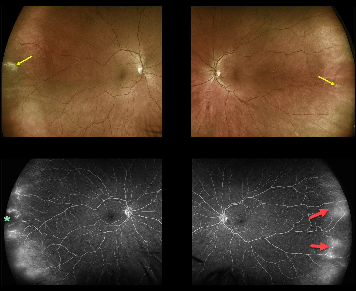
This healthy, asymptomatic 40YO female presented with 20/25 vision bilaterally.
Optos ultra-widefield (UWF) imaging showed far temporal peripheral probable lattice lesions and subtle telangiectasia. The vascular changes, including capillary dropout and telangiectasia, were more evident on fluorescein angiography (FA).
Learning Points:
In 2016, Shah et al described common peripheral retinal vascular findings on Optos UWF FA in otherwise healthy adults (58 eyes, 31 patients) undergoing imaging for unrelated macular pucker or choroidal nevi (Retina 2016;36:1087-1092).
Findings included vessels crossing the horizontal raphe (45%), right-angle vessels (71%), terminal networks (78%), absence of capillary detail (98%), ground glass hyperfluorescence (88%), drusen (34%), and microaneurysms (41%). One or more of these findings were present in most eyes.
Another study by Lu et al. found that normal peripheral retinas typically exhibit granular background fluorescence, with or without a mottled fluorescent band (AJO 2017;173:84-90).


