

Persistent Fetal Vasculature and Hylaoid
<< Back to Cases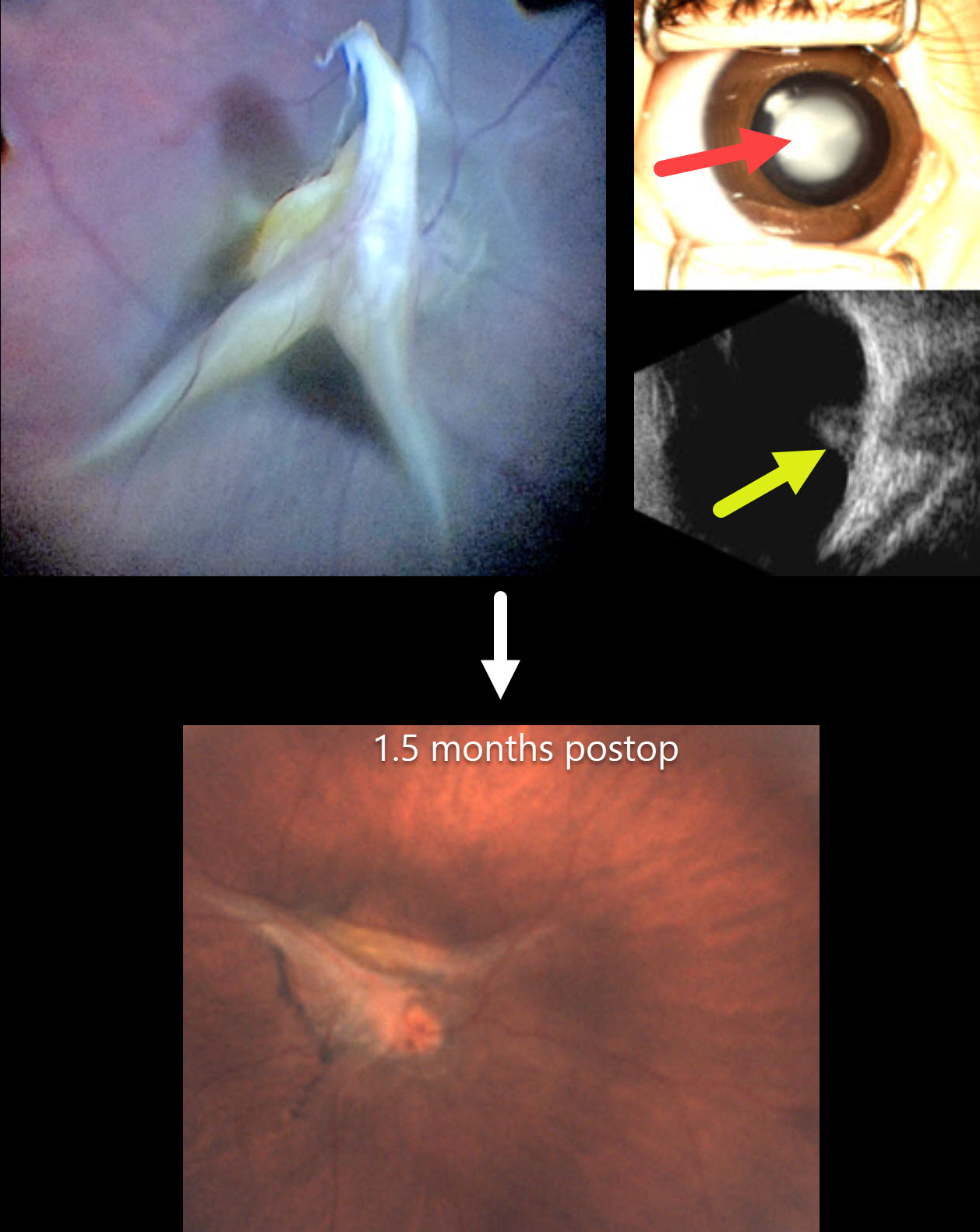
PERSISTENT FETAL VASCULATURE
European VitreoRetina Society (EVRS), Gökçen Deniz and Şengül Özdek
Originally posted on @retina.rocks December 19, 2025
This healthy 4-month-old girl, born full-term, presented with leukocoria in her right eye that her parents recently noticed.
Preoperatively, there was a white central cataract with a clear peripheral zone. B-scan ultrasonography showed a highly reflective stalk that extended from the disc to the periphery with a tent-like elevation over the disc. The left eye was normal.
At surgery, the lens was aspirated with vitrectomy to release all traction. Although the stalk overlying the nerve was trimmed, persistent blood flow was noted as stalk pulsations. The retina remained attached 1.5 months postoperatively.
This case was submitted by the European VitreoRetina Society (EVRS), Gökçen Deniz, and Şengül Özdek .
Learning Points:
Persistent fetal vasculature (PFV), previously recognized as persistent hyperplastic primary vitreous (PHPV), represents a failure of regression of fetal vessels. It is a common cause of leukocoria and subsequent amblyopia. There is no identifiable genetic association.
In PFV, the arterial blood supply from the disc to the anterior segment that is supposed to regress after birth does not. This results in a remnant of the fetal hyaloid artery remaining abnormally patent. The arterial flow from this remnant can cause cataract, intraocular hemorrhages, and retinal detachment, as the abnormal vessel continues to be supplied by systemic arterial pressure.
The main surgical pearl of this case is that, when you have a clear zone around the central opacity of PFV, such eyes usually do not have peripheral retinal extensions through the ciliary body, which allows safe surgical entry through the pars plicata. One still needs to check internally that the incisions are anterior to the peripheral retina, and limbal entry is needed.
PERSISTENT FETAL VASCULATURE
Gokcen Deniz Gulpinar Ikiz and Sengül Özdek
Originally posted on @retina.rocks September 16, 2024
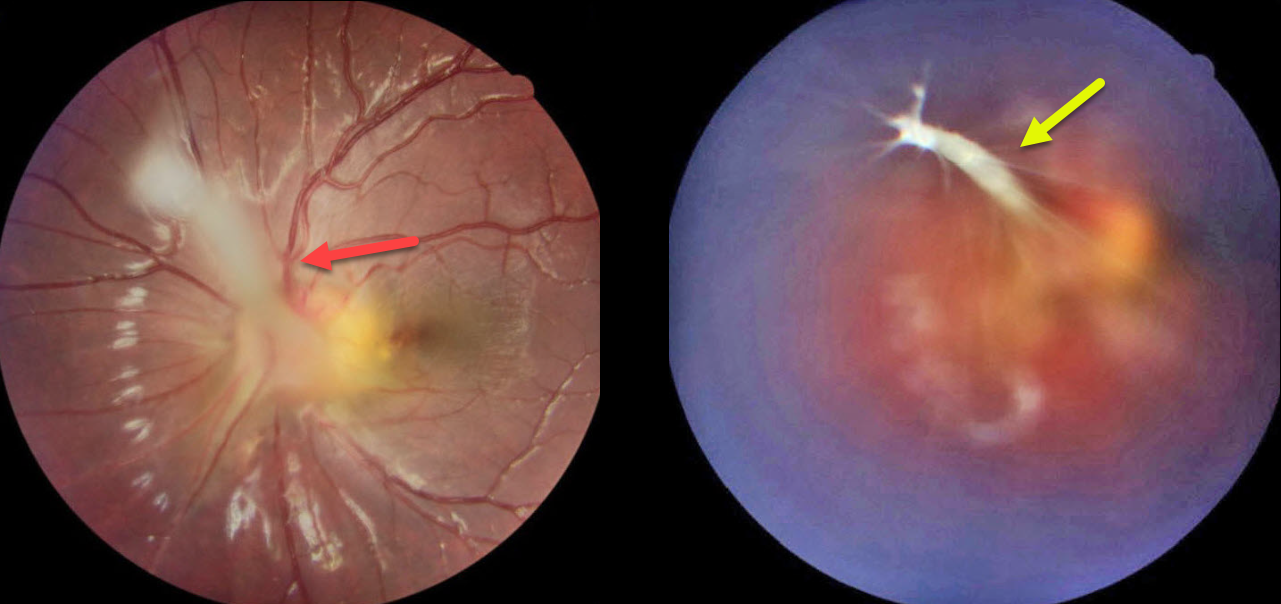
This 7YO girl presented with blurred vision in her esotropic left eye, which had previously been treated with patching of her normal right eye. Vision was 20/20 OD and 20/50 OS. Refraction was +0.25 +0.25 x180 OD and +1.00-1.50 x60 OS.
Color imaging of her left eye shows a fibrotic stalk that extends from the optic nerve head superonasally, terminating in the mid-vitreous in a spider web configuration. The macula is dragged towards the nerve, causing a shallow nasal foveal traction detachment. Lens-sparing vitrectomy will be performed in the near future.
Learning Points:
Persistent fetal vasculature (PFV), previously recognized as persistent hyperplastic primary vitreous (PHPV), represents a failure of regression of fetal vessels. It is a common cause of leukocoria and subsequent amblyopia. There is no identifiable genetic association.
Characteristically, the affected eye is microphthalmic with a shallow chamber predisposed to secondary angle closure glaucoma, a varying degree of cataract, and retrolental and/or posterior fibrovascular membranes that can cause retinal traction. If diagnosed early in life, PFV can be surgically treated with a combined lensectomy and vitrectomy. Anterior-type PFV is associated with better vision than combined or posterior types, and younger age at presentation correlates with improved vision after treatment.
PERSISTENT FETAL VASCULATURE
Emma Oreškovič and Nataša Drača
Originally posted on @retina.rocks January 16, 2024
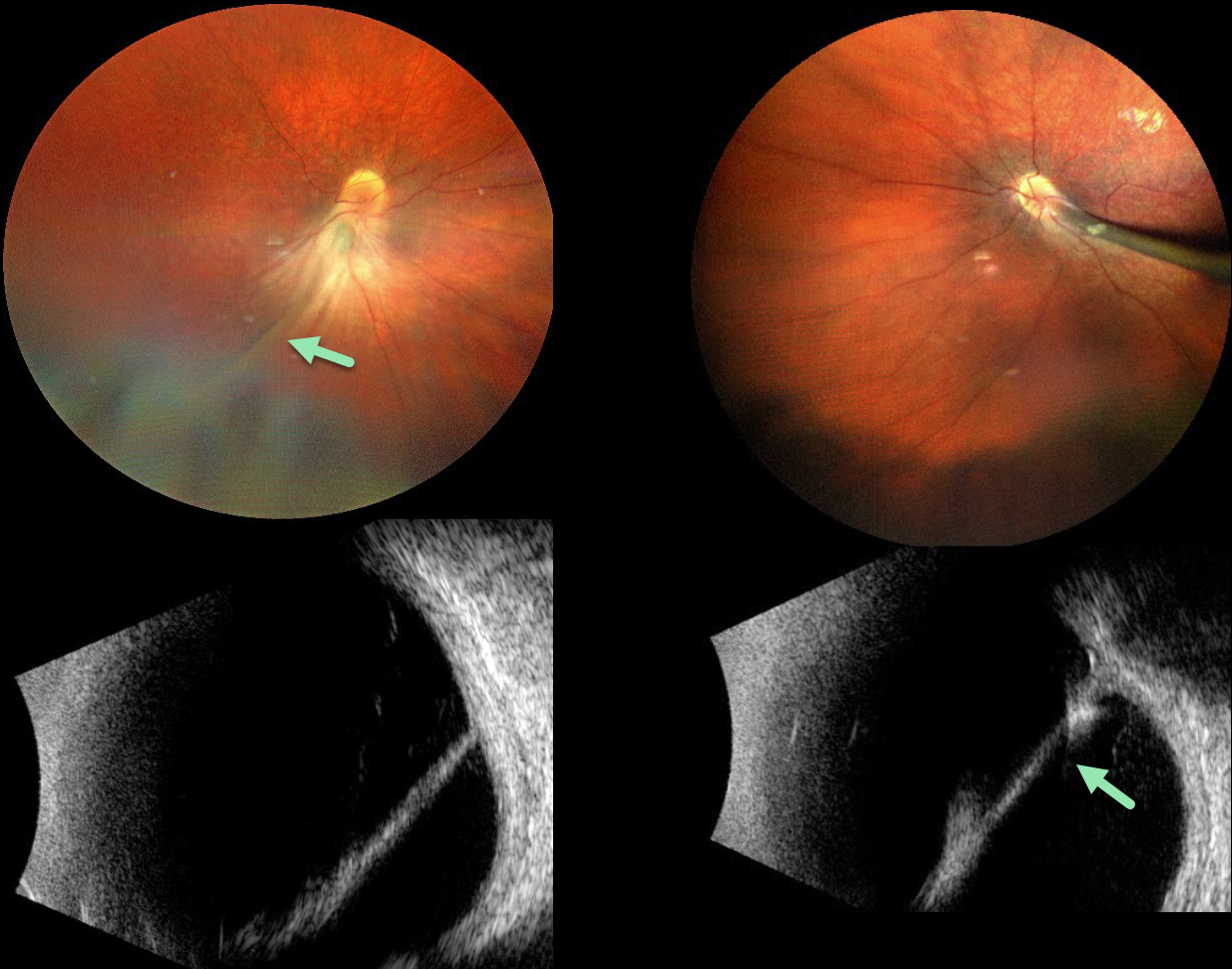
The 9YO girl presented with a known history of persistent fetal vasculature (PFV) since age 4. She was the product of a normal, full-term pregnancy. Vision was 20/400 OU. She also has congenital nystagmus.
Color photography shows bilateral fibrous stalks extending from the optic disc to the posterior lens capsule. The stalks are hyperreflective on B-scan ultrasonography. Falciform folds are also observed. Each retina was otherwise completely attached. She was referred to a pediatric retinal specialist for further management.
Learning Points:
Persistent fetal vasculature (PFV), previously recognized as persistent hyperplastic primary vitreous (PHPV), represents a failure of regression of fetal vessels. It is a common cause of leukocoria and subsequent amblyopia. There is no identifiable genetic association.
Characteristically, the affected eye is microphthalmic with a shallow chamber predisposed to secondary angle closure glaucoma, a varying degree of cataract, and retrolental and/or posterior fibrovascular membranes that can cause retinal traction.
If diagnosed early in life, PFV can be surgically treated with a combined lensectomy and vitrectomy. Anterior-type PFV is associated with better vision than combined or posterior types, and younger age at presentation correlates with improved vision after treatment.
For a review of PFV signs and symptoms, see Goldberg, AJO 1997;124:587-626.
CHOROIDAL COLOBOMA + PERSISTENT FETAL VASCULATURE
Lesia Khrystych
Originally posted on @retina.rocks May 22, 2023
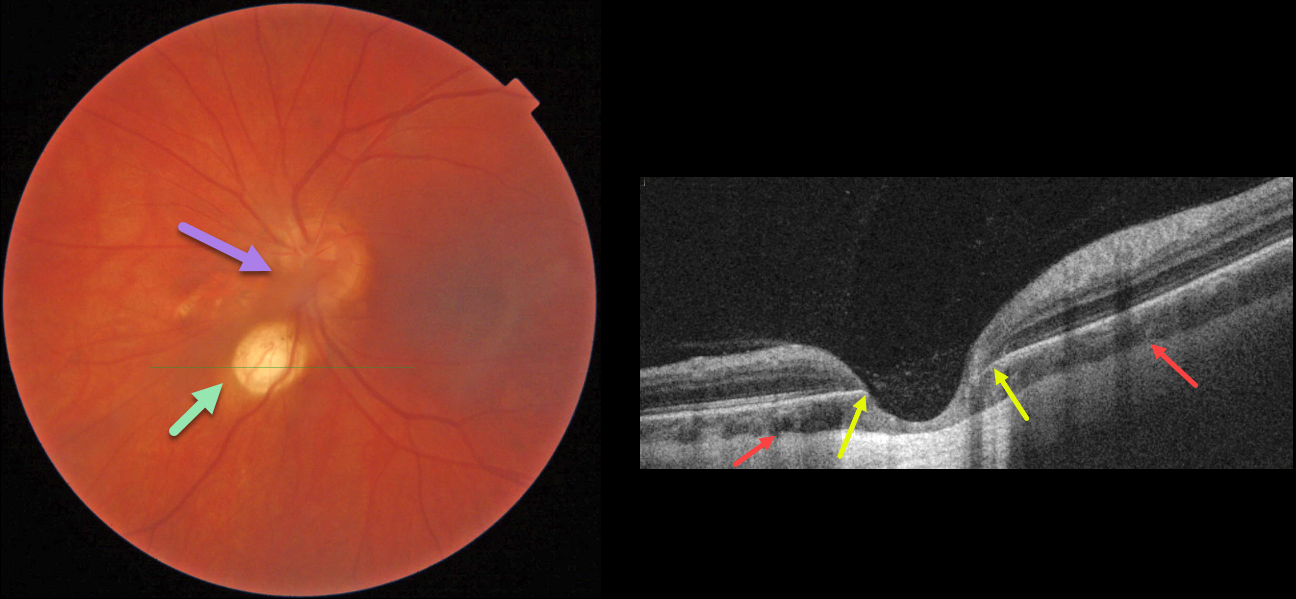
This 35YO female has a history of decreased vision in her left eye since childhood. Vision was 20/20 in her right eye and stable at 20/200 in her left eye. Anterior segments were normal except for a posterior polar cataract OS.
Color imaging of the left posterior pole shows a fairly round, one-disc-diameter depigmented lesion just inferonasal to the disc. An area of non-specific pigmentary changes is noted just nasal to the nerve, along with persistent fetal vasculature (hyaloid artery).
OCT through the depigmented lesion shows a choroidal coloboma. The underlying sclera is intact and slightly bowed posteriorly. Within the coloboma, there is loss of the outer retinal layers with draping of the inner retina against the bare sclera. The RPE is also absent within the coloboma.
Learning Points:
Choroidal colobomas result from a failure of closure of the optic vesicle and choroidal fissure during normal fetal development, but can also be caused by congenital toxoplasmosis and Zika virus. They can be solitary or multiple, and the visual potential depends on the extent of optic nerve and macular involvement. Patients with choroidal colobomas are at an increased risk for retinal detachment and macular neovascularization.
Embryologically, the hyaloid artery is vital to the proper development of the anterior segment, and usually regresses at about 10 weeks of gestation; however, it may persist in some cases. In severe cases, the eye may be microphthalmic with leukocoria and retinal traction. These eyes are typically amblyopic. A genetic association has not been identified.
PERSISTENT FETAL VASCULATURE
Originally posted on @retina.rocks August 23, 2022
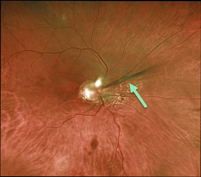
This 48YO male gave a history of blindness in his right eye since birth. Vision was no light perception. The anterior chamber was shallow with a stalk of tissue radiating from the nerve to the back of his crystalline lens. The macula was hypoplastic. The left eye was completely normal.
Learning Points:
Persistent fetal vasculature (PFV) is a failure of regression of fetal vessels and a common cause of leukocoria and subsequent amblyopia. There is no identifiable genetic association.
Characteristically, the affected eye is microphthalmic with a shallow chamber, a varying degree of cataract, and retrolental and/or posterior fibrovascular membranes that can cause retinal traction.
If diagnosed earlier in life, PFV can be surgically treated with a combined lensectomy and vitrectomy.
We are observing our patient, and it is unclear why he has no light perception vision, given the relatively mild structural findings.
PERSISTENT HYALOID ARTERY
Originally posted on @retina.rocks November 29, 2021
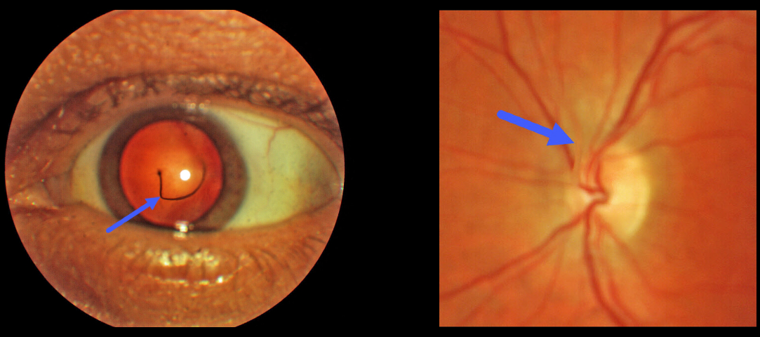
This 44YO male was referred for an asymptomatic vitreous floater. Upon examination, a large translucent strand was noted posterior to the lens, connecting the posterior capsule of the lens to the optic nerve.
Learning Points:
The hyaloid vascular system, embryologically located within the hyaloid canal, provides the developing anterior segment with nutrients. The hyaloid artery usually regresses around 10 weeks of gestation, but in some cases, it may not fully regress.
An anterior remnant may remain on the posterior capsule, which is known as a Mittendorf Dot. Posterior remnants, attached to the optic disc, are otherwise known as Bergmeister’s papilla.


