

Pigmented Paravenous Chorioretinal Atrophy
<< Back to Cases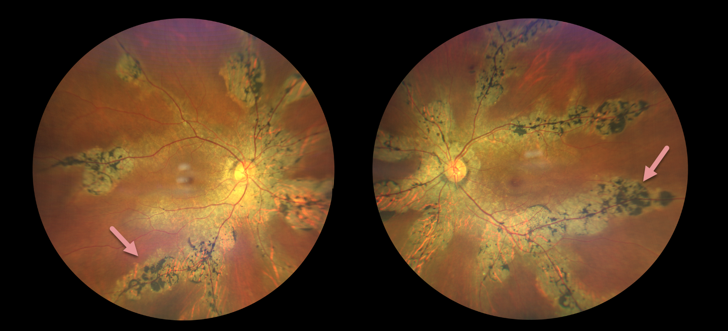
PIGMENTED PARAVENOUS CHORIORETINAL ATROPHY
Brahim KhourI, Oriana Gomez, Sebastian Polo, and Axel Ramos Nuñez Cazares
Originally posted on @retina.rocks August 14, 2026
This healthy 55YO female presented with 2 years of mild bilateral vision loss. Family history was negative. Vision was 20/20 OD and 20/40 OS.
Color photography shows bilateral paravenous chorioretinal scarring with intraretinal pigment migration.
Learning Points:
Pigmented paravenous chorioretinal atrophy (PPCRA) is a rare condition characterized by paravenous pigment clumps with peripapillary and radial zones of retinal pigment epithelial (RPE) atrophy (Lee et al, AJO 2021;224:120-132). It is bilateral, often asymmetric, with various causes, including genetic and pseudo-PPCRA (inflammatory, infectious, or unknown). Patients may be asymptomatic or present with symptoms such as nyctalopia, while central visual acuity is often relatively preserved in the absence of macular involvement. The underlying pathophysiology remains incompletely understood, although primary choroidal and RPE abnormalities have been proposed to contribute to the characteristic retinal changes.
PIGMENTED PARAVENOUS CHORIORETINAL ATROPHY
Will Gibson
Originally posted on @retina.rocks December 26, 2024
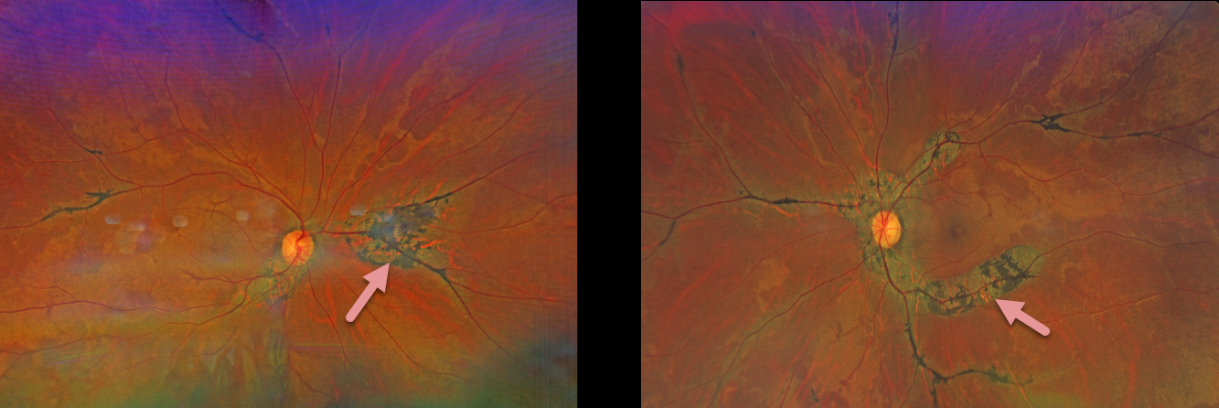
This 67YO male presented with asymptomatic retinal findings. There was no family history of ocular disease. Vision was 20/20 OU.
Color photography shows bilateral paravenous chorioretinal scarring with intraretinal pigment migration.
Learning Points:
Pigmented paravenous chorioretinal atrophy (PPCRA) is a rare condition characterized by paravenous pigment clumps with peripapillary and radial zones of RPE atrophy. It is bilateral, often asymmetric, with various causes, including genetic and pseudo-PPCRA (inflammatory, infectious, or unknown). Patients often have symptoms, usually nyctalopia, but most retain good vision, especially those without macular involvement. The underlying pathophysiology is likely a primary choroidopathy, which then causes overlying RPE and retinal changes. The chorioretinal changes are best visualized with fundus autofluorescence, which was unfortunately unavailable for our patient.
PIGMENTED PARAVENOUS CHORIORETINAL ATROPHY
Originally posted on @retina.rocks January 14, 2022
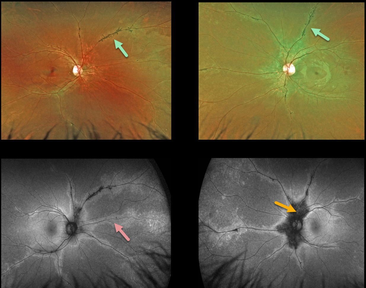
This 24YO female presented with 20/20 vision bilaterally and no visual symptoms. Optos ultrawidefield imaging shows normal central maculas with intraretinal pigment migration and depigmented RPE along the retinal veins. These changes extend from the peripapillary retina into the retinal peripheries.
Fundus autofluorescence (FAF) best highlights these changes with peripapillary hypo-FAF and paravenous hyper-FAF.
Our patient underwent Spark genetic testing. Results showed a pathogenic TNEM216 variant, which is associated with autosomal recessive Joubert Syndrome. However, she had absolutely no signs or symptoms of this syndrome.
Learning Points:
Pigmented paravenous chorioretinal atrophy (PPCRA) is a rare condition characterized by paravenous pigment clumps with peripapillary and radial zones of RPE atrophy.
It is bilateral, often asymmetric, with various causes, including genetic and pseudo-PPCRA (inflammatory, infectious, or unknown).
Patients tend to have some symptoms, usually nyctalopia, but most retain good vision, especially those without macular involvement.
The underlying pathophysiology is likely a primary choroidopathy that causes overlying RPE and retinal changes. The chorioretinal changes are best visualized with FAF. For a great clinical review, see Lee et al, AJO 2021;224:120-132.
PIGMENTED PARAVENOUS CHORIORETINAL ATROPHY
Originally posted on @retina.rocks September 15, 2020
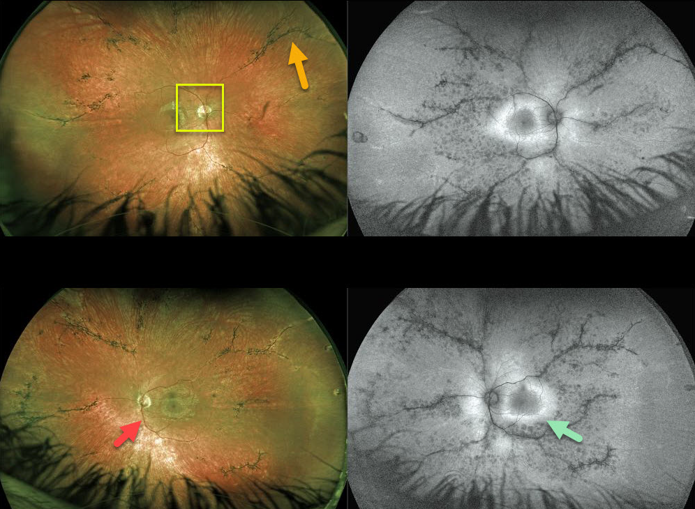
This 16YO patient with pigmented paravenous chorioretinal atrophy has excellent central vision of 20/20 OU but with significant nyctalopia.
Posterior segment findings include attenuated retinal vessels, bone spicules (intraretinal migration of the RPE along retinal capillaries), and optic disc pallor.
The bone spicules in this patient are primary perivascular, indicating the likely diagnosis of pigmented paravenous chorioretinal atrophy (PPCRA).
Fundus autofluorescence for this patient shows a ring of hyperautofluorescence around each central macula. This ring identifies at-risk retina for future degeneration and can help monitor the progression of this disease.
Learning Points:
Pigmented paravenous chorioretinal atrophy (PPCRA) is a rare condition characterized by paravenous pigment clumps with peripapillary and radial zones of RPE atrophy. It is bilateral, often asymmetric, with various causes, including genetic and pseudo-PPCRA (inflammatory, infectious, or unknown). Patients tend to have some symptoms, usually nyctalopia, but most retain good vision, especially those without macular involvement. The underlying pathophysiology is likely a primary choroidopathy that causes overlying RPE and retinal changes. The chorioretinal changes are best visualized with fundus autofluorescence.


