

Polypoidal Choroidal Vasculopathy (PCV)
<< Back to Cases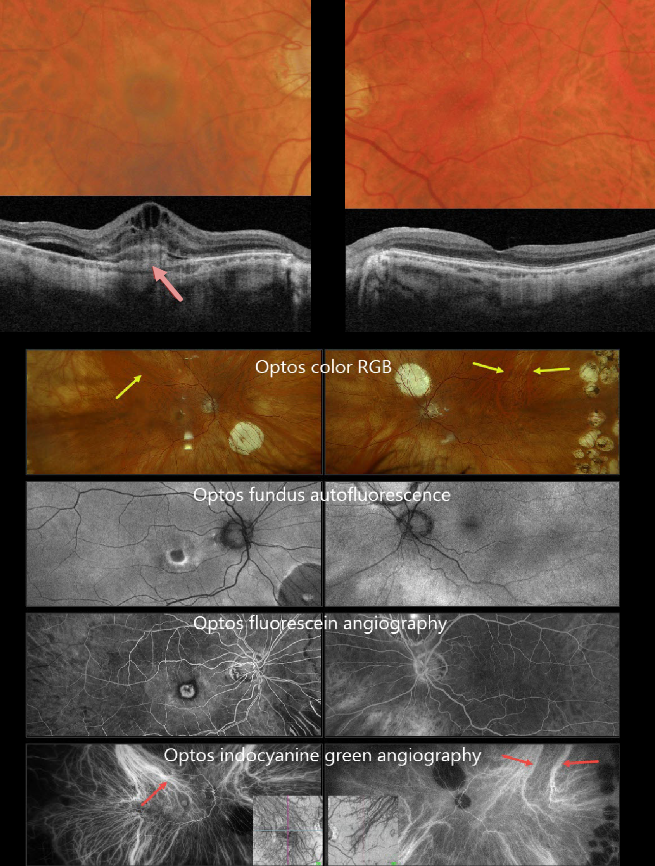
POLYPOIDAL CHOROIDAL VASCULOPATHY
Originally posted on @retina.rocks July 8, 2026
This 71YO white male underwent successful bilateral vitrectomies for rhegmatogenous retinal detachments by us years earlier. He had since moved out of state and was recently diagnosed with neovascular AMD in his right eye, which was treated with an initial anti-VEGF injection one month earlier. After moving back to our area, vision was 20/400 OD and 20/20 in his normal OS.
Triton color photography shows a poor foveal reflex with a surrounding subretinal pigment ring. Barely visible, smaller drusen are noted more peripherally. Swept-source OCT shows a probable type 1 (below the RPE) macular neovascularization (MNV) with overlying subretinal hyperreflective material, intraretinal and subretinal fluid. There are multiple smaller drusen in his left macula. Although the choroid appears thinned bilaterally, the vessels that are present mostly span the entire choroidal thickness.
Optos fundus autofluorescence (FAF) shows the MNV to be hypo-FAF with a rim of surrounding hyper-FAF (image 2). The MNV is hyperfluorescent on fluorescein angiography, surrounded by a rim of blockage from the subretinal pigment. The choroid is markedly abnormal on indocyanine green angiography, revealing that each macula is drained by the superotemporal choroidal vortex. These dilated choroidal vessels are also visible on color RGB imaging.
Learning Points:
Polypoidal choroidal vasculopathy (PCV) is a variant of type 1 macular neovascularization. PCV is likely distinct from age-related macular degeneration, occurring in a different population (more common in African Americans and Asians) and characterized by a relative lack of drusen and a thicker choroid. PCV presents differently in Caucasians, with multiple phenotypes that can include coexisting drusen, the presence or absence of a branching choroidal neovascular network, or background central serous chorioretinopathy findings (Sirks et al., Ophthalmology Retina 2025;9:994-1004).
PCV is part of the pachychoroid spectrum, more recently termed venous overload choroidopathy, which appears to be caused by relative vortex outflow resistance (Spaide et al., Progress Retinal Eye Research 2022;86;100973). The choroidal vortex system usually respects the horizontal meridian; anastomoses that cross it, as occurred in our patient, are characteristic of these eyes.
Historically, these lesions were believed to be somewhat resistant to anti-VEGF monotherapy and often required combined treatment with photodynamic therapy. More recent data from the PLANET study suggest that Eylea monotherapy is also effective (AJO 2019;204:80-89).
Given the lack of response to initial anti-VEGF therapy, intravitreal Eylea was given, followed by PDT 1 week later. Six weeks following PDT, vision remained stable, but the OCT showed marked improvement. We will continue to follow him closely.
POLYPOIDAL CHOROIDAL VASCULOPATHY
Originally posted on @retina.rocks March 18, 2026
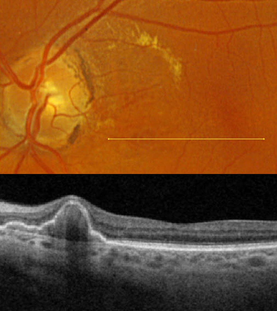
This 67YO female presented for a second opinion regarding possible age-related macular degeneration (AMD). She was without acute visual complaints. Vision was 20/25 in her normal OD and 20/30 OS.
Optos color RGB imaging shows macula lipid in the superonasal macula. Several red subretinal nodules are present inferior to the lipid. Triton swept-source OCT through these lesions shows a nodular and somewhat multi-lobulated RPE detachment (PED). We will follow her closely for any increase in exudation.
Learning Points:
At the Macula Society meeting in 1983 and at the Academy of Ophthalmology meeting in 1984, Brucker et al presented 7 patients with multiple recurrent sub-RPE and subretinal hemorrhages, which they termed the posterior uveal bleeding syndrome. This was followed by multiple authors reporting similar findings (Stern et al AJO 1985;100:560-569, Kleiner et al Retina 1990;10:9-17, and Yannuzzi et al Retina 1990;10:1-8). Dr. Gass, who was assigned as the reviewer for Yannuzzi’s manuscript titled “Idiopathic Polypoidal Choroidal Vasculopathy,” took issue with calling these lesions ‘polyps’ (Alexander J Brucker, personal communication 2/12/26), which are defined by Stedman’s Medical Dictionary as growths that protrude from a mucous membrane. When Yannuzzi challenged Dr. Gass for a better term, he could not come up with one; Yannuzzi’s terminology won out, and the rest is history. In our patient’s case, the clinical red ‘polyps’ corresponded to peaked, multilobulated RPE detachments on OCT.
PCV is likely distinct from age-related macular degeneration, occurring in a different population (more common in African Americans and Asians), with a relative lack of drusen and a thicker choroid. In our experience, many patients with ‘wet AMD’ actually have PCV or pachychoroid neovascularization.
POLYPOIDAL CHOROIDAL VASCULOPATHY WITH EXCISION OF SUBRETINAL BLOOD
The European VitreoRetinal Society (EVRS), Anjana Mirajkar, Akansha Sharma, Vaidehi Sathaye, Iam Pramodsuman and Manish Nagpal
Originally posted on @retina.rocks February 27, 2026
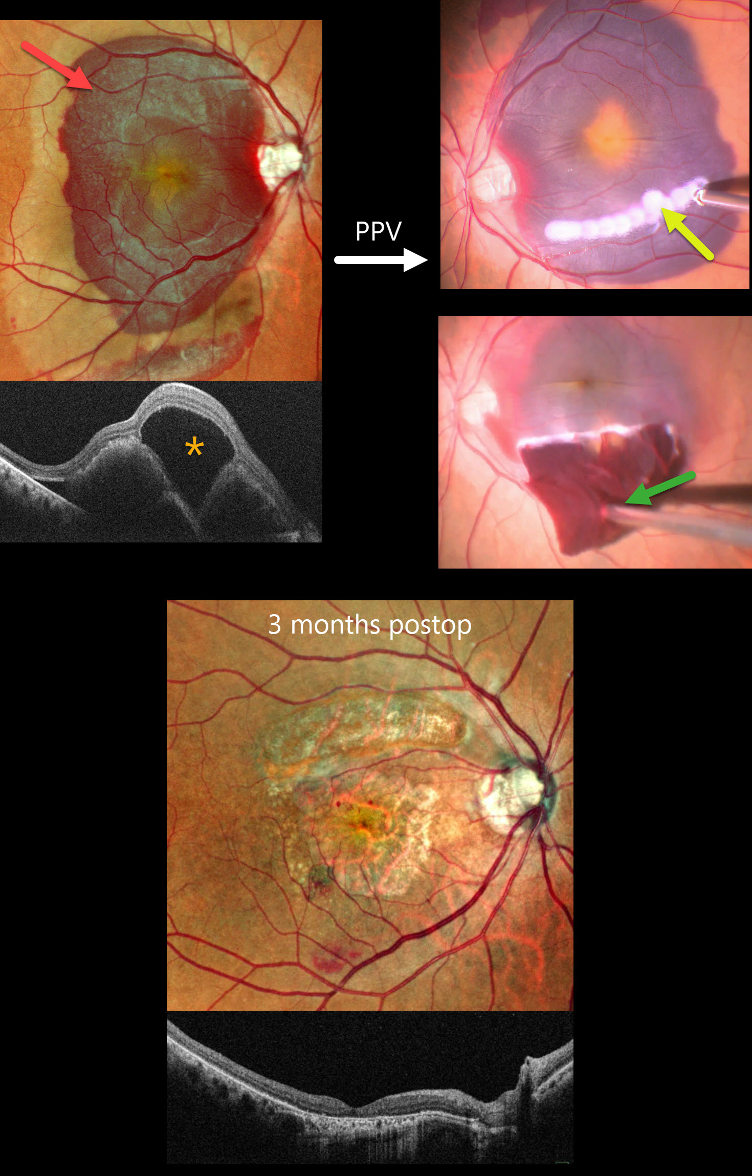
This 64YO female presented with 2 months of vision loss in her right eye. Vision was counting fingers OD and 20/30 in her normal OS.
Pseudocolor SLO imaging shows a large submacular hemorrhage. OCT scanning shows a central large bacillary layer detachment vs subretinal fluid overlying the elevated subretinal blood. Preoperative intravitreal tissue plasminogen activator (tPA) was injected, followed 2 days later by vitrectomy, a smile incision with excision of the subretinal clot, and gas tamponade.
Three months postoperatively, dry macular scarring is noted, and vision improved to 20/200.
Learning Points:
Common causes of submacular hemorrhage include neovascular age-related macular degeneration, polypoidal choroidal vasculopathy (PCV, our patient), trauma, and retinal arterial macroaneurysms. Multiple treatment options are available, including observation, anti-VEGF therapy, pneumatic displacement with positioning, and pars plana vitrectomy.
Surgery for thick submacular hemorrhage is often recommended due to the fear of possible toxicity from heme byproducts or fibrin formation (Toth et al, Arch Ophthalmol 1991;109:723-729). However, it is uncertain whether surgical results are better than anti-VEGF monotherapy (Gabrielle et all, Ophthalmology 2023;130:947-957). The value of adding TPA to any of these surgical options is also uncertain (Chotikkakamthorn et al, Retina 2025;45:394-401).
DOMINANTLY INHERITED RADIAL BASAL LAMINAR DRUSEN WITH POSSIBLE POLYPOIDAL CHOROIDAL VASCULOPATHY
Malvika Singh and Manish Nagpal
Originally posted on @retina.rocks June 25, 2025
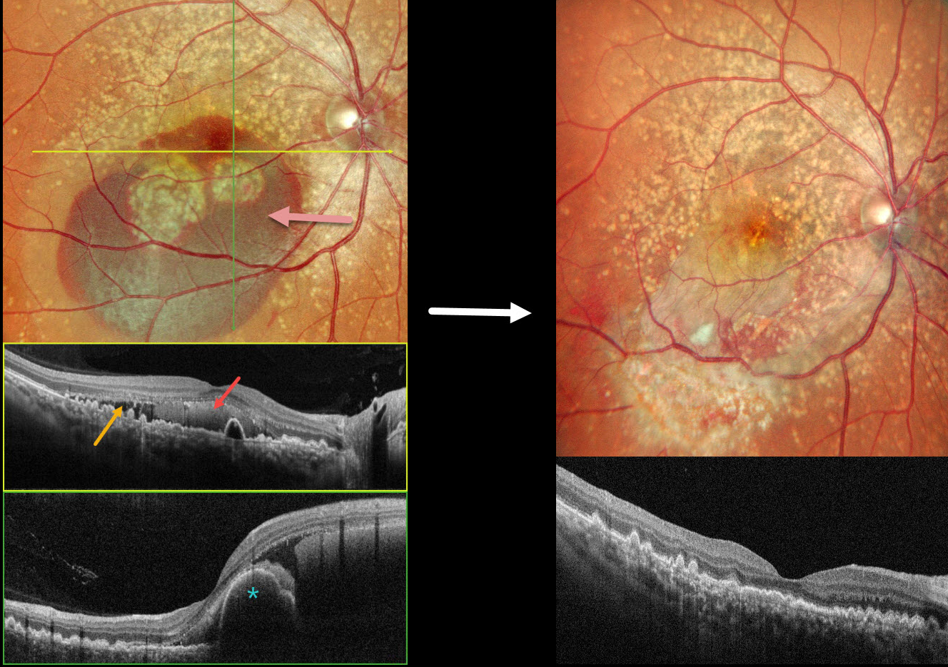
This 49YO female presented with 3 days of decreased vision in her OD. Vision was 20/70 OD and 20/15 OS. There was no family history of eye disease.
Pseudocolor SLO imaging shows extensive mixed and confluent drusen extending through each macula into the midperipheries. Inferior submacular blood extends into the right foveal center. OCT scanning shows extensive conically shaped drusen. The subretinal blood in her right eye is hyperreflective, with additional temporal subretinal fluid. A bilobed nodular RPE detachment is present within the nasal blood.
She underwent pars plana vitrectomy, fluid-air exchange, and intravitreal Avastin. One month postoperatively, vision improved to 20/15 OD with resolved fluid and trace residual blood.
Learning Points:
Doyne’s honeycomb macular dystrophy, also known as dominantly inherited radial basal laminar drusen or Malattia Leventinese, is a rare macular disorder caused by a mutation in the EFEMP1 gene on chromosome 2p16. The EFEMP1 protein is a member of the fibulin family of extracellular matrix glycoproteins. The defective protein creates an abnormally thickened RPE basement membrane.
Centrally large, nodular, and confluent drusen are noted, along with a temporal radiating pattern of smaller cuticular drusen. Later, there may be variable amounts of RPE hyperplasia and fibrous metaplasia. Macular neovascularization, as in this case, may also develop. Our patient’s pre-op right OCT was suggestive of polypoidal choroidal vasculopathy, although this is not a known association with Doyne’s.
POLYPOIDAL CHOROIDAL VASCULOPATHY
Originally posted on @retina.rocks May 14, 2025
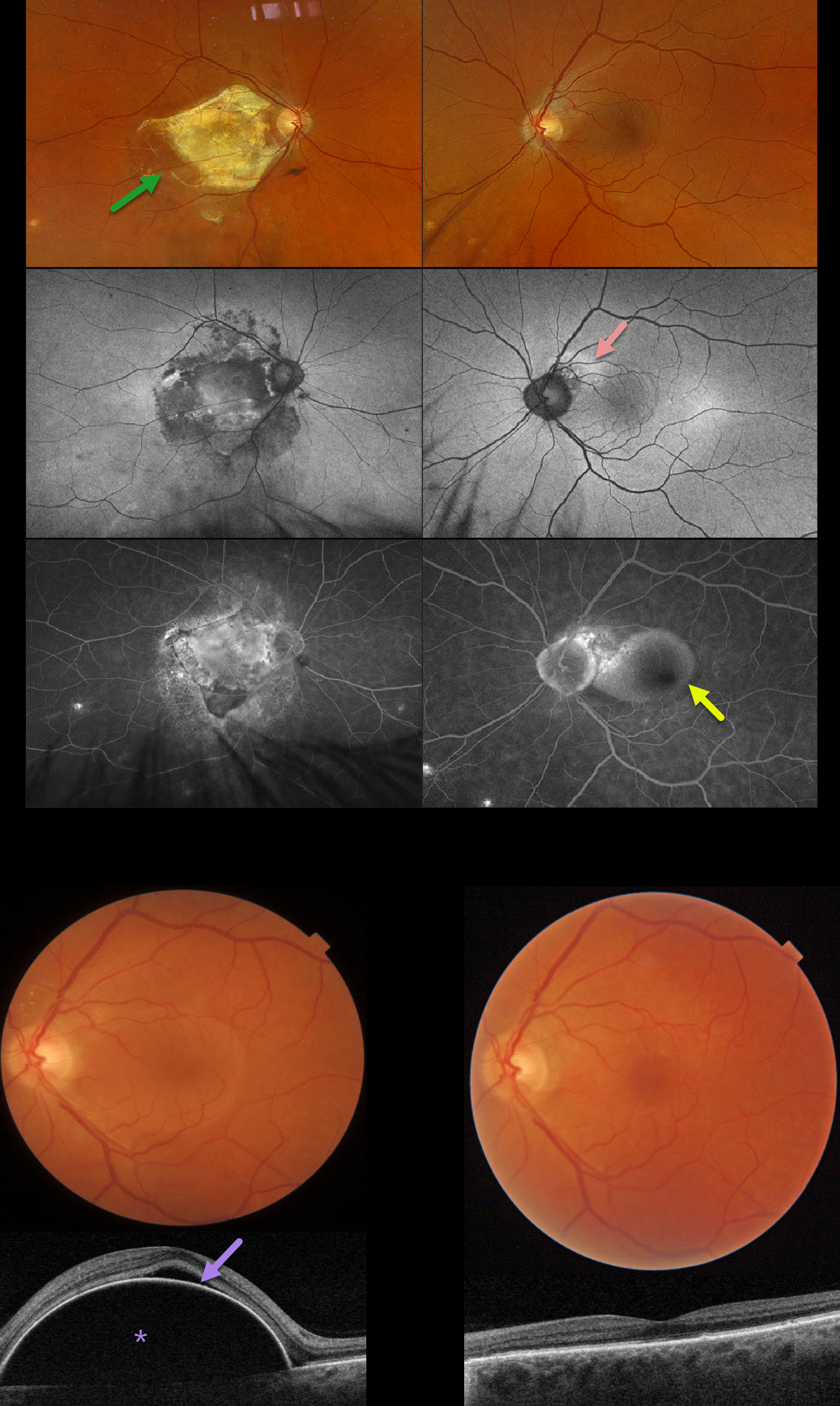
This 70YO male presented with 1 week of vision loss in his left eye. He has a long history of polypoidal choroidal vasculopathy (PCV). He received prior anti-VEGF injections OD, but his vision subsequently decreased to counting fingers from a fibrotic disciform scar due to poor compliance with office visits. Several years earlier, he developed a peripapillary polyp in his left eye that was resistant to Eylea monotherapy but responded well to combination treatment with photodynamic therapy (PDT).
Optos color RGB imaging shows a fibrotic disciform scar OD and a retinal pigment epithelial detachment (PED) OS. Fundus autofluorescence (FAF) shows variable hyper-FAF OS in the region of the prior polyp. Ill-defined angiographic leakage is noted superotemporal to the left nerve, with an uneven-filling PED. Triton swept-source OCT confirms a large and highly elevated PED with some overlying subretinal fluid.
Given the prior excellent response to combination treatment, intravitreal Eylea followed by PDT to the peripapillary notch was applied. Six weeks later, vision improved to 20/25, and the PED completely resolved with an almost normal OCT.
Learning Points:
Polypoidal choroidal vasculopathy (PCV) is a variant of type 1 macular neovascularization. PCV is likely distinct from age-related macular degeneration, occurring in a different population (more common in African Americans and Asians) and characterized by a thicker choroid and a relative lack of drusen. Historically, these lesions were believed to be somewhat resistant to anti-VEGF monotherapy, often requiring combined treatment with photodynamic therapy. More recent data from the PLANET study suggest that Eylea monotherapy is also effective (AJO 2019;204:80-89). However, our patient was resistant to monotherapy and responded remarkably well to combination therapy.
Originally described by Gass (Retina 1984;4:205-220), a notched PED is a sign of an occult MNV. The notch represents a type 1 MNV, which tethers the overlying RPE from the surrounding more highly elevated PED. Hydrostatic leakage from the MNV may contribute to the larger PED.
POLYPOIDAL CHOROIDAL VASCULOPATHY
Originally posted on @retina.rocks May 21, 2024
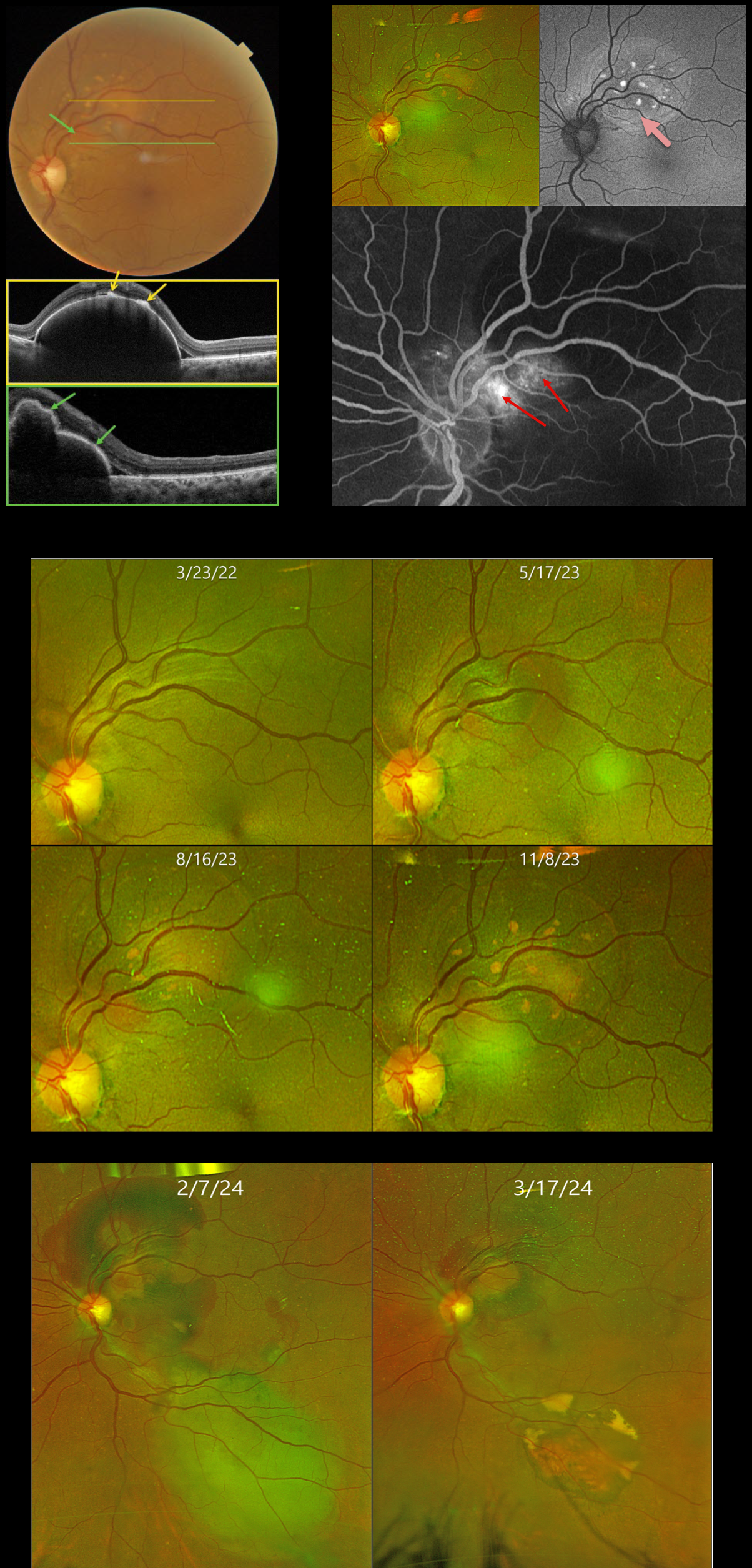
This 59YO female was initially referred to us on 3/23/22 with a 1-year history of vision loss in her right eye. Vision was counting fingers OD and 20/25 OS.
Over the past 10 months, a gradually enlarging RPE detachment developed in the left superonasal macula. On 11/8/23, Triton swept-source OCT confirms a highly elevated RPE detachment. The orange lipofuscin deposits seen on color imaging appear as hyperreflective lesions on OCT. Scanning through the reddish polyp shows a multi-lobulated elevation of the RPE. The lipofuscin deposits markedly hyper-autofluoresce. The large variably staining/leaking polyp creates a notch along the inferonasal edge of the PED.
Due to the patient’s history of a large macular hemorrhage resulting in counting fingers vision in her fellow eye, half-fluence photodynamic therapy (PDT) was applied to the polyp in her left eye. However, one month following PDT on 2/27/24, there was increased blood around the treated superior lesion, with new blood inferiorly.
Multiple full-fluence PDT applications were then applied to the old and new lesions. Six weeks later, the patient’s fundus was dramatically improved, with near-complete resolution of all blood and fluid. Vision was 20/20, and we will continue to follow her closely.
Learning Points:
Polypoidal choroidal vasculopathy (PCV) is a variant of type 1 macular neovascularization. PCV is likely distinct from age-related macular degeneration, occurring in a different population (more common among African Americans and Asians), with a relative lack of drusen and a thicker choroid. The sub-RPE vessels were classically described using indocyanine green angiography, although newer literature suggests that OCT is likely just as effective at diagnosing these vascular polyps (Permadi et al., Int J Retina Vitreous 2022;8;14).
Historically, these lesions were believed to be somewhat resistant to anti-VEGF monotherapy, often requiring combined treatment with photodynamic therapy. Newer data from the PLANET study suggest that Eylea monotherapy is also successful (AJO 2019;204:80-89).
Originally described by Gass (Retina 1984;4:205-220), a notched PED is a sign of an occult MNV. The notch represents a type 1 MNV, which tethers the overlying RPE from the surrounding more highly elevated PED. Hydrostatic leakage from the MNV may contribute to the larger PED.
POLYPOIDAL CHOROIDAL VASCULOPATHY
Originally posted on @retina.rocks November 27, 2023
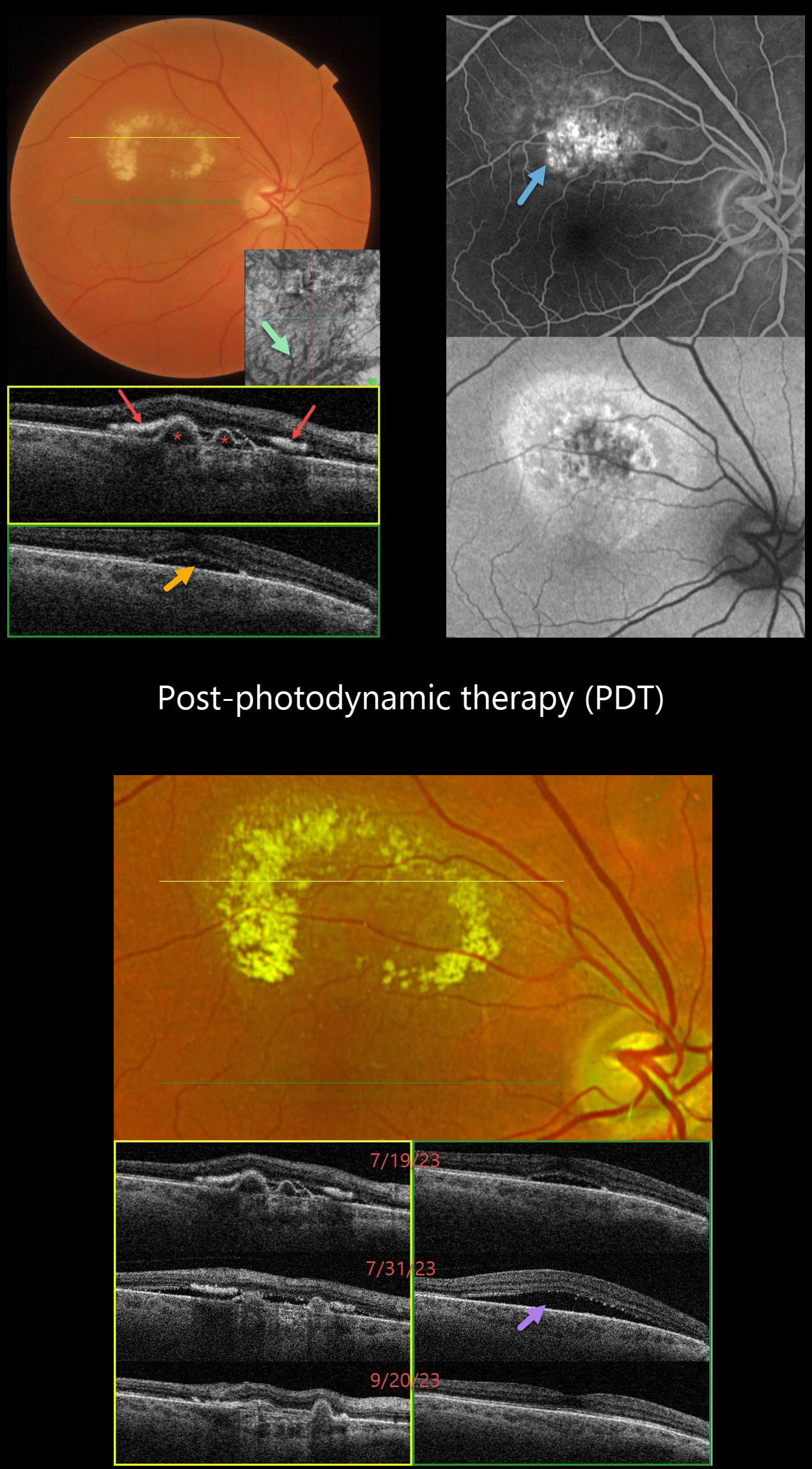
This 66YO female complained of 2 weeks of vision loss in her right eye. Vision was 20/40 OD and 20/20 in her normal OS.
Color imaging shows a circinate area of lipid in the superior macula. Triton swept-source OCT through the superior macula shows several sub-RPE polyps with hyper-reflective subretinal lipid. Scanning through the central macula shows shallow foveal subretinal fluid. Dilated inferior choroidal vessels are noted on en face OCT imaging.
Optos fluorescein angiography shows superior macular staining and leakage, with variable hyper-autofluorescence surrounding this region. Intravitreal Avastin was injected on 7/19/23, followed by photodynamic therapy (PDT) on 7/26/23.
She returned 5 days following PDT on 7/31/23 with increased blurriness. Vision remained at 20/40, although increased SRF was noted on OCT. Observation was recommended.
On 9/20/23, vision improved to 20/30, all SRF resolved, and the polyps were flattening.
Learning Points:
Polypoidal choroidal vasculopathy (PCV) is a variant of type 1 (below the RPE) macular neovascularization. It can be treated with either anti-VEGF monotherapy or combined with PDT to achieve better polyp regression and reduced treatment burden.
Transient PDT-induced increased exudative SRF sometimes develops immediately following treatment (Manayath et al, Retina 2020;40:135-144) but resolves spontaneously, as in our patient.
PACHYCHOROID SPECTRUM
Originally posted on @retina.rocks November 22, 2022
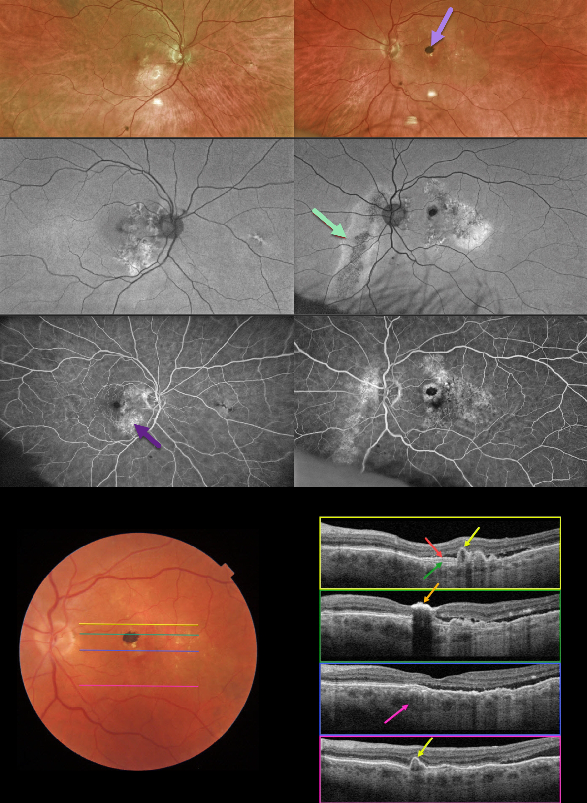
This 78YO male was referred for asymptomatic fundoscopic changes. Vision was 20/25 OD and 20/30 OS. He had a history of laser treatment in his left eye decades earlier.
Optos imaging shows bilateral areas of pigmentary loss in each macula, extending inferiorly. A hyperpigmented laser scar is noted in the left superior macula.
Fundus autofluorescence (FAF) shows variable hypo- and hyper-FAF, along with a prominent gutter extending from the left nerve inferonasally.
Fundus fluorescein angiography (FFA) shows window defects within these areas, with blockage from the hyperpigmented laser scar OS.
Swept-source OCT shows numerous findings. Scanning superior to the laser scar shows several choroidal polyps. A shallow elevation of the RPE exposes the underlying Bruch’s membrane. Scanning through the laser scar shows intense hyperreflectivity with posterior shadowing. Scanning through the macular center shows temporal subretinal fluid, along with a thickened choroid. Finally, scanning in the inferior macula shows an additional polyp.
Learning Points:
This case has an overwhelming amount of features from the pachychoroid spectrum, which includes idiopathic central serous retinopathy (ICSC), pachychoroid neovasculopathy, polypoidal choroidal vasculopathy, pachychoroid pigment epitheliopathy, peripapillary pachychoroid syndrome, and choroidal excavation.
The thermal laser treatment many years ago may have been for pachychoroid neovasculopathy, a recently described phenotype (see Pang and Freund, Retina 2015;35:1-9) found in the pachychoroid spectrum. These lesions are particularly common in patients with ICSC, and often remain silent as a flat, irregular RPE elevation on OCT without exudative complications.
Our patient likely had further progression of their pachychoroid disease, resulting in multiple polyps, which are found in polypoidal choroidal vasculopathy (PCV). PCV is a variant of type 1 macular neovascularization, also in the pachychoroid spectrum. PCV is likely distinct from age-related macular degeneration, occurring in a different population (more common in African Americans and Asians), with a relative lack of drusen, and the presence of a thicker choroid.
In addition to the findings above, the patient had a large gutter in his left eye, a common finding in ICSC. Gutters form from pathologies with chronic leakage. Conditions that form gutters other than ICSC include choroidal hemangiomas, choroidal nevi, and uveal melanoma.
For more information on the pachychoroid spectrum, see Cheung et al, Eye 2019;33:14-33 for a great pachychoroid disease review.
POLYPOIDAL CHOROIDAL VASCULOPATHY
Akansha Sharma and Manish Nagpal
Originally posted on @retina.rocks June 20, 2022
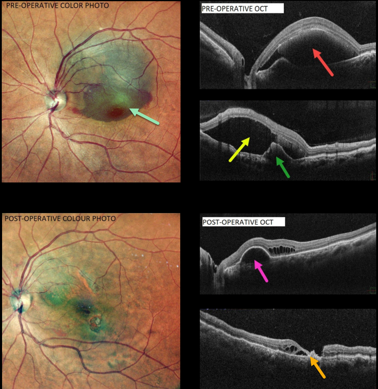
This 60 YO female presented with counting fingers vision from a large central and superior submacular hemorrhage.
OCT scanning shows variable subretinal blood and subretinal fluid along with an RPE detachment. There are no drusen.
Pars plana vitrectomy with drainage of the subretinal blood was performed. TPA was not used. Postoperatively, there is virtually no blood. OCT scanning shows no subretinal blood or fluid. Variable outer retinal cystic changes and atrophy are noted, along with a residual RPE detachment. Vision was 20/120.
Learning Points:
Polypoidal choroidal vasculopathy (PCV) is a variant of type 1 macular neovascularization. PCV is likely distinct from age-related macular degeneration, occurring in a different population (more common in African Americans and Asians), with a relative lack of drusen and a thicker choroid.
The sub-RPE vessels were classically characterized using indocyanine green angiography, although recent literature suggests that OCT is just as effective at diagnosing these vascular polyps. Historically, these lesions were believed to be somewhat resistant to anti-VEGF monotherapy and often required combined treatment with photodynamic therapy. More recent data from the PLANET study suggest that Eylea monotherapy is also effective (AJO 2019;204;80-89).
POLYPOIDAL CHOROIDAL VASCULOPATHY
Originally posted on @retina.rocks April 6, 2022
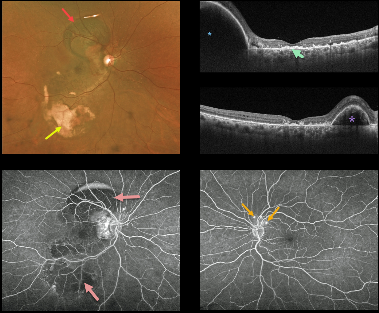
This 59YO female patient presented with variable mostly peripheral submacular blood in her right eye and counting fingers vision. Optos imaging shows the extent of the subretinal blood, which is dark superiorly and yellow inferiorly.
Triton swept-source OCT shows variable outer retinal and subretinal hyperreflective material from resolved submacular blood, and this is seen clinically as the coarse subretinal pigment most evident in the inferotemporal macula. A large RPE detachment (PED) is seen superiorly in the vertical scan, with a smaller PED nasally in the horizontal scan.
Fluorescein angiography shows mostly blockage from the subretinal blood. Several small asymptomatic polyps are noted superior to the left nerve.
Learning Points:
Polypoidal choroidal vasculopathy (PCV) is a variant of type 1 macular neovascularization. PCV is likely distinct from age-related macular degeneration, occurring in a different population (more common in African Americans and Asians), with a relative lack of drusen and a thicker choroid.
The sub-RPE vessels were classically characterized using indocyanine green angiography, although recent literature suggests that OCT is just as effective at diagnosing these vascular polyps.
Historically, these lesions were believed to be somewhat resistant to anti-VEGF monotherapy, often requiring combined treatment with photodynamic therapy.
More recent data from the PLANET study suggest that Eylea monotherapy is also effective (AJO 2019;204:80-89).
POLYPOIDAL CHOROIDAL VASCULOPATHY
Originally posted on @retina.rocks February 8, 2021
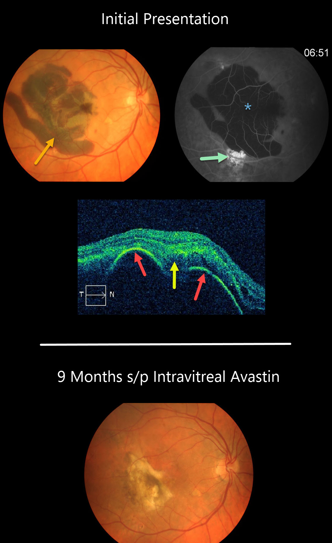
This patient presented with a large acute submacular hemorrhage in the right eye.
The blood caused diffuse blockage on fluorescein angiography, and the source of the blood may have been from the area of subretinal hyperfluorescence inferiorly.
OCT scanning shows a large retinal pigment epithelium (RPE) detachment with notched thumb-like projections with subretinal hyperreflectivity from the blood.
Our patient’s subretinal blood resolved following several intravitreal Avastin injections.
Learning Points:
Polypoidal choroidal vasculopathy (PCV) is a variant of type 1 macular neovascularization. PCV is likely distinct from age-related macular degeneration, occurring in a different population (more common in African Americans and Asians) and characterized by a relative lack of drusen and a thicker choroid.
The sub-RPE vessels were classically characterized using indocyanine green angiography, although recent literature suggests that OCT is just as effective at diagnosing these vascular polyps. Historically, these lesions were believed to be somewhat resistant to anti-VEGF monotherapy and often required combined treatment with photodynamic therapy. More recent data from the PLANET study suggest that Eylea monotherapy is also effective (AJO 2019;204;80-89).


