

Posterior Vitreous Detachment (PVD)
<< Back to Cases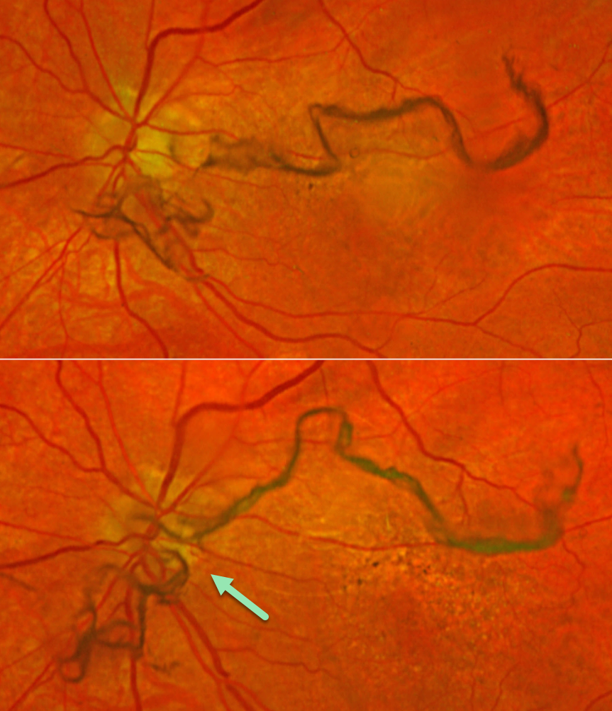
POSTERIOR VITREOUS DETACHMENT
Originally posted on @retina.rocks March 24, 2026
This 73YO female presented with several days of new floaters in her left eye. Vision was 20/15 bilaterally. Optos color RGB imaging shows extremely prominent mobile string-like posterior hyaloid condensations floating over the disc and macula. There were no retinal breaks or detachment.
Learning Points:
The clinical diagnosis for a complete posterior vitreous detachment (PVD) remains challenging and can be established with biomicroscopy when a Weiss ring, representing avulsed peripapillary glial tissue, is visualized. OCT can be helpful in supplementing the clinical exam (Wagley et al, Retina 2021;41:2296-2300).
An acute PVD is the moment of truth for when a retinal break or detachment develops. The historical risk of a break is 10-15%, although a large retrospective study from Kaiser Permanente found this risk to be significantly lower at 5% for a retinal break and 4% for retinal detachment (Ophthalmology 2022;129:67-72). The presence of vitreous pigment or blood and lattice, and a history of a retinal break or detachment in the fellow eye, increases this risk.
POSTERIOR VITREOUS DETACHMENT
Originally posted on @retina.rocks February 14, 2025
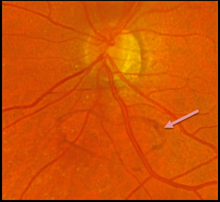
This 61YOF put a smile on our face by presenting with this asymptomatic heart-shaped Weiss ring just inferior to her left nerve.
HAPPY VALENTINE’S DAY!
POSTERIOR VITREOUS DETACHMENT (PVD)
Originally posted on @retina.rocks August 5, 2022
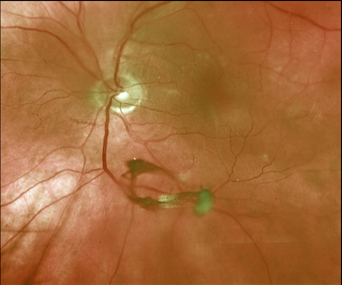
This 67YO male presented with a symptomatic posterior vitreous detachment (PVD). A prominent Weiss ring is beautifully captured in this Optos image.
His peripheral retina was normal, and he was asked to return in a few months unless he developed new flashes, floaters, or vision loss.
Learning Points:
The clinical diagnosis for a complete PVD can be established with biomicroscopy when a Weiss ring, representing avulsed peripapillary glial tissue, is visualized. OCT can be helpful in supplementing the clinical exam (see Wagley et al, Retina 2021;41:2296-2300).
An acute PVD is the moment of truth for when a retinal break or detachment develops. The historical risk for a break is at least 10-15%, although a recent large retrospective study from Kaiser Permanente found this risk to be significantly lower at 5% for a retinal break and 4% for retinal detachment (Ophthalmology 2022;129;67-72).
The presence of vitreous pigment or blood and lattice, and a history of a retinal break or detachment in the fellow eye, increases this risk.
RHEGMATOGENOUS RETINAL DETACHMENT
Originally posted on @retina.rocks November 2, 2021
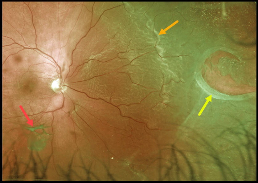
This patient presented with a macula-on rhegmatogenous retinal detachment (RRD) from a large nasal retinal tear. The tear has a rolled posterior edge, indicating possible early proliferative vitreoretinopathy.
The hydration outer retinal folds at the posterior edge of the detachment are one of the key clinical findings that help differentiate a rhegmatogenous from exudative and traction retinal detachments.
The causative posterior vitreous detachment is seen floating above the retina just inferior to the macula.
Learning Points:
The normal neurosensory retina remains attached to the RPE due to numerous physiological mechanisms, including the inner (retinal vascular endothelium) and outer (tight junctions at the apex of the RPE cells) blood retinal barriers, hyperosmotic choroid passively drawing fluid into the subretinal space, RPE pump, interphotoreceptor matrix, and friction between the RPE microvilli and the photoreceptor outer segments.
Rhegmatogenous retinal detachment (RRD) occurs when liquefied vitreous, entering the subretinal space through a retinal break, overwhelms the forces that favor retinal detachment.
Repairing RRD involves finding and closing all breaks, essentially denying the liquid vitreous access to the subretinal space. The break(s) can be closed internally with a gas bubble with pneumatic retinopexy or vitrectomy surgery, or externally by sewing a piece of silicone material to the eye wall (scleral buckle). This indents, or buckles, the sclera up against the retina break.


