

Presumed Solitary Circumscribed Retinal Astrocytic Proliferation (PSCRAP)
<< Back to Cases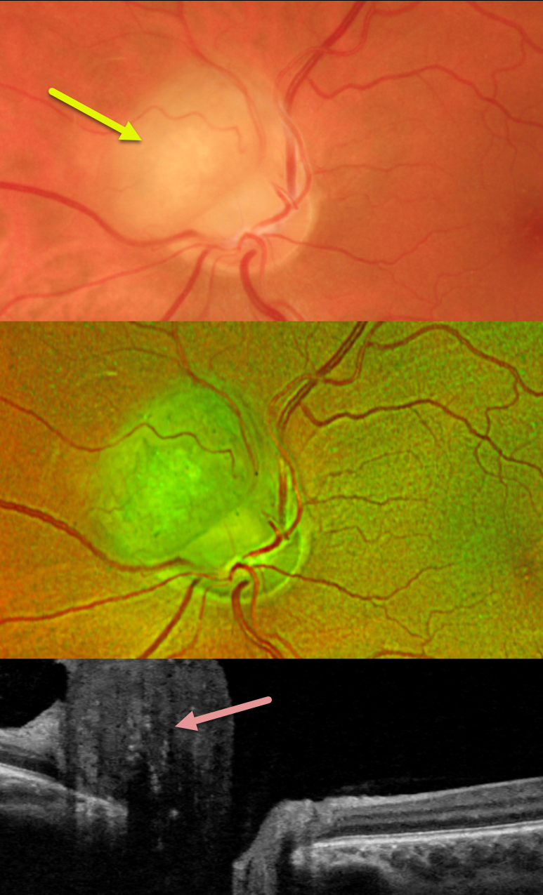
PSCRAP (PRESUMED SOLITARY CIRCUMSCRIBED RETINAL ASTROCYTIC PROLIFERATION)
Mattie Adams
Originally posted on @retina.rocks October 7, 2024
This healthy 41YO female was referred for an asymptomatic lesion in her left eye. Vision was 20/25 in her normal right eye and 20/25 in her left eye. Ocular examinations were normal except for a round nodular amelanotic elevated lesion abutting (and possibly involving) the left superonasal optic nerve. Swept-source OCT shows diffuse hyperreflectivity of the lesion with posterior shadowing.
Learning Points:
pSCRAP (presumed solitary circumscribed retinal astrocytic proliferation) was originally described by Shields et al as a unique lesion found in adulthood that resembles a retinal astrocytoma or retinoma (Arch Ophthalmology 2011;129:1189-1194). Although originally felt to arise from the inner retina, it is now thought to be either a deep retinal glial lesion or a form of RPE fibrous metaplasia (Shields et al, Retinal Cases & Brief Reports 2017;11:18-23).
pSCRAP (PRESUMED SOLITARY CIRCUMSCRIBED RETINAL ASTROCYTIC PROLIFERATION)
Originally posted on @retina.rocks July 26, 2021
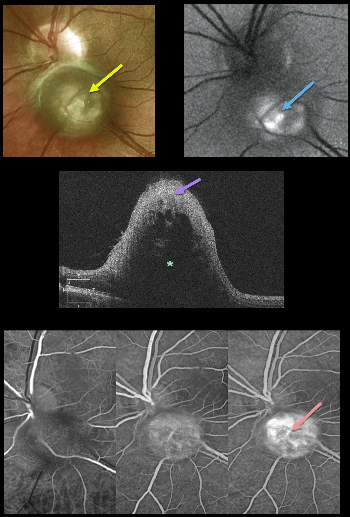
This healthy 28YO male was referred for an asymptomatic lesion in his left eye. Vision was 20/25 OU. Ocular examination was normal bilaterally except for a round nodular amelanotic elevated lesion abutting (and possibly involving) the left inferior optic nerve.
The lesion shows some central hyperautofluorescence. OCT shows marked reflectivity involving the inner retina with posterior shadowing. Optos fluorescein angiography shows probable intrinsic vascularity of the lesion.
Learning Points:
PSCRAP (presumed solitary circumscribed retinal astrocytic proliferation was originally described by Shields et al as a unique lesion found in adulthood that resembles a retinal astrocytoma or retinoma (Arch Ophthalmology 2011;129;1189-1194).
Although originally felt to arise from the inner retina, it is now felt to be either a deep retinal glial lesion or a form of RPE fibrous metaplasia (Shields et al, Retinal Cases & Brief Reports 2017;11:18-23).


