

Diabetic Retinopathy Proliferative (PDR) Neovascularization
<< Back to Cases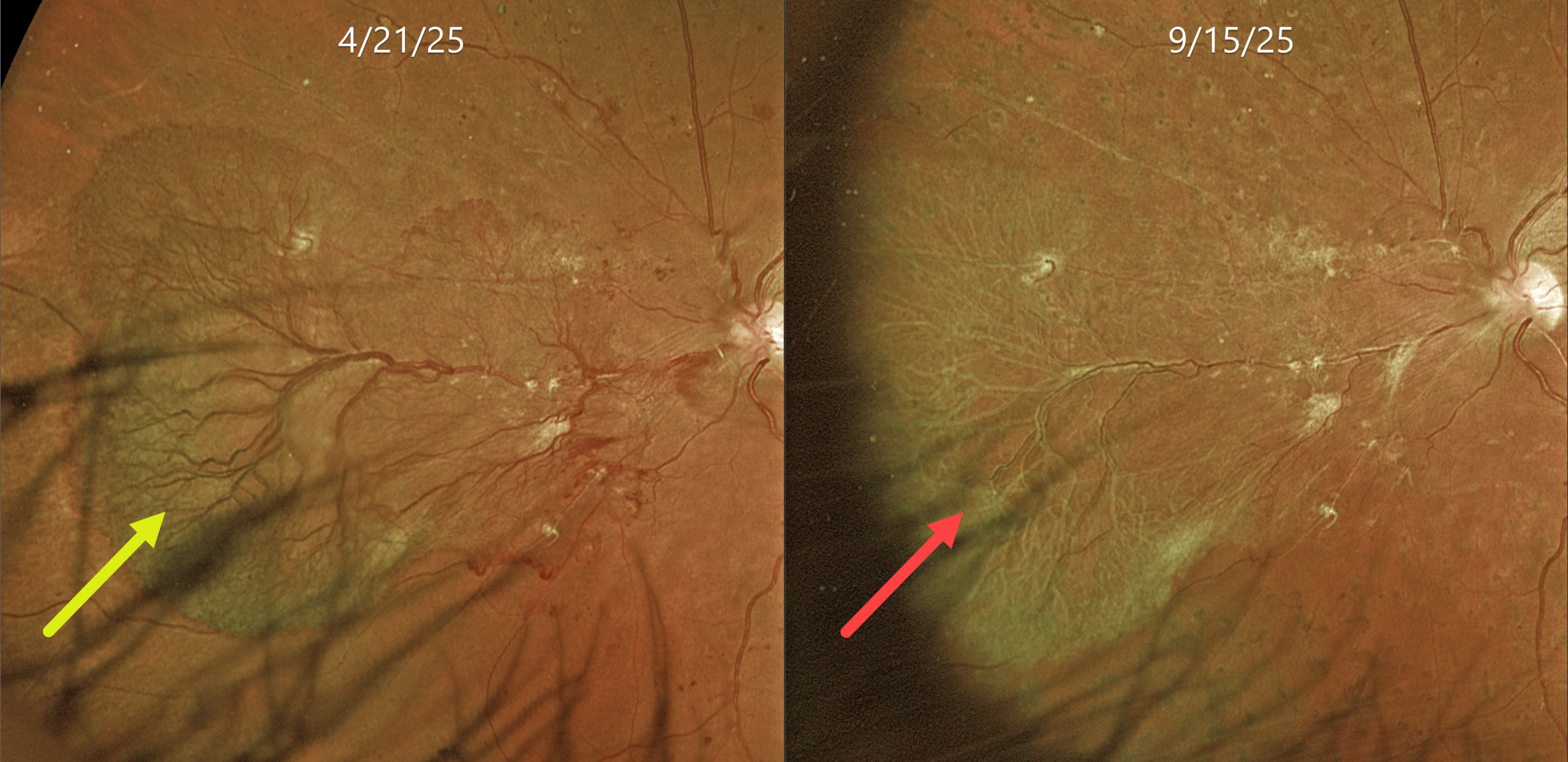
PROLIFERATIVE DIABETIC RETINOPATHY
Originally posted on @retina.rocks June 24, 2026
This 53YO female with a known history of type 2 diabetes was referred for asymptomatic proliferative disease (PDR) in her left eye. Vision was 20/50.
Optos color RG imaging shows a large sea fan-shaped area of succulent and flat neovascularization extending from the optic nerve into the nasal midperiphery. Panretinal photocoagulation (PRP) was applied in three sessions over several months.
Learning Points:
An attached vitreous provides the scaffold for neovascularization to grow onto and spread. Retinal neovascularization grows from the inner retinal surface and proliferates along the outer posterior hyaloid face (Vaz-Pereira et al, Retina 2017;37:1287-1296). Following PRP or anti-VEGF therapy for PDR, retinal neovascularization ‘involutes’, often morphing from a fine capillary network into larger vessels with fibrosis. In the process, the contracting neovascularization and vitreous can cause increasing traction. Rapid tissue contraction with traction retinal detachment is an uncommon but well-described phenomenon known as the ‘crunch’ syndrome (Survey of Ophthalmology 2021;66:926-932).
We were quite concerned about the possibility of a crunch in our patient and discussed it prior to starting treatment. Five months following her initial exam, the neovascularization completely fibrosed without traction. Vision was 20/40. We are continuing to follow her closely.
VERY SEVERE PDR AND DME FOLLOWING PATIENT LOST TO FU
Sowparnika Basavaraju
Originally posted on @retina.rocks June 16, 2026
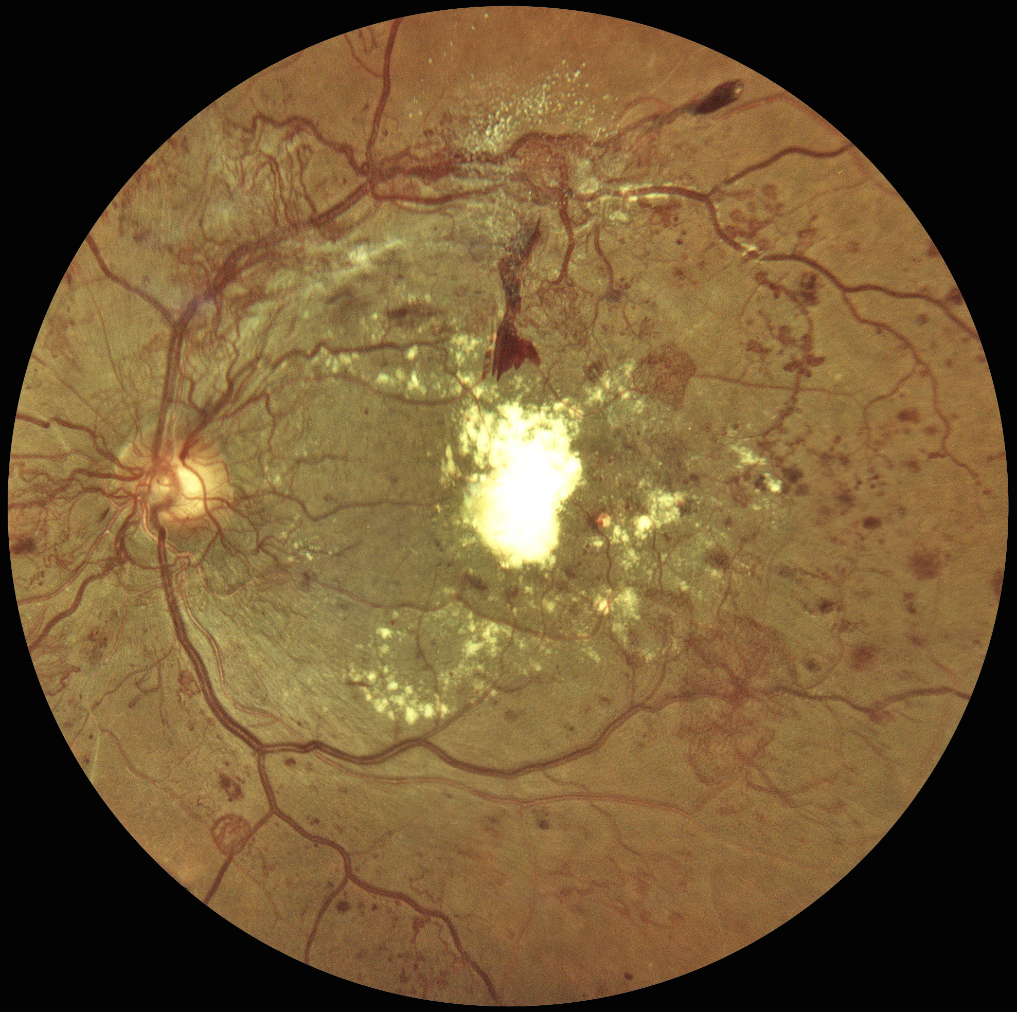
This 48YO male with a history of poorly controlled type 2 DM was seen 4 years earlier for an eye examination elsewhere but was immediately lost to follow-up. He presented to us with hand-motion vision in his left eye.
Color fundus photography shows florid disc and peripheral neovascularization variably coating the posterior pole. A large plaque of subfoveal lipid is also noted.
Learning Points:
These findings would have been caught earlier if he had not been lost to follow-up, which, unfortunately, is quite common among diabetic patients (Obeid et al, Ophthalmology 2018;125:1386-1392; Gao et al, Ophthalmology Retina 2019;3:230-236; Green et al, AJO 2020;216:18-27; Ophthalmology Retina 2025;9:1167-1174, etc, etc).
At this stage, our patient will not recover central vision due to the subfoveal lesion, and our hope is to prevent further vision loss. Anti-VEGF therapy, panretinal photocoagulation, and macular photocoagulation were suggested. He is at significant risk for traction retinal detachment as the neovascularization involutes (crunch), and we will therefore follow him very closely.
ETHAMBUTOL TOXICITY WITH CONCOMITANT DIABETIC RETINOPATHY
Ayushi Gupta and Vishal Agrawal
Originally posted on @retina.rocks May 4, 2026
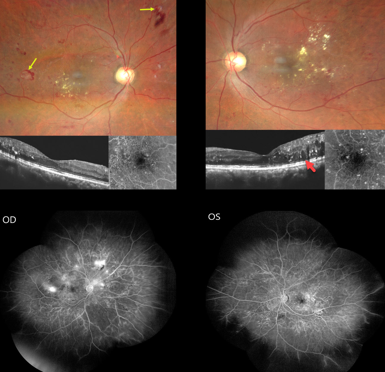
This 54YO male presented for a second opinion with several weeks of progressive bilateral painless vision loss. He gave a history of active pulmonary tuberculosis, diagnosed about 6 months earlier, which was being treated with antitubercular therapy (ATT) including ethambutol. There was also a 5-year history of well-controlled type 2 diabetes. Vision was 20/200 OU.
Fundus photography shows bilateral non-central diabetic macular edema (DME) with mostly temporal lipid. The central macula looks fairly normal bilaterally on OCT, and fundus fluorescein angiography (FFA) shows relatively preserved foveal avascular zones. Retinal neovascularization (NV) with some associated preretinal blood is noted OD. Widefield FFA confirms scattered retinal NV OD with moderate bilateral capillary nonperfusion. Disc OCT showed a normal retinal nerve fiber layer, and 30-2 visual fields showed severe generalized depression (not shown).
Learning Points:
Our patient was previously seen by other specialists who felt that diabetic retinopathy was the sole reason for his complaints and findings, despite the lack of central foveal fluid or ischemia. Our patient reminds us of 2 dictums in medicine: 1) we try to explain all findings by a single disease, and 2) that a patient can have as many diseases as they so desire. In our patient’s case, we felt that ethambutol better explained his clinical picture.
Ethambutol is one of the agents used as part of ATT, a multi-drug regimen for treating active tuberculosis. About 1-3% of patients can develop drug-induced optic neuropathy, most commonly following 2 months of therapy but rarely developing within days of starting treatment. Symptoms include decreased visual acuity, scotoma, color blindness, and visual defects in one or both eyes.
Although the FDA recommends baseline visual acuity and color vision testing, followed by monthly color discrimination testing during therapy, there are no studies demonstrating any benefit of such screening. Ethambutol should be discontinued if toxicity is suspected. Although vision loss is often thought to be permanent, a recent systematic review showed significant improvement in vision, color vision, and visual field loss with discontinuation of the drug (Sabhapandit et al., Indian J Ophthalmol 2023;71:729-735).
We contacted our patient’s pulmonologist, who stoppe therapy immediately. Anti-VEGF therapy followed by panretinal photocoagulation was recommended.
PROLIFERATIVE DIABETIC RETINOPATHY WITH ASTEROID HYALOSIS
The European VitreoRetinal Society (EVRS), Akansha Sharma, Manish Nagpal, and Vaidehi Sathaye
Originally posted on @retina.rocks April 27, 2026
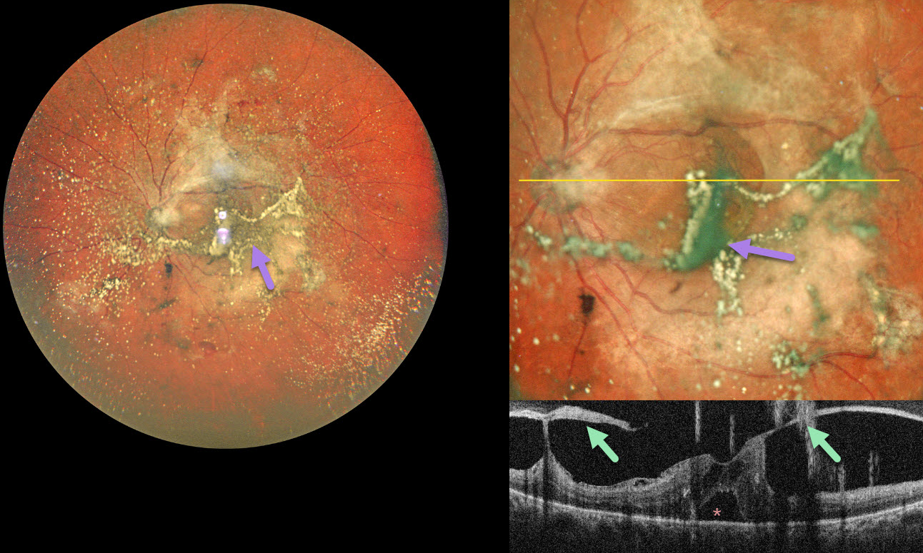
This 55YO diabetic male presented with 2 months of bilateral vision loss. Vision was 20/80 OD and 20/120 OS.
Pseudocolor SLO ultrawidefield imaging of his left eye shows moderate asteroid hyalosis, with larger condensed stands of material obscuring the underlying macula. Spectral domain OCT scanning shows complex variable vitreous traction on the disc and macula with a small foveal detachment. The asteroid particles appear as hyperreflective vertical lines resembling falling rain.
Learning Points:
Examining the fundus in a patient with dense asteroid, as in this diabetic patient, can be extremely challenging. Fluorescein angiography and OCT are great modalities because they can visualize the retina when the clinical exam can’t. In our patient’s case, the OCT revealed foveal traction, indicating the need for vitrectomy surgery.
PROLIFERATIVE DIABETIC RETINOPATHY (PDR) WITH PROGRESSIVE NV DESPITE FULL PRP
Shishir Verghese
Originally posted on @retina.rocks March 4, 2026
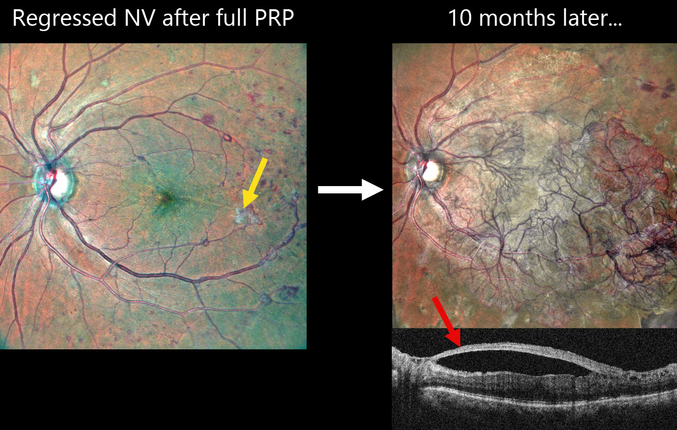
This 60YO female with a history of type 2 diabetes underwent full panretinal photocoagulation (PRP) in her left eye. Following laser, MultiColor SLO imaging shows small residual areas of variably fibrosed retinal neovascularization. Vision was 20/30.
Ten months later, she presented complaining of 2 months of decreased vision in this eye. Vision was 20/60. MultiColor SLO imaging shows retinal neovascularization covering the posterior pole. On OCT, the fovea is remarkably attached with the hyperreflective neovascularization elevated along the partially detached posterior hyaloid.
Learning Points:
Although PRP is often considered a ‘one and done’ procedure, this is not always the case (Gonzalez et al, Ophthalmology 2021;128:1448-1457). Our patient is a perfect example of why these patients require lifelong monitoring even if their disease initially appears quiescent.
Given the aggressive neovascular proliferation, intravitreal anti-VEGF was given, followed by vitrectomy 5 days later. Although PRP is often considered a ‘one and done’ procedure, this is not always the case (Gonzalez et al, Ophthalmology 2021;128:1448-1457). Our patient is a perfect example of why these patients require lifelong monitoring even if their disease initially appears quiescent.
Given the aggressive neovascular proliferation, intravitreal anti-VEGF was given, followed by vitrectomy 5 days later.
RECURRENT DIABETIC NEOVASCULARIZATION TREATED WITH SERIAL EYLEA
Michael Colucciello
Originally posted on @retina.rocks February 16, 2026
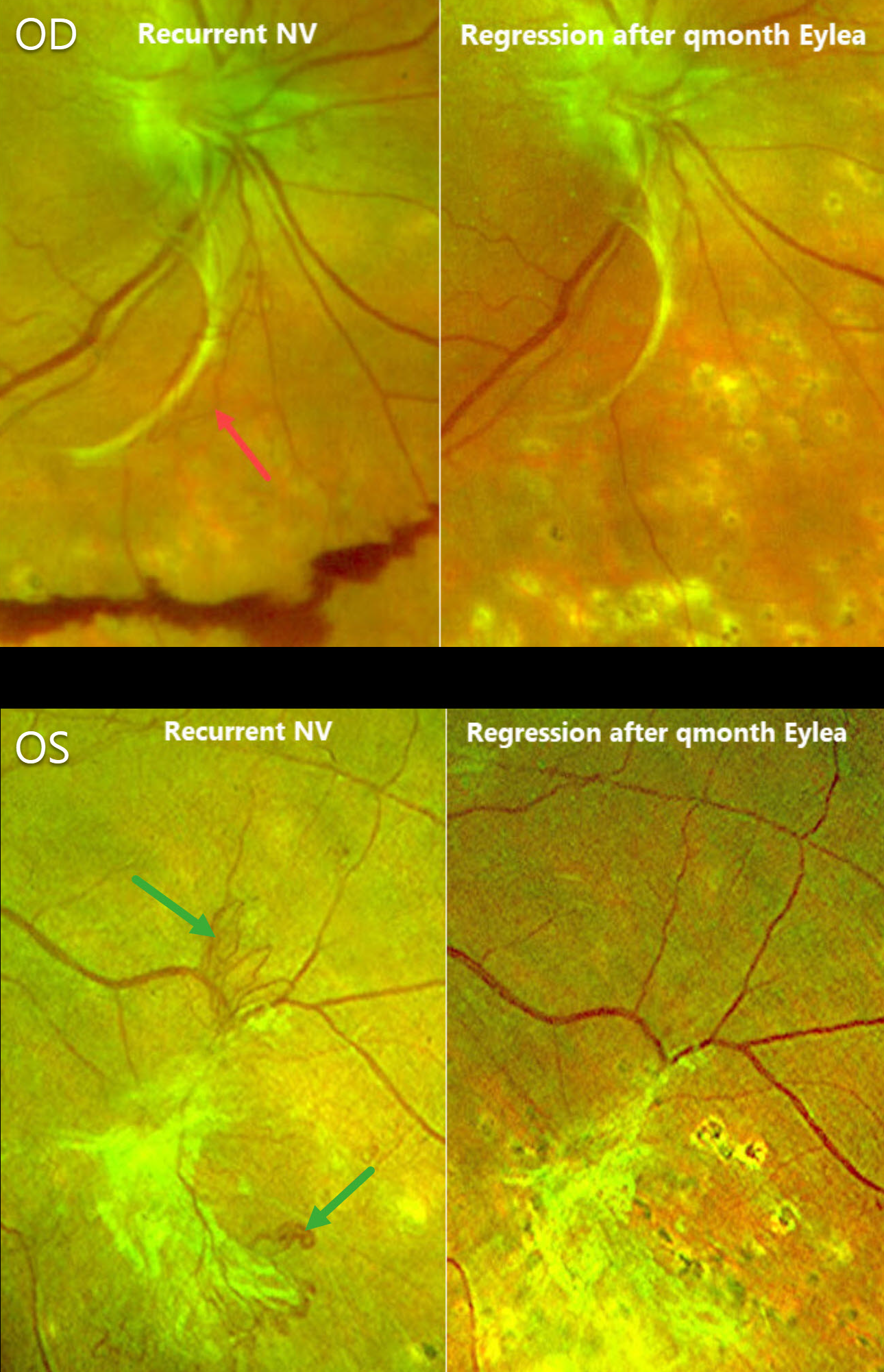
This 71YO female underwent prior full panretinal photocoagulation (PRP) bilaterally for proliferative diabetic retinopathy (PDR). She initially responded well with involution of all neovascularization. She presented for a routine follow-up with 20/25 vision OU.
Optos color RG imaging OD shows fibrosed disc neovascularization extending inferiorly with areas of active red neovascularization and inferior preretinal blood. The left eye shows fibrotic NV in the inferotemporal midperiphery along with some adjacent active vessels. Serial monthly bilateral Eylea injections were given, and 16 weeks later, the active NV fully regressed bilaterally.
Learning Points:
Although PRP is often considered a ‘one and done’ procedure, this is not always the case (Gonzalez et al, Ophthalmology 2021;128:1448-1457). Our patient is a perfect example of why these patients require lifelong monitoring even if their disease initially appears quiescent.
Prior to anti-VEGF therapy, our patient would have been treated with either additional fill-in PRP (putting her at risk for iatrogenic peripheral field loss) or possibly vitrectomy for increasing blood. Few currently advocate anti-VEGF injections as the sole treatment for PDR per the DRCR Protocol S. However, regarding adjuvant therapy to laser, we find these injections helpful for stabilizing macular edema or NV before starting PRP (Alsoudi et al, JAMA Ophthalmology 2024;142:972-975) and for those eyes needing supplemental treatment for active disease despite prior laser (Park et al, Eye 2021;35:3056-3063).
PROLIFERATIVE DIABETIC RETINOPATHY NVD VS RETINAL ARTERIOVENOUS MALFORMATION
Ayushi Gupta and Vishal Agrawal.
Originally posted on @retina.rocks November 12, 2025
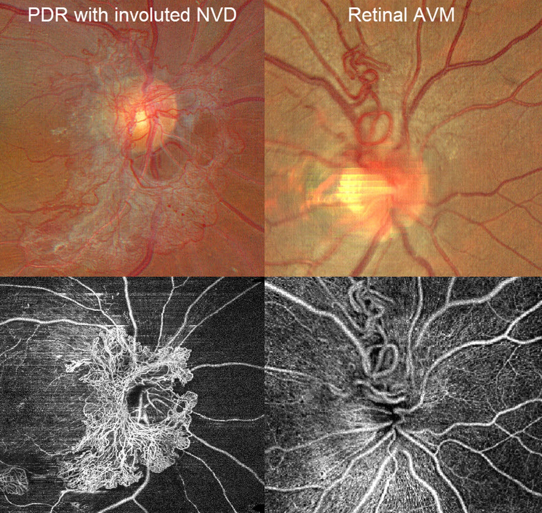
A patient with disc neovascularization (NVD) due to proliferative diabetic retinopathy (PDR) following full panretinal photocoagulation is compared with a patient with an asymptomatic retinal arteriovenous malformation (AVM).
Color photography of the PDR patient shows diffuse flat NVD with variable tight capillary networks of new vessels, admixed with larger vessels and fibrosis. OCT angiography (OCTA) confirms flow signals within these vessels, along with diffuse capillary loss superior, nasal, and inferior to the nerve. Color photography and OCTA of the retinal AVM show a tangle of arterioles and veins extending superiorly from the nerve. Fluorescein angiography, if performed, would show leakage from the NVD but not from the AVM.
TRACTIONAL RETINAL DETACHMENT
Originally posted on @retina.rocks January 29, 2025
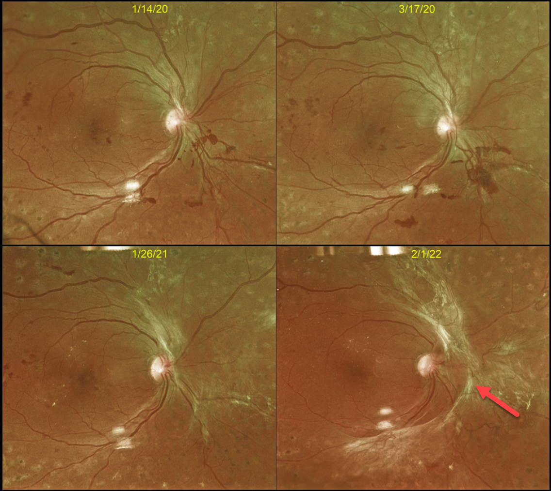
This 43YO male with type 2 diabetes initially responded beautifully in 2016 to full panretinal photocoagulation (PRP) for proliferative diabetic retinopathy (PDR). However, he presented on 3/17/20 with some new preretinal blood (Optos color RG image), and fill-in PRP was applied.
Over the course of 2 years, progressive fibrotic neovascularization with extramacular traction developed. Despite the traction, vision remained at 20/40, and careful observation was recommended. We were hesitant to add further laser or anti-VEGF therapy for fear of causing increased traction and ‘crunch’ (Tan et al, Surv Ophthalmology 2021;66:926-932). When last examined on 4/3/24, the retinal findings remained unchanged, and vision was 20/30 (not shown).
Although PRP is often a ‘one and done’ procedure, this is not always the case (Gonzalez et al, Ophthalmology 2021;128:1448-1457). Our patient is a perfect example of why these patients require lifelong monitoring even if their disease appears quiescent.
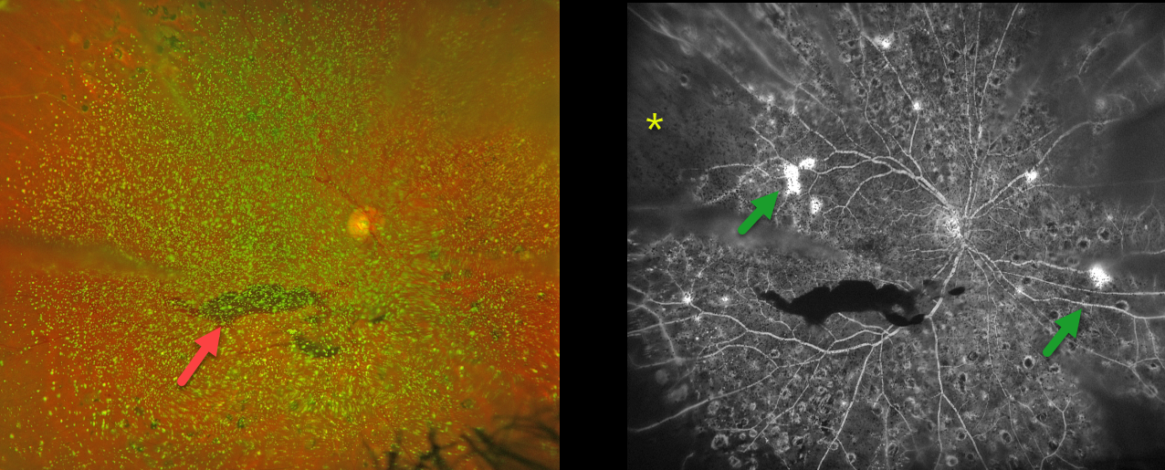
PROLIFERATIVE DIABETIC RETINOPATHY (PDR) WITH ASTEROID HYALOSIS
Originally posted on @retina.rocks October 16, 2024
This 50YO female with a history of type 2 diabetes was referred for new floaters. Several years earlier, we began panretinal photocoagulation (PRP) for proliferative diabetic retinopathy (PDR), but she had been lost to follow-up.
Optos color RG imaging shows dense asteroid hyalosis with a hazy inferior preretinal hemorrhage. Fluorescein angiography (FA) shows significant retinal ischemia, scattered retinal neovascularization, and partial PRP scarring.
Examining the fundus with dense asteroid, as in this diabetic patient, can be extremely challenging. FA and OCT are great modalities because they can visualize the retina when the clinical exam can’t. Optos FA was critical in confirming proliferative retinopathy with peripheral ischemia in our patient.
We were fortunately able to complete the PRP despite the vitreous opacities.
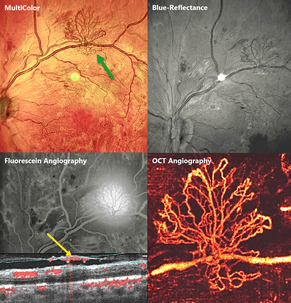
PROLIFERATIVE DIABETIC RETINOPATHY (PDR)
Seif Anwar
Originally posted on @retina.rocks October 2, 2024
This 23YO male presented with bilateral proliferative diabetic retinopathy (PDR). Vision was 20/60 OD and 20/80 OS. Multimodal imaging of his left eye highlights an area of retinal neovascularization along the superotemporal arcade.
Which modality do you feel best images the neovascularization? Or are they all complementary?
MultiColor imaging shows the lacy active red vessels overlying the orange-red RPE and choroid. These vessels are better highlighted in the Blue-Reflectance image, since they appear black on a grey background. They profoundly leak on fluorescein angiography. En face OCT angiography (OCTA) beautifully shows the fine neovascular details. Finally, B-scan OCTA shows the neovascular vessels growing along the back surface of the partially detached posterior hyaloid.
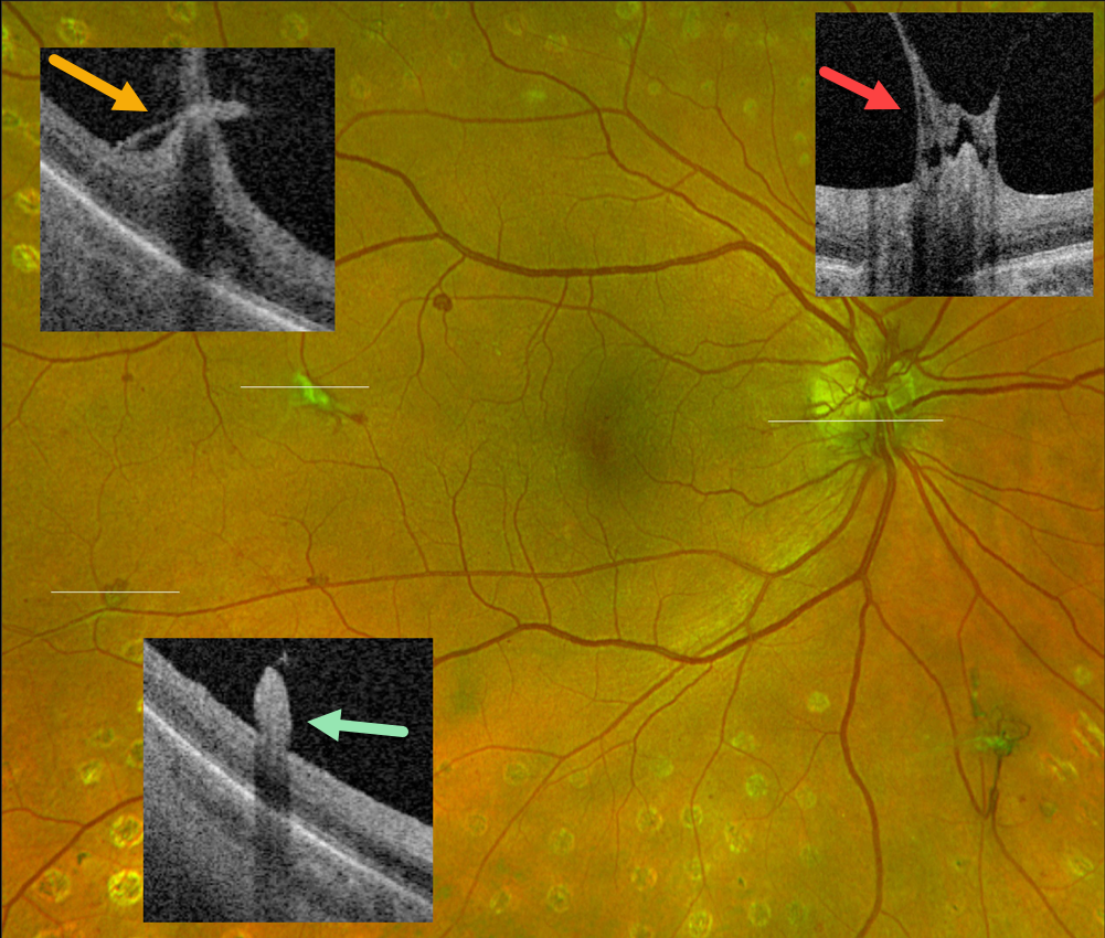
PROLIFERATIVE DIABETIC RETINOPATHY (PDR)
Originally posted on @retina.rocks August 27, 2024
This 55YO female with type 2 diabetes has involuted proliferative diabetic retinopathy (PDR) following full panretinal photocoagulation.
Optos color RG imaging shows residual disc (NVD) and peripheral retinal neovascularization (NVE). Optos Silverstone swept-source OCT through disc and temporal macular neovascularization shows active vitreous traction on the residual vessels. There is no traction on small, reddish NVE in the distal inferotemporal macula. Continued observation was recommended.
Learning Points:
An attached vitreous provides the scaffold for neovascularization to grow onto and spread. Retinal neovascularization grows from the inner retinal surface and proliferates along the outer posterior hyaloid face (Vaz-Pereira et al, Retina 2017;37:1287-1296). This tethering to a partially detached and contracting hyaloid is what causes tractional retinal detachment and vitreous hemorrhage. Thus, although an eye with a complete posterior vitreous detachment can develop small buds of neovascularization along the retinal surface (as seen in our patient’s inferotemporal NVE), the detached hyaloid is protective against the growth of large sheets of fibrovascular tissue.
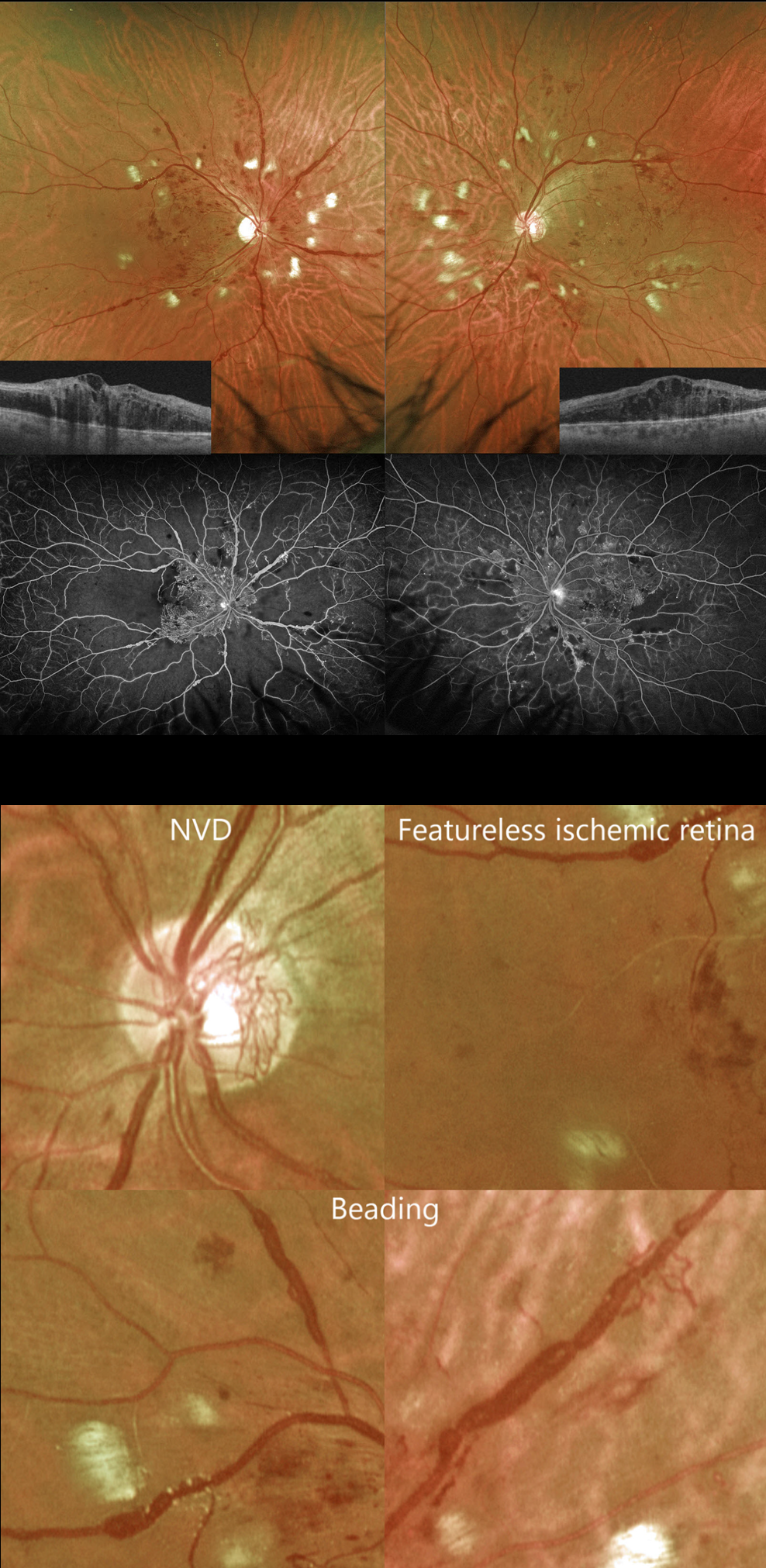
PROLIFERATIVE DIABETIC RETINOPATHY (PDR)
Originally posted on @retina.rocks August 1, 2024
This 41YO male with type 2 diabetes complained of vision loss for 5 months. He was recently hospitalized for severe systemic hypertension. Vision was counting fingers OD and 20/60 OS.
Optos color RG imaging shows bilateral venous beading and extensive cotton wool spots. Diffuse central edema is noted on Triton swept-source OCT, and severe ischemia is seen on fluorescein angiography.
But on closer inspection, the severe ischemia is evident clinically. The retina between the larger vessels is featureless, and many of the veins are sausaged and beaded. Once these clinical features are recognized, the angiographic findings can usually be predicted.
There is a relative lack of severe retinal hemorrhages and neovascularization (only disc neovascularization is present). The extensive cotton wool spots are more hypertensive than diabetic changes but indicate ischemia of the nerve fiber layer.
The patient subsequently underwent full bilateral panretinal photocoagulation and anti-VEGF therapy.
PROLIFERATIVE DIABETIC RETINOPATHY (PDR)
Originally posted on @retina.rocks July 15, 2024
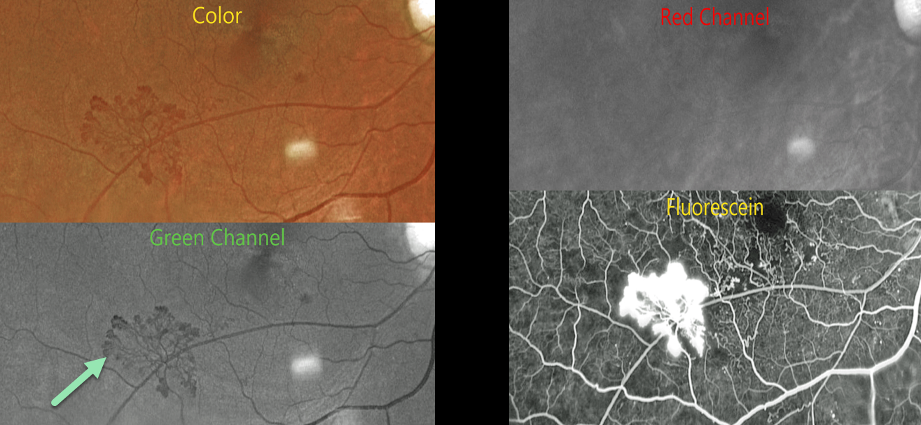
This 54YO male with proliferative diabetic retinopathy (PDR) and retinal neovascularization was evaluated with Optos multimodal imaging.
Optos color RG imaging shows active neovascularization in the inferotemporal macula.
The neovascularization is best seen in the green channel, which isolates the RPE and neurosensory retina. The blood vessels virtually disappear with the red channel, which is best for viewing the choroid. Finally, the vessels profusely leak on fluorescein angiography.
Learning Points:
Optos RG color images are generated from a red (635nm) and green (532nm) laser. The absence of a blue channel results in their greenish tint, which, at first, many clinicians find somewhat unsettling. However, much information is contained in these images once one understands how they are constructed. As shown in our patient, these images allow isolation of the neurosensory retina/RPE and choroid and often reveal more information than is visible in a true-color image.
Red neovascularization on an orange background (color image) is much more difficult to see than black neovascularization on a grey background (green channel). Recently, Optos introduced a blue channel to its California model, which, in our opinion, now offers the best of all worlds: stunning true-color images along with the ability to view each channel separately.
PROLIFERATIVE DIABETIC RETINOPATHY (PDR)
Originally posted on @retina.rocks January 29, 2024
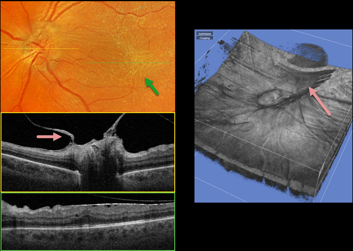
This 58YO male with type 2 diabetes has long-involuted proliferative diabetic retinopathy (PDR) following full panretinal photocoagulation (PRP). He has no visual complaints, and vision is 20/50 OS.
Optos color RGB imaging shows a diffuse macular pucker with striae extending nasally towards the nerve and temporally into the distal macula.
A condensation of elevated fibrosed tissue, representing a partially detached Weiss’ ring, surrounds the optic nerve. This traction is clearly evident in Triton swept-source OCT (image 1, middle) and in the 3D reconstruction (image 2). A moderate epiretinal membrane extends throughout the macula (image 1, bottom).
Observation was recommended, and vitrectomy with membrane peeling will be performed if the patient becomes more symptomatic.
PROLIFERATIVE DIABETIC RETINOPATHY (PDR)
Originally posted on @retina.rocks January 25, 2024
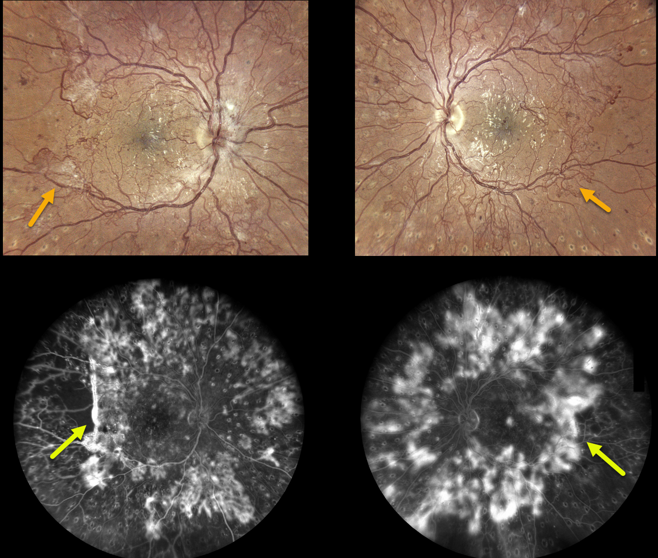
This 28YO female with type 1 diabetes recently received bilateral full panretinal photocoagulation (PRP), completing treatment several months earlier.
Despite this, active neovascularization encircles the major arcades, extending into the midperipheries, clinically and angiographically. She was visually asymptomatic with vision of 20/25 OD and 20/30 OS. There was no edema or traction on OCT (not shown).
After extensive discussion with our patient, we decided against fill-in PRP or anti-VEGF therapy for fear of possibly causing contraction of the neovascularization and traction detachment. We will follow her closely and recommend vitrectomy if vitreous hemorrhage or traction develops.
What would you do?
Learning Points:
Before PRP, patients with proliferative diabetic retinopathy (PDR) routinely went blind, and one of the treatments actually included pituitary ablation (for an amazing historical perspective on diabetic laser photocoagulation, see Goldberg and Jampol, Ophthalmology 1987;94:741-746).
Following PRP, the retinal neovascularization usually morphs from active (tight small red vascular networks) into involuted (residual larger vessels, fibrosed tissue, or occasionally complete vessel disappearance) vasculature.
Once considered a “one and done” treatment, more recent studies show that patients may still need additional laser, anti-VEGF injections, or vitrectomy (see Gonzalez et al, Ophthalmology 2021;128:1448-1457).
PROLIFERATIVE DIABETIC RETINOPATHY (PDR)
Originally posted on @retina.rocks January 15, 2024
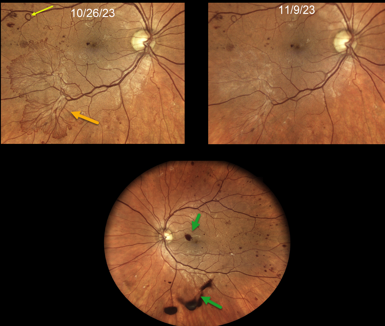
This 37YO female with type 1 diabetes presented with recent floaters in her left eye. It had been over 5 years since her last eye examination. Vision was 20/30 OD and 20/50 OS.
Colour photography shows a large flat area of active retinal neovascularisation in the inferotemporal macula of the right eye. Some foveal preretinal blood and scattered small retinal neovascularisation are seen in her left eye.
Panretinal photocoagulation (PRP) was recommended for her left eye. The decision for how to treat the right eye was more complex. After a long talk with the patient, we gave an intravitreal Avastin injection in her right eye.
She failed to show for her visit one week later, but thankfully came in 2 weeks following the injection. Vision was 20/30 OD, and the neovascularisation in the right macula had nearly completely regressed. PRP was started.
Learning Points:
Intravitreal anti-VEGF is commonly used before diabetic vitrectomies to cause rapid involution of the vascularized preretinal tissue and control intraoperative bleeding during membrane dissection. These injections are given within a week of surgery, as neovascularisation can contract, increasing traction. This uncommon yet well-known phenomenon is called the ‘crunch’ syndrome (Tan et al, Survey of Ophthalmology 2021;66:926-932).
We also sometimes like to use anti-VEGF injections to stabilize these more succulent retinal neovascularizations before proceeding with PRP. In our experience, anti-VEGF therapy causes a more rapid and complete involution than we see with PRP.
Of course, the flip side is an increased risk of ‘crunch’, so these patients need to understand the possibility of early vitrectomy and be followed quite closely afterward.
PROLIFERATIVE DIABETIC RETINOPATHY (PDR)
Originally posted on @retina.rocks January 3, 2024
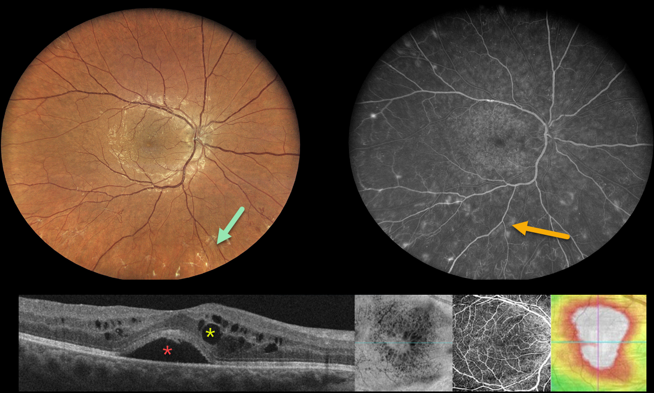
This 38YO diabetic female was referred for diabetic retinal changes. Vision was 20/50 OD and 20/40 OS. Multimodal imaging shows the often subtle proliferative changes with various perspectives of her center-involved diabetic macular edema (CI-DME).
Widefield color imaging shows minimal retinal hemorrhages with a foveal cyst. Upon closer inspection, subtly fibrosed retinal neovascularization is noted, particularly inferiorly. Fluorescein angiography shows numerous buds of retinal neovascularization outside the macula.
Multiple OCT images are shown below, including a B-scan, which shows subfoveal fluid with cysts in both the inner and outer nuclear layers. En face imaging shows variably sized radiating cysts. OCT angiography is fairly normal with a preserved foveal avascular zone. Finally, a thickness map shows diffuse thickening.
Learning Points:
These presentations can be very humbling for us clinicians and remind us of the importance of peripheral retinal examination and ultra-widefield imaging with fluorescein angiography in screening for proliferative disease. Widefield OCT angiography may also prove to be an equally effective yet less invasive screening tool (Al-Khersan et al., AJO 2021;224:292-300).
PROLIFERATIVE DIABETIC RETINOPATHY (PDR)
Sehrish Momin and Haroon Tayyab
Originally posted on @retina.rocks July 31, 2023
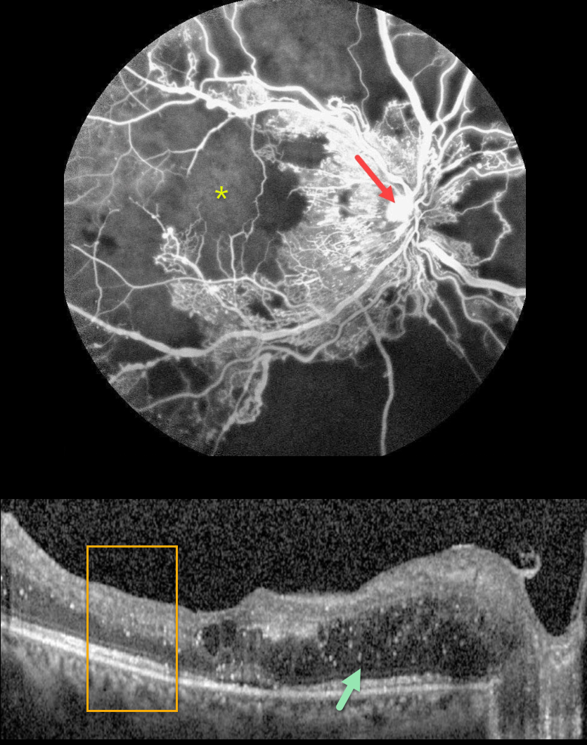
This 27YO female with a history of uncontrolled type 1 diabetes presented with bilateral vision loss. Vision was 20/40 OD and 20/200 OS.
Fluorescein angiography shows severe capillary loss that extends through the central macula. The remaining vessels are telangiectatic, especially bordering the areas of frank loss, and an area of leaking disc neovascularization is noted.
OCT scanning shows mostly nasal edema of the outer nuclear layer with numerous suspended hyperreflective particles (hyperreflective foci, HRF), along with more central cysts and trace subretinal fluid. The inner retinal layers have lost some of their normal hyper- and hypo-reflective structures (disorganization of the retinal inner layers, DRIL). Similar findings were noted in her left eye (images not shown).
Monthly anti-VEGF therapy was recommended although the patient was subsequently lost to follow-up.
Learning Points:
Anti-VEGF therapy has become the standard treatment for central-involved diabetic macular edema (CI-DME), with the vast majority of eyes showing improvements in macular thickness and vision.
Although we expect our patient’s edema to improve if she returns for treatment, we are more guarded regarding her final acuity since the capillary loss is permanent. Eyes with CI-DME and baseline central DRIL have a worse visual prognosis (Sun et al, JAMA Ophthalmol 2014;123:1309-1316).
HRF are small dot-like retinal opacities found in some eyes with diabetic macular edema. Although their pathogenesis is debated, they likely represent extravasated lipoproteins that can later become clinically evident lipid exudates (Ganne et al, Indian J Ophthalmol 2021;69:3208-3217).
Although HRF numbers decrease with treatment, it is unclear whether they serve as a biomarker for predicting visual outcome (Huang et al, Ophthalmology Retina 2022;6:814-827).
PROLIFERATIVE DIABETIC RETINOPATHY (PDR)
Mattie Adams
Originally posted on @retina.rocks May 30, 2023
This 59YO female was referred for proliferative diabetic retinopathy (PDR). She noted floaters in her right eye for several months. There was a 20-year history of type 2 diabetes, which was currently insulin-dependent. Vision was 20/25 bilaterally.
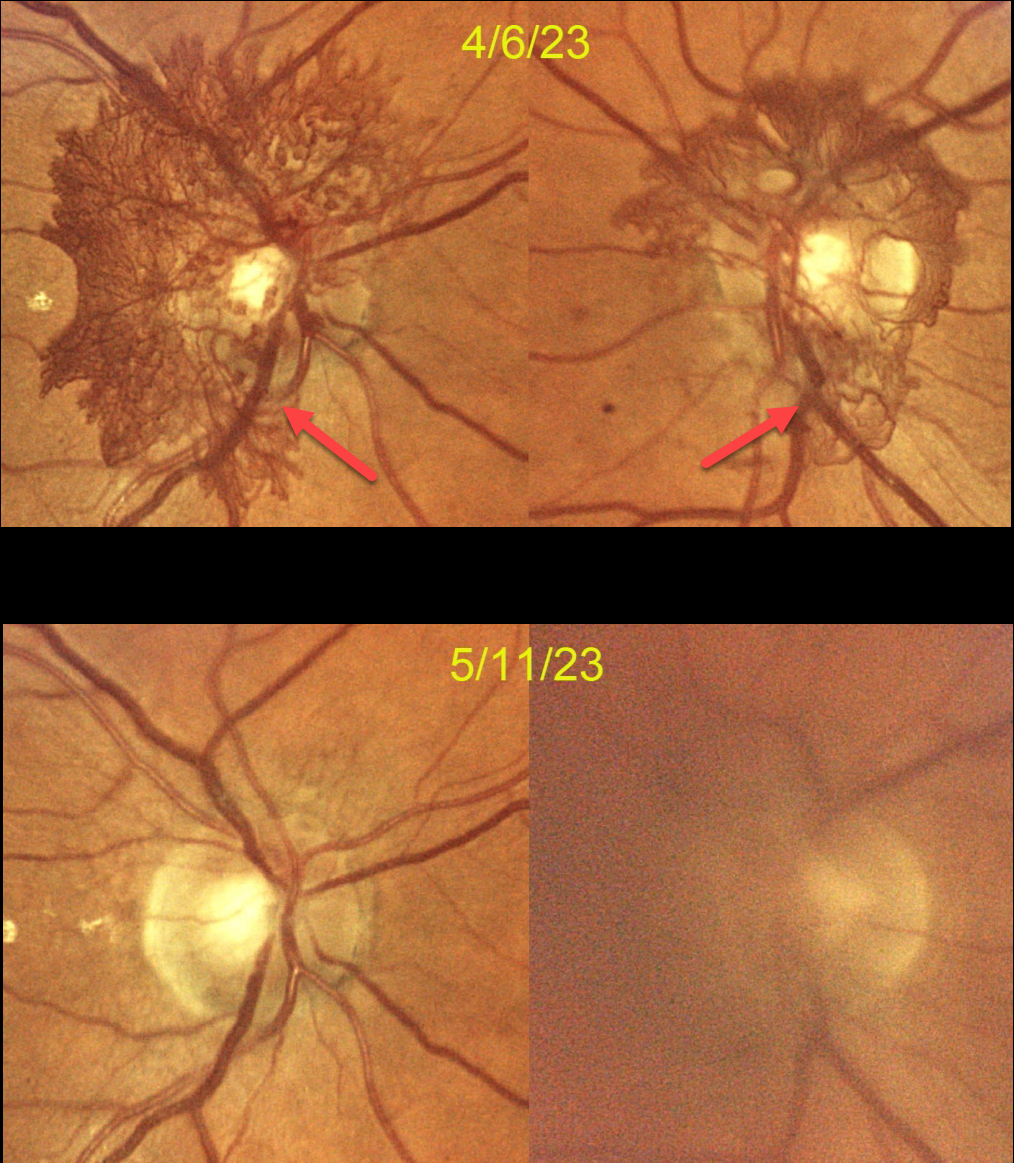
Color photography dated 4/6/23 shows florid bilateral disc neovascularization (NVD) secondary to previously undiagnosed PDR. Bilateral Avastin injections were given that day. She returned two weeks later with several days of blurred vision OS. Vision was 20/60 in this eye due to a new moderate vitreous hemorrhage.
She returned on 5/11/23, subjectively better, with 20/25 OD and 20/30 OS. The vitreous hemorrhage in her left eye was much improved, and the NVD had dramatically resolved in each eye. Panretinal photocoagulation (PRP) will be started bilaterally.
Learning Points:
PRP has been around since the 1960s and still remains the mainstay for treating proliferative disease. Although the majority of treatment-naive retinal neovascularizations will respond to PRP, in our experience, this type of succulent NVD rarely regresses significantly with laser alone.
As in this case, we prefer to first stabilize the neovascularization with anti-VEGF therapy, and then add PRP as a more permanent solution to prevent reproliferation.
PROLIFERATIVE DIABETIC RETINOPATHY (PDR)
Originally posted on @retina.rocks March 16, 2023
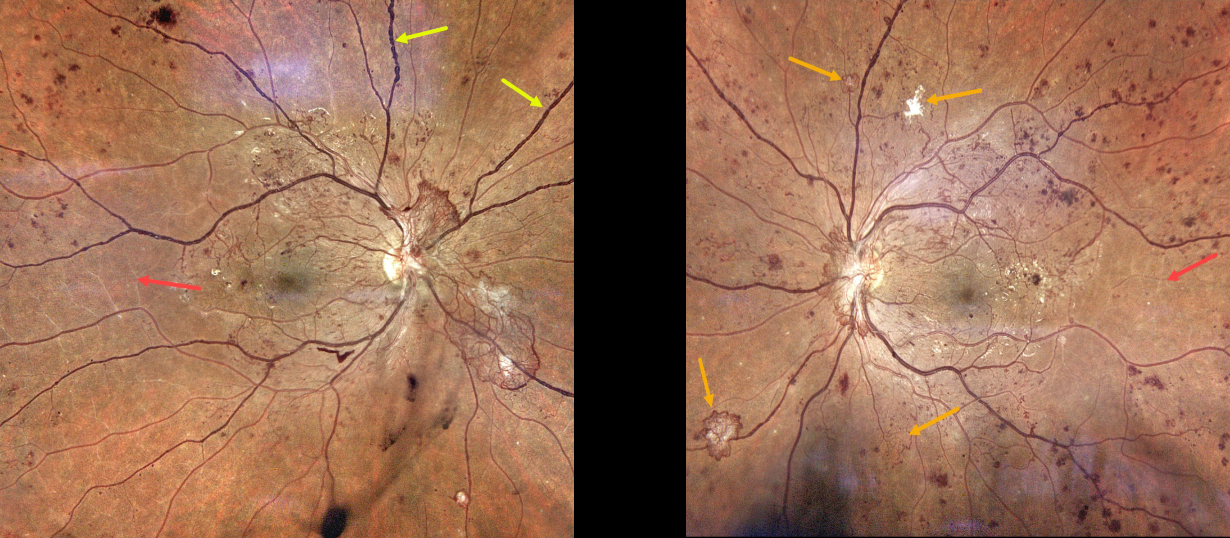
This 28YO male with a history of type 1 diabetes was referred for asymptomatic retinal findings. He noticed some recent floaters in his right eye. Vision was 20/50 OU.
Color photography shows severe bilateral proliferative diabetic retinopathy. The right eye has a large area of active disc neovascularization, venous beading, and white vessels temporally indicating severe ischemia. There is some mild inferior vitreous blood.
The left eye has a smaller area of succulent NVD along with scattered smaller areas of variably active and fibrosed peripheral neovascularization. Clinical ischemic retina is also noted. Panretinal photocoagulation will be started bilaterally.
PROLIFERATIVE DIABETIC RETINOPATHY (PDR)
Originally posted on @retina.rocks March 1, 2023
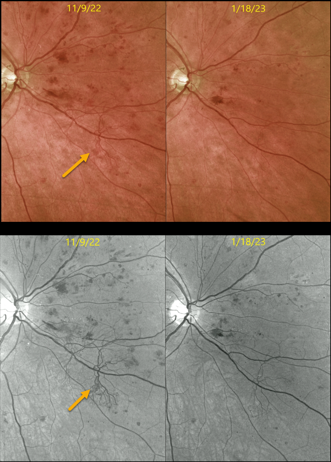
This 56YO male with type 2 diabetes presented on 11/9/22 with 20/30 vision OD with both central-involved diabetic macular edema (CI-DME) and proliferative diabetic retinopathy. Optos color and green-channel imaging show a large patch of retinal neovascularization.
Two monthly intravitreal injections of Avastin were administered for the CI-DME. On 1/18/23, vision improved to 20/25, and the edema completely resolved. The retinal neovascularization dramatically regressed. Macular laser for the injection treatment burden will be performed, followed by panretinal photocoagulation (PRP).
Learning Points:
The DRCR.net’s Protocol S explicitly compared PRP to Lucentis for the primary treatment of proliferative diabetic retinopathy (JAMA 2015;314(20):2137-2146). At 2 years, vision was equivalent between both treatment groups, and both treatments were equally effective in controlling neovascularization.
However, Lucentis-treated eyes experienced less peripheral visual field loss, developed macular edema less often, and needed fewer vitrectomies.
A secondary analysis of Protocol S suggested that Lucentis monotherapy was cost-effective for PDR+vision-impairing CI-DME but not for PDR without CI-DME (JAMA Ophthalmology 2019;137:1424-1432).
The role of intravitreal therapy vs PRP as the primary management for proliferative disease is intriguing, but in our practice, we find it difficult to justify, given the well-tolerated, long-lasting effects of laser therapy, contrasted with the risk of poor patient follow-up and the expense and treatment burden of anti-VEGF therapy.
PROLIFERATIVE DIABETIC RETINOPATHY (PDR)
Originally posted on @retina.rocks December 6, 2022
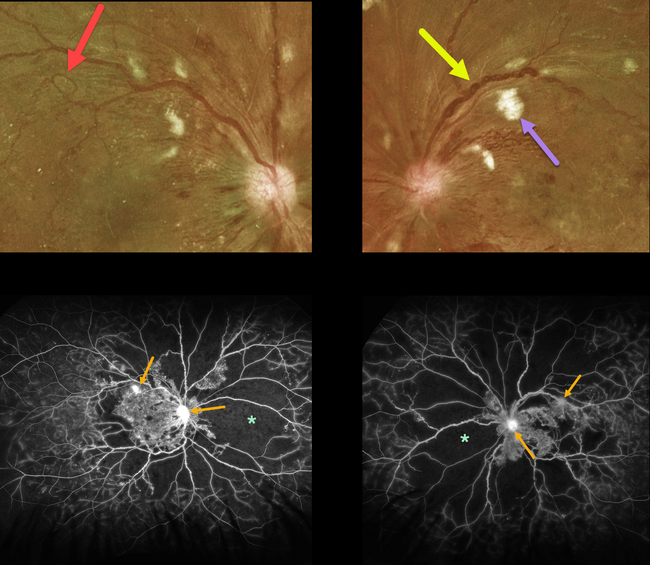
This 33YO diabetic male presented with vision of 20/30 OD and 20/100 OS.
Optos color imaging shows bilateral severe posterior pole retinal hemorrhages and nerve fiber layer infarcts. Florid disc neovascularization is noted bilaterally. Marked venous beading is noted in each eye, and a large venous loop is noted in his right eye. The peripheral retina is also markedly ischemic.
Fluorescein angiography shows marked severe ischemia. The venous beading is much more global angiographically, with extensive venous staining of the irregularly beaded veins. Despite the ischemia, just a few areas of neovascularization are noted in each eye.
Vision is good OD since the foveal avascular zone is relatively normal, and vision is poor OS due to foveal ischemia.
Panretinal photocoagulation was recommended for each eye.
PROLIFERATIVE DIABETIC RETINOPATHY (PDR)
Veronika Matello and Barbara Parolini
Originally posted on @retina.rocks November 14, 2022
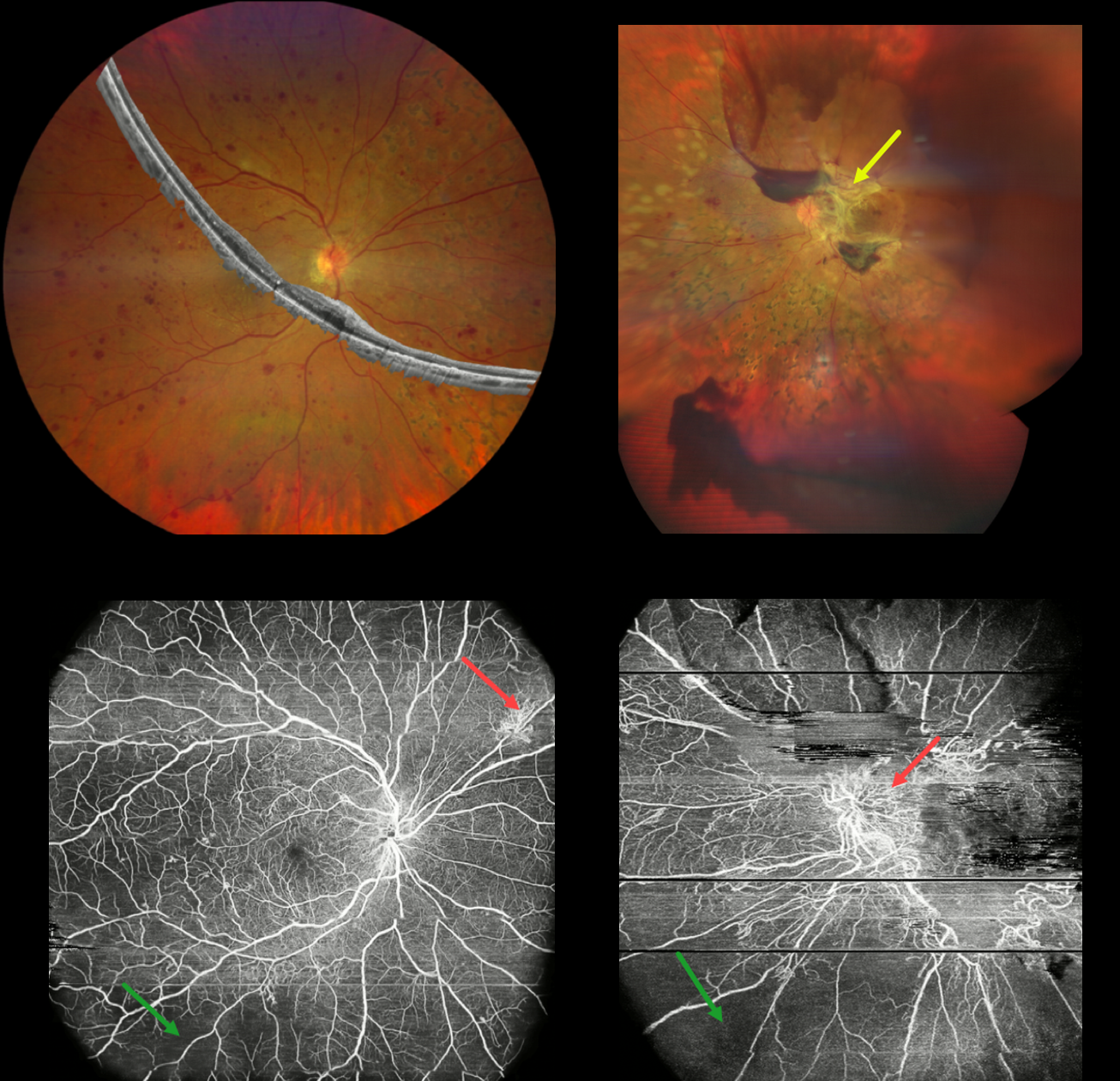
This 43YO female has a 16-year history of type 1 diabetes, and presented with 3 years of progressive vision loss in her left eye. Vision was 20/20 OD and 20/400 OS.
Composite widefield color imaging and OCT of her right eye show moderate retinal hemorrhages, nasal panretinal photocoagulation scarring, and no edema. Widefield OCT angiography shows peripheral ischemia with superonasal midperipheral neovascularization.
Multimodal imaging of her left eye shows fibrotic neovascularization around the nerve and macula. OCT angiography shows the neovascularization and peripheral retinal ischemia.
Learning Points:
Ultra-widefield imaging with fluorescein angiography is becoming increasingly important in screening for proliferative disease, especially since about 70% of angiographic nonperfusion lies outside the posterior pole (Silva et al, DRCR Protocol AA, Retina 2022;42:1302-1310). Widefield OCT angiography may also prove to be an equally effective yet less invasive screening tool (Al-Khersan et al., Am J Ophthalmol. 2021;224:292-300).
PROLIFERATIVE DIABETIC RETINOPATHY (PDR)
Originally posted on @retina.rocks November 3, 2022
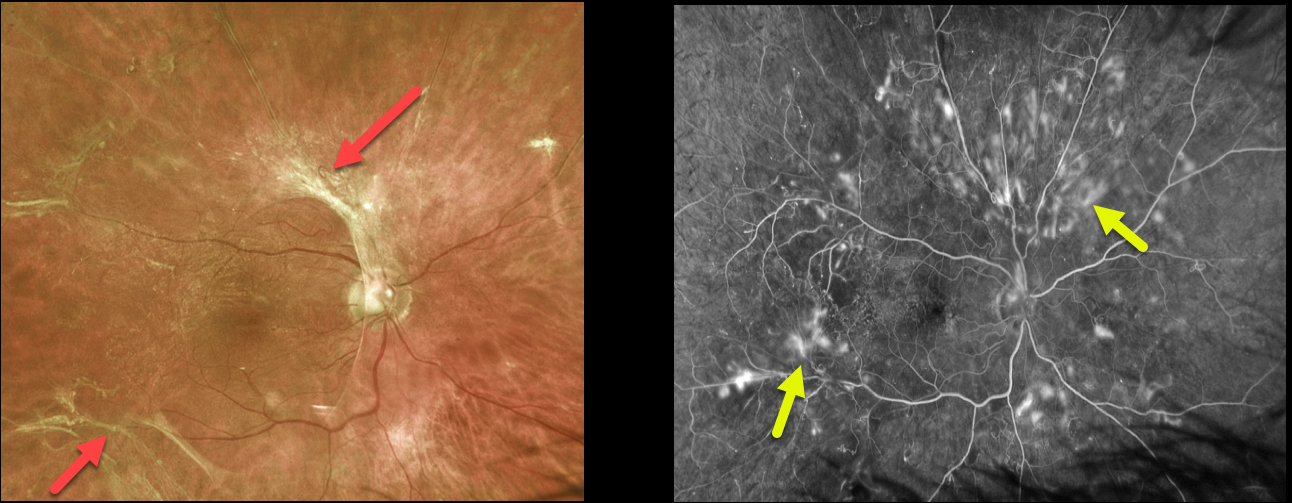
This 58YO male presented with asymptomatic retinal changes in his right eye. Vision was 20/25 OD.
The left eye was phthisical. He had a history of type 2 diabetes.
Optos color imaging shows fibrosed retinal neovascularization scattered around the posterior pole. Fluorescein angiography shows scattered retinal neovascularization.
We were concerned that panretinal photocoagulation could further contract the neovascular tissue, possibly increasing traction and leading to vision loss. We were also hesitant to recommend primary vitrectomy since he is monocular, has excellent vision, and minimal macular traction.
We are therefore following him closely with observation and will likely recommend primary vitrectomy for increased traction. What would you recommend?
PROLIFERATIVE DIABETIC RETINOPATHY (PDR)
Originally posted on @retina.rocks October 12, 2022
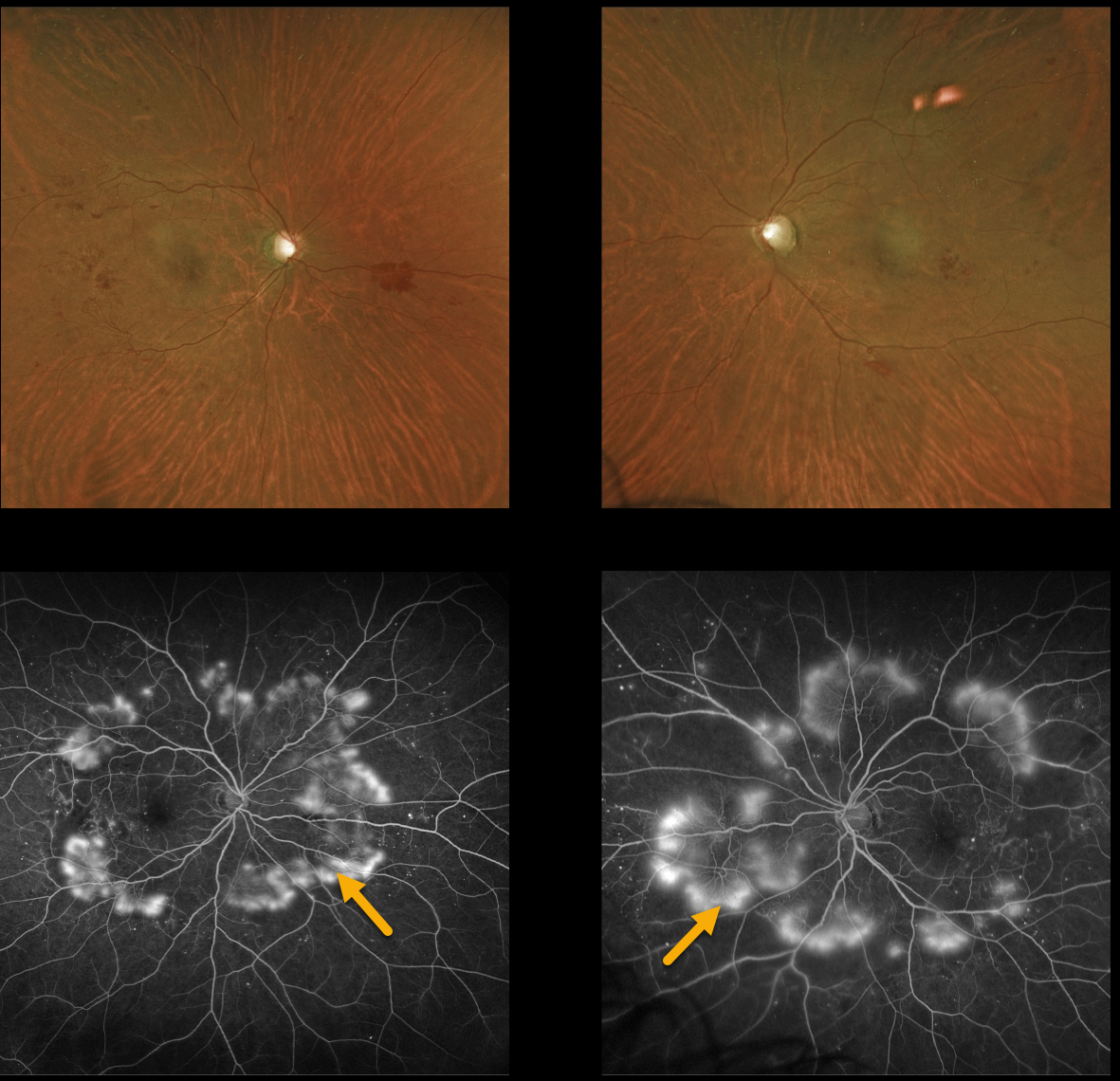
This 51YO female with type 2 diabetes presented with severe peripheral proliferative diabetic retinopathy (PDR).
Despite having extensive angiographic neovascularization encircling each posterior pole, these findings are remarkably less obvious on color imaging.
Learning Points:
These types of presentations are very humbling to us clinicians and remind us of the importance of peripheral retinal examination and ultra-widefield imaging with fluorescein angiography in screening for proliferative disease.
Widefield OCT angiography may also prove to be an equally effective yet less invasive screening tool (Al-Khersan et al., Am J Ophthalmol 2021;224:292-300).
PROLIFERATIVE DIABETIC RETINOPATHY (PDR)
Originally posted on @retina.rocks August 17, 2022
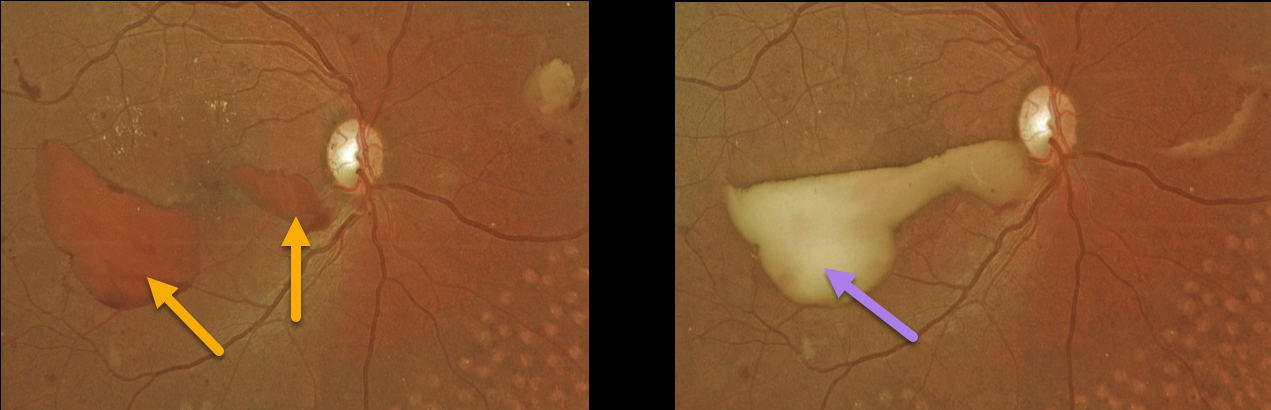
This 37YO female presented with recent vision loss in her right eye. She had a history of prior laser treatment elsewhere several years ago. Vision was 20/30 OD and 20/25 OS.
Red preretinal blood was seen in the inferotemporal right macula with partial PRP scarring. One month following a single Avastin injection, the devitalized preretinal blood was yellowing. Bilateral PRP is planned.
Learning Points:
PRP has been around since the 1960s and still remains the mainstay for treating proliferative disease. Often considered a “one and done”, patients can still develop break-through hemorrhage or progressive neovascular growth and traction.
Intravitreal anti-VEGF injections are often helpful, but some will require vitrectomy.
In our patient’s case, the new preretinal blood was most likely due to her having been incompletely treated rather than a PRP failure.
PROLIFERATIVE DIABETIC RETINOPATHY (PDR)
Originally posted on @retina.rocks June 30, 2022
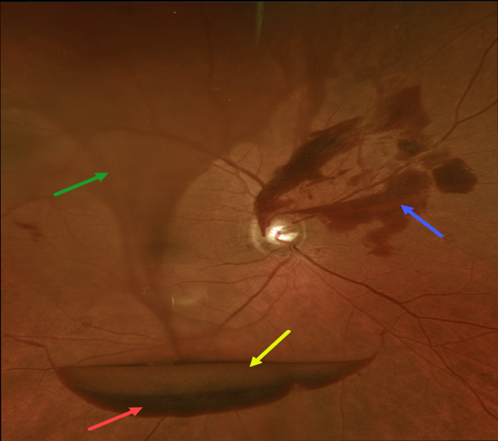
The preretinal blood in this 70YO male with proliferative diabetic retinopathy (PDR) has multiple layers.
The red arrow highlights subhyaloid blood, while the yellow arrow shows the yellowing, devitalized subhyaloid blood just above. The blue arrow shows subhyaloid blood emanating from disc neovascularization. Last but not least, the twister of blood within the vitreous gives the appearance of a tornado literally shaking up all of this blood.
Panretinal photocoagulation was started.
PROLIFERATIVE DIABETIC RETINOPATHY (PDR)
Originally posted on @retina.rocks June 16, 2022
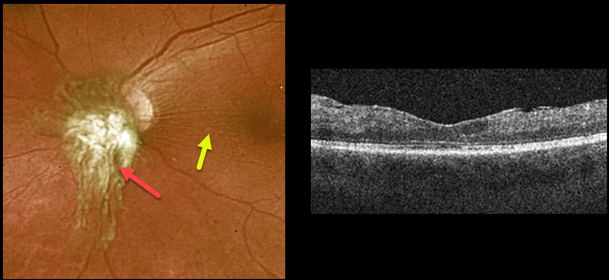
This 26YO female presented with bilateral disc and peripheral retinal neovascularization from proliferative diabetic retinopathy. Vision was 20/50 OD and 20/200 OS.
Panretinal photocoagulation was started in each eye. She was then lost to follow-up for a year, when she returned with a somewhat unusual elevated patch of fibrosed disc neovascularization in her left eye. Some mild nasal macular traction is noted in this image and on OCT scanning.
Some peripheral vitreous hemorrhage was also present in her left eye (not shown). Vision was 20/50 in her stable OD and counting fingers OS.
The PRP was completed in each eye. The fibrosed NVD OS will not resolve and hopefully won’t cause further macular traction.
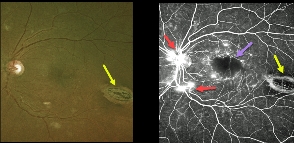
DIABETIC MACULAR ISCHEMIA WITH TORPEDO MACULOPATHY
Originally posted on @retina.rocks June 9, 2022
This 45YO female with type 2 diabetes presented with bilateral proliferative diabetic retinopathy (PDR). Vision in her left eye was 20/200 due to severe foveal ischemia, which is best seen on fluorescein angiography. Leaking disc and peripheral neovascularization are also seen angiographically. The macula was also edematous on OCT scanning (not shown).
An incidental finding completely unrelated to her diabetic retinopathy was an oval-shaped chorioretinal lesion in the distal inferotemporal macula known as torpedo maculopathy.
Although anti-VEGF therapy was started in her left eye for the macula edema, we doubt vision will significantly improve due to the foveal ischemia.
Learning Points:
Gass originally described torpedo maculopathy as a solitary hypopigmented nevus of the RPE (Arch Ophthalmology 1992;1358-1359). These benign, unilateral lesions are teardrop-shaped and almost always located in the horizontal meridian of the temporal macula.
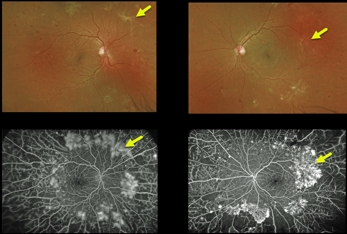
PROLIFERATIVE DIABETIC RETINOPATHY (PDR)
Originally posted on @retina.rocks May 27, 2022
This 29YO male with type 1 diabetes presented with severe peripheral proliferative diabetic retinopathy (PDR). Despite having extensive peripheral ischemia and neovascularization, the posterior poles were remarkably almost normal.
Learning Points:
These types of presentations are very humbling for us clinicians and remind us of the importance of peripheral retinal examination and ultra-widefield imaging with fluorescein angiography in screening for proliferative disease.
Widefield OCT angiography may also prove to be an equally effective yet less invasive screening tool (Al-Khersan et al., Am J Ophthalmol 2021;224:292-300).
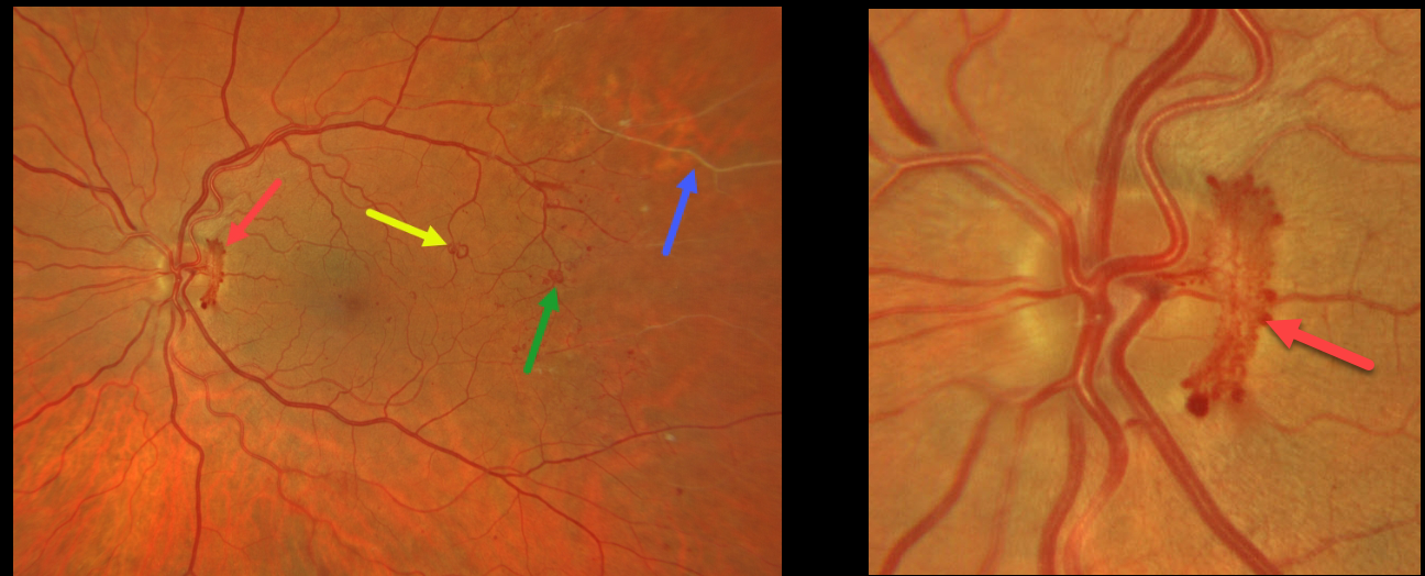
PROLIFERATIVE DIABETIC RETINOPATHY (PDR)
Originally posted on @retina.rocks April 8, 2022
This patient presented with some beautifully imaged findings for proliferative diabetic retinopathy (PDR).
A wide-angle photograph shows active disc neovascularization (NVD), a venous loop, a few small patches of distal macular peripheral neovascularization (NVE), and distal retinal ischemia. A magnified image of the nerve shows the NVD.
Although venous loops are not generally included in the clinical classification of diabetic retinopathy, they are universally seen in severely ischemic eyes.
This patient is currently undergoing panretinal photocoagulation.
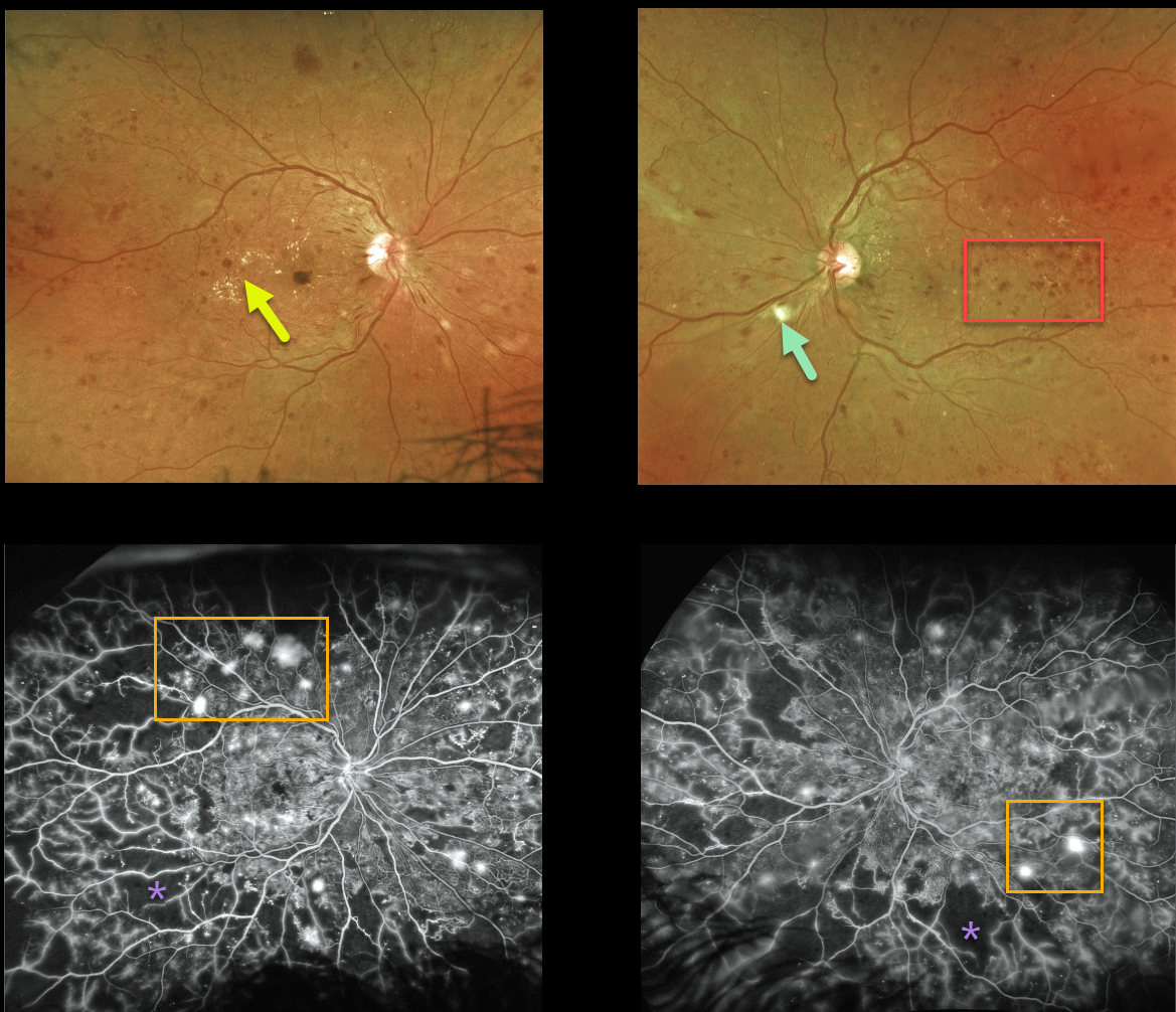
PROLIFERATIVE DIABETIC RETINOPATHY (PDR)
Originally posted on @retina.rocks March 23, 2022
This 41YO female presented with bilateral proliferative diabetic retinopathy (PDR).
Optos ultra-widefield color imaging shows extensive bilateral retinal hemorrhages, along with hard exudates and cotton wool spots.
Fluorescein angiogram shows extensive scattered retinal neovascularization with more peripheral retinal ischemia.
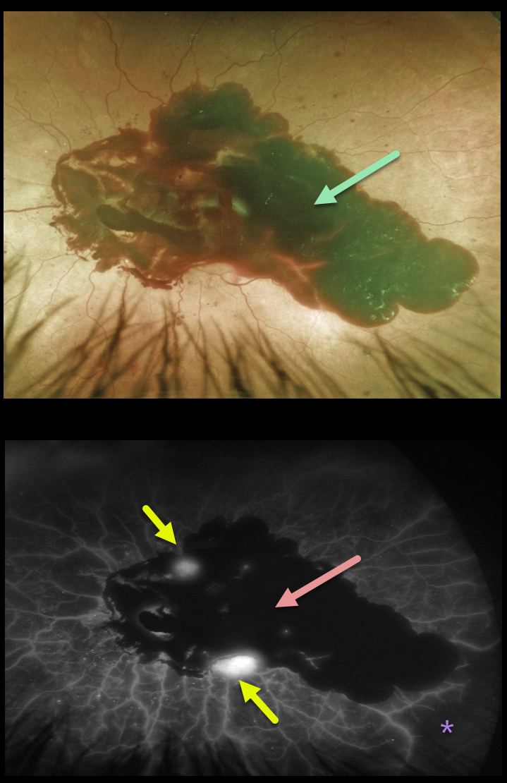
PROLIFERATIVE DIABETIC RETINOPATHY (PDR)
Originally posted on @retina.rocks February 1, 2022
This 69YO male with type 2 diabetes presented with severe acute vision loss in his left eye from a large posterior pole subhyaloid hemorrhage.
The hemorrhage blocks on fluorescein angiography, with a few areas of leaking neovascularization and severe peripheral ischemia.
He was initially treated with intravitreal Avastin, and over the coming months, panretinal photocoagulation (PRP) was performed.
However, he later developed a large vitreous hemorrhage and will likely need vitrectomy surgery.
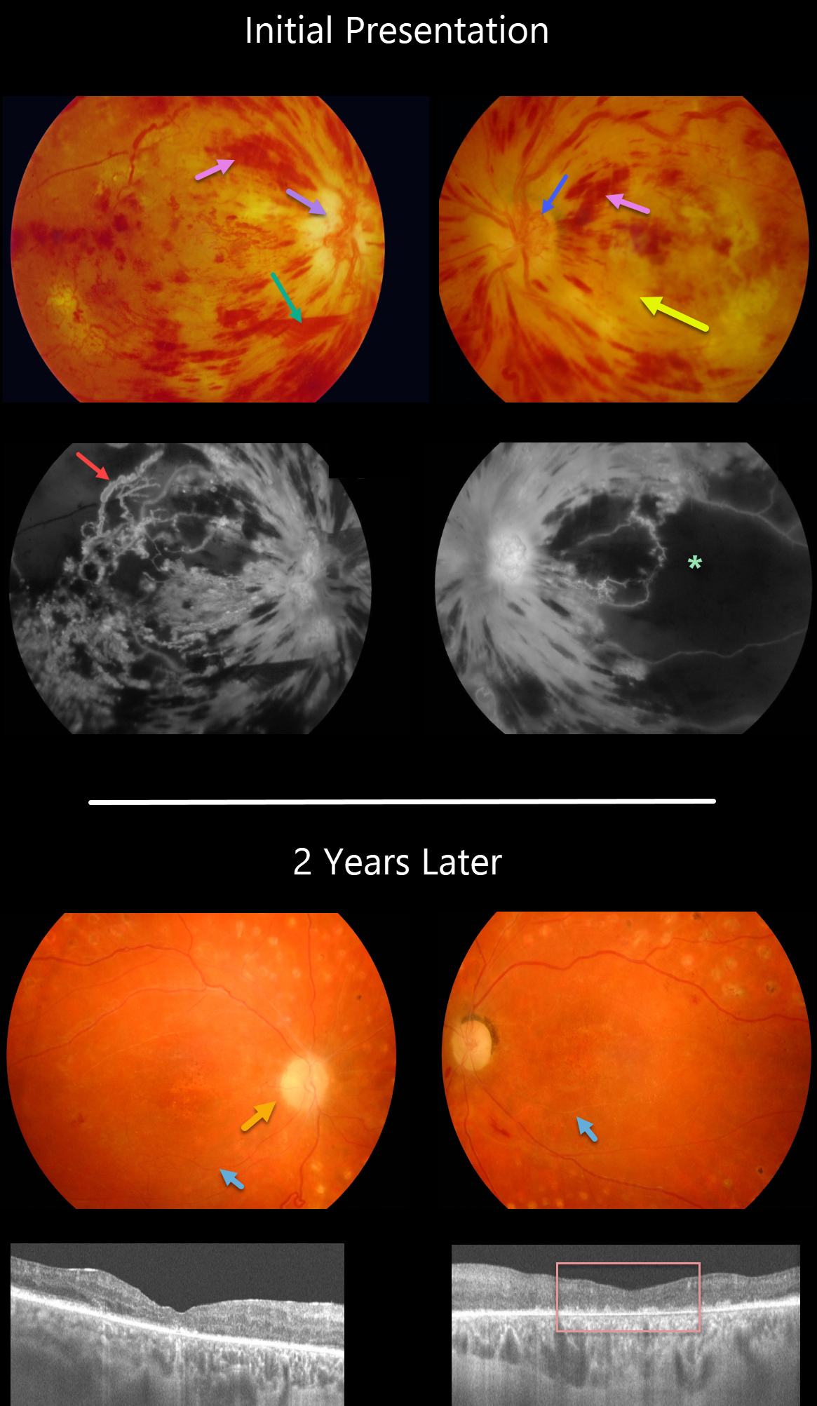
PDR + HYPERTENSIVE RETINOPATHY
Originally posted on @retina.rocks November 30, 2021
This 30YO female presented in 2008 with acute severe bilateral vision loss. She had a history of well-controlled type 1 diabetes.
There were severe bilateral retinal hemorrhages with white, ischemic maculas. A small preretinal hemorrhage was present just inferior to the right nerve. The right optic nerve was swollen. Disc neovascularization was noted in her left eye.
Fluorescein angiography showed severe macular ischemia, especially in her left eye, with near total obliteration of the normal macular vascular anatomy. Prominent staining venous beading was present in the right superotemporal macula.
Her blood pressure was severely elevated. Following emergent hypertensive control, bilateral anti-VEGF therapy was given, followed by panretinal photocoagulation.
Two years later, vision returned to 20/40 OD and 20/400 OS. The optic nerves were pale, the retinal vessels were narrowed and sheathed, and the foveas had dry pigmentary changes.
OCT scanning showed thinned, atrophic maculae with disorganized inner retinal layers (DRIL).
Learning Points:
Malignant hypertension, defined as blood pressure above 180 systolic and/or 120 diastolic, is a life-threatening medical emergency. Eye doctors are in a unique position to often diagnose this condition. Patients will often present with bilateral optic nerve swelling, flame-shaped retinal hemorrhages, and, with more chronic disease, lipid precipitates in the nasal macular outer plexiform layer.
Hypertensive retinopathy can have a similar appearance to diabetic retinopathy, and patients (like this one) can even present with signs of both.
Over time, patients with hypertensive retinopathy will develop near complete regression of all findings following systemic blood pressure treatment and control.
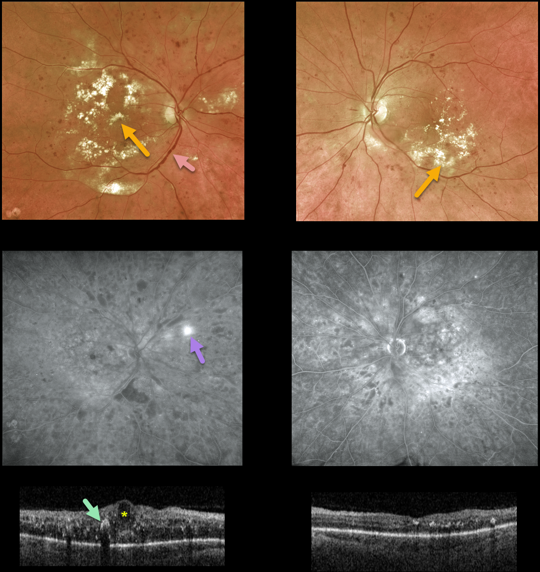
DIABETIC MACULAR EDEMA (DME)
Originally posted on @retina.rocks October 14, 2021
This 59YO male with a history of type 2 diabetes presented with bilateral diabetic macular edema. Vision was counting fingers OD and 20/30 OS.
There was extensive bilateral macular lipid, particularly in his right eye. Venous beading and sausaging were noted OD, indicating severe retinal ischemia.
Fluorescein angiography confirmed severe ischemia with peripheral temporal retinal neovascularization.
Triton swept-source OCT scanning shows severe central-involved diabetic macular edema (CI-DME) with scattered hyperreflective lipid flecks OD, non-central-involved diabetic macular edema (NCI-DME), and milder lipid OS.
Macular laser for the NCI-DME was recommended for his left eye. Monthly anti-VEGF injections were recommended for the CI-DME in his right eye. Remarkably, vision improved to 20/80 so far after a single injection.
Learning Points:
Anti-VEGF injections are the mainstay of treatment for CI-DME. It is expected that these injections will also stabilize, or possibly reverse, this patient’s proliferative disease.
We also plan to begin panretinal photocoagulation once the central edema resolves. There are multiple ways we could have approached his treatment plan. What would you do and why?
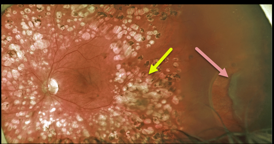
RHEGMATOGENOUS RETINAL DETACHMENT
Originally posted on @retina.rocks September 22, 2021
This 50YO male presented for a routine diabetic follow-up. A new asymptomatic peripheral retinal tear causing a rhegmatogenous retinal detachment was noted anterior to full panretinal photocoagulation (PRP) scarring.
We will often demarcate asymptomatic retinal detachments with laser photocoagulation (Vrabec and Baumel, Ophthalmology 2000;107:1063-1067). This patient essentially had laser demarcation before his retinal detachment, but the end result was the same, with his detachment being contained by the laser scarring.
Learning Points:
We opted to treat this eye with vitrectomy, but one could argue that this detachment could also be closely followed since it was contained by the prior laser.
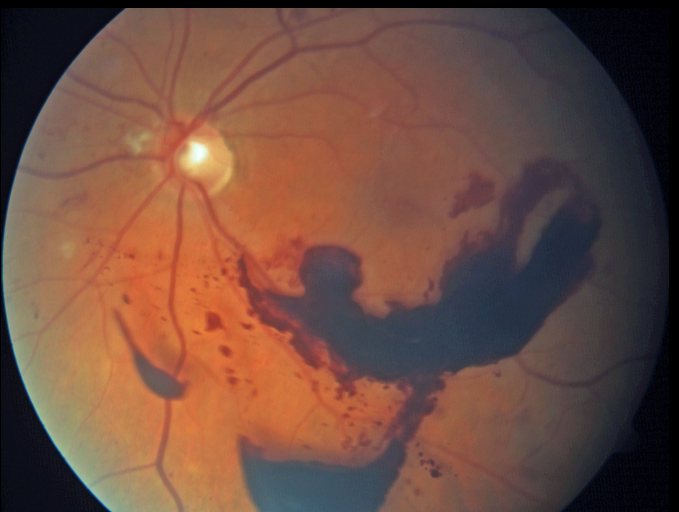
MERMAID MACULOPATHY
Originally posted on @retina.rocks September 8, 2021
This 55YO female with a history of proliferative diabetic retinopathy presented with a symptomatic preretinal hemorrhage. Vision was 20/50.
You don’t need to have an overly active imagination to see this rare finding of mermaid maculopathy.
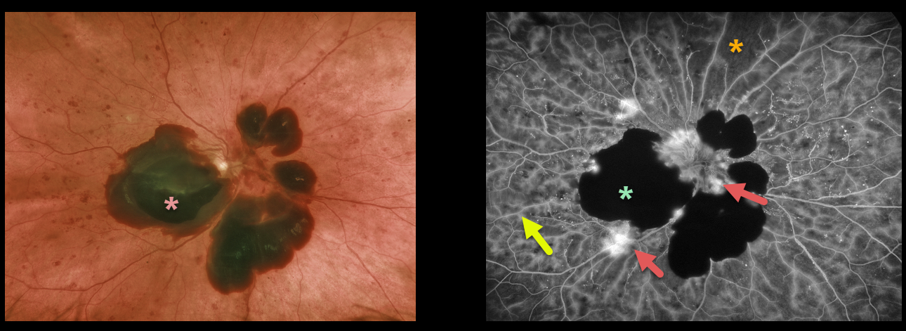
PROLIFERATIVE DIABETIC RETINOPATHY (PDR)
Originally posted on @retina.rocks August 10, 2021
This 53YO female with type 2 diabetes presented with acute vision loss from previously undiagnosed proliferative diabetic retinopathy.
Optos color imaging shows patches of mostly subhyaloid blood in the macula and peripapillary retina.
Fluorescein angiography beautifully exhibits various angiographic characteristics for hypo- and hyperfluorescence. We can see both blocking and filling defects, in addition to diffuse vascular staining and leakage of the disc and adjacent to the subhyaloid blood.
Learning Points:
Angiographically, hypofluorescence is classified as either a blockage or a filling defect. Blood, which masks the underlying retinal and choroidal fluorescence, causes blockage. There is diffuse capillary loss and ischemia, appearing as glassy-dark areas of hypofluorescence throughout, representing filling defects. Filling defects are virtually never as dark as areas of blockage.
Angiographic hyperfluorescence is classified into window defects, pooling, staining, leakage, and neovascularization, and our case exhibits several of these features.
The endothelial cells of the larger vessels in areas of capillary dropout become ischemic, leading to breakdown of the inner retinal barrier. The larger vessels, therefore, often show late staining in these regions as fluorescein dye leaks into and stains the vessel wall. Vascular staining is evident throughout the entire imaged fundus.
The new blood vessels growing on the disc and adjacent to the subhyaloid blood lack endothelial tight junctions, which is why neovascularization shows profound angiographic leakage.
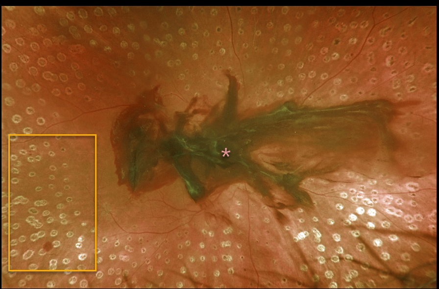
PROLIFERATIVE DIABETIC RETINOPATHY (PDR)
Originally posted on @retina.rocks June 28, 2021
Despite full panretinal photocoagulation, this 57YO patient with proliferative diabetic retinopathy developed an acute premacular hemorrhage. Her vision was 20/300 in the right eye.
Learning Points:
Panretinal photocoagulation has been around since the 1960s and still remains the mainstay for treating proliferative disease. Often considered a “one and done,” patients can still develop breakthrough hemorrhages.
Intravitreal anti-VEGF injections are often helpful, but some will require vitrectomy.
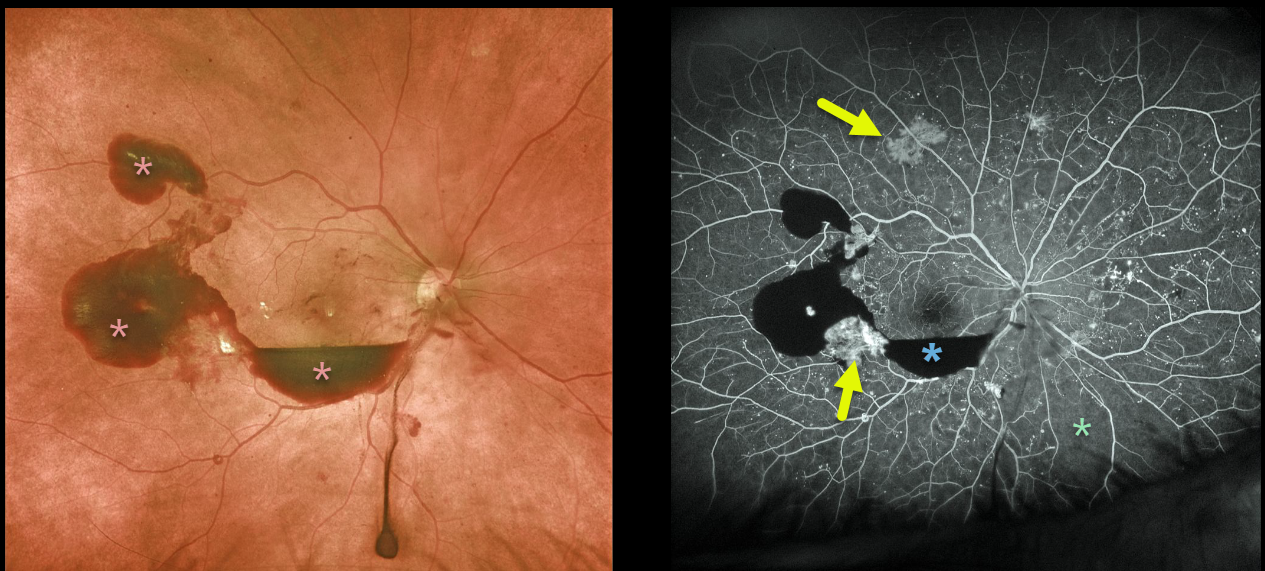
PROLIFERATIVE DIABETIC RETINOPATHY (PDR)
Originally posted on @retina.rocks May 17, 2021
The 58 yo male with proliferative diabetic retinopathy presented with preretinal blood. Optos fluorescein angiography shows blockage from this blood with scattered areas of midperipheral neovascularization and peripheral ischemia, especially inferonasally.
Learning Points:
Angiographically, hypofluorescence is classified as either a blockage or a filling defect. Blood, which masks the underlying retinal and choroidal fluorescence, causes blockage. There is diffuse capillary loss and ischemia, appearing as glassy-dark areas of hypofluorescence throughout, representing filling defects. Filling defects are virtually never as dark as areas of blockage.
Angiographic hyperfluorescence is classified into window defects, pooling, staining, leakage, and neovascularization. The new blood vessels adjacent to the subhyaloid blood and in the mid-periphery lack endothelial tight junctions, which is why neovascularization shows profound angiographic leakage.
PROLIFERATIVE DIABETIC RETINOPATHY (PDR)
Originally posted on @retina.rocks April 29, 2021
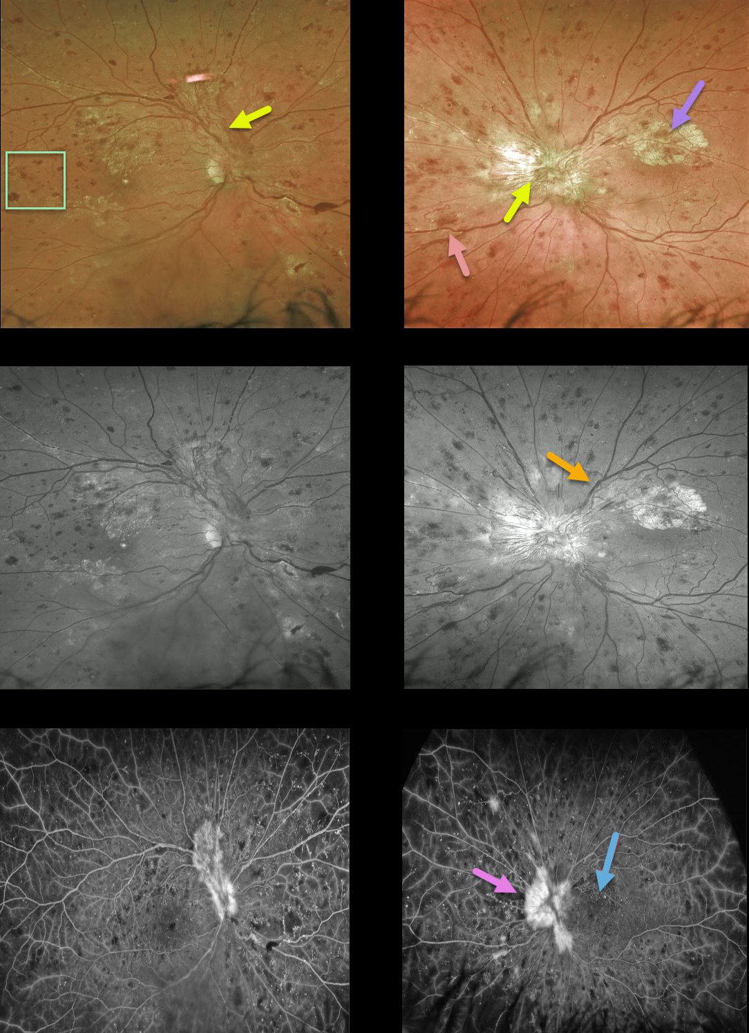
This 40YO man presented with bilateral proliferative diabetic retinopathy (PDR). Vision was 20/60 OD and 20/200 OS.
Optos color and green channel imaging show prominent bilateral fibrovascular disc neovascularization, obscuring the left disc.
A few smaller areas of more peripheral neovascularization are present nasally in the OS. Severe retinal hemorrhages are noted in all quadrants; the retinal veins are beaded OU superiorly, along with some perivascular lipid superotemporally OD and nasally OS. The retinal vascular abnormalities are best highlighted with the green channel image.
There also appears to be an area of fibrosed neovascularization in the superior left macula. However, these findings are visible beneath the retinal vessels, indicating they are subretinal and composed of lipid. Lipid is usually angiographically invisible but, when dense, can obscure the underlying choroidal fluorescence, as in this case.
Fluorescein angiography shows leakage from the neovascularization, diffuse vascular staining, and mostly peripheral retinal nonperfusion.
PROLIFERATIVE DIABETIC RETINOPATHY (PDR)
Originally posted on @retina.rocks April 2, 2021
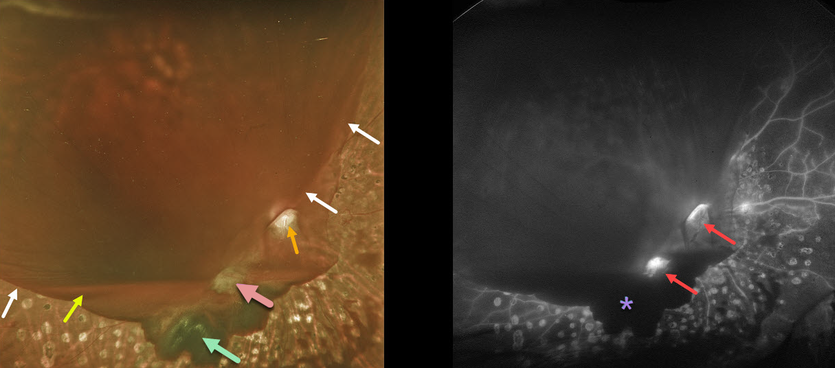
This diabetic patient developed a symptomatic hemorrhage in his right eye despite partial panretinal photocoagulation (PRP) treatment. The blood in our patient highlights the various layers of vitreous separation.
The reason why the far inferior and nasal retina and PRP scarring are visible is that the hyaloid is completely attached here. The hyaloid is shallowly elevated inferiorly, which gives a scalloped appearance to the dark red subhyaloid blood.
Elsewhere, the hyaloid is completely detached and highly elevated, allowing blood to pool in this space (white arrows mark the junction between the attached and detached hyaloid). A layer of blood pools inferiorly at the base of the hyaloid separation.
The optic disc and fibrosed neovascularization are well visualized because the vitreous remains tethered, preventing blood from accumulating.
Fluorescein angiography highlights areas of neovascularization and the blockage of the underlying retina by various layers of blood.
Learning Points:
At birth, the vitreous is diffusely adherent to the entire retina. But over the years, the vitreous begins to separate, first in the midperiphery, then in the peripheral macula, then the central macula, and finally the optic nerve.
For a great review of posterior vitreous detachment (PVD) evolution and complications, see Johnson AJO 2010;149:371-382.
Retinal neovascularization arises from the inner retina and extends onto the back surface of the vitreous; without this scaffold, it will not develop. A complete PVD is therefore protective against posterior segment retinal neovascularization.
Since the vitreous in the posterior pole is most adherent around the optic nerve and major arcades, this is where diabetic neovascularization most commonly develops. It is also why the vitreous often remains tethered to these structures as the proliferation progresses.
The posterior hyaloid continues to detach and contract, as does the neovascularization, which can cause vitreous hemorrhage and traction retinal detachment.
PROLIFERATIVE DIABETIC RETINOPATHY (PDR)
Originally posted on @retina.rocks February 25, 2021
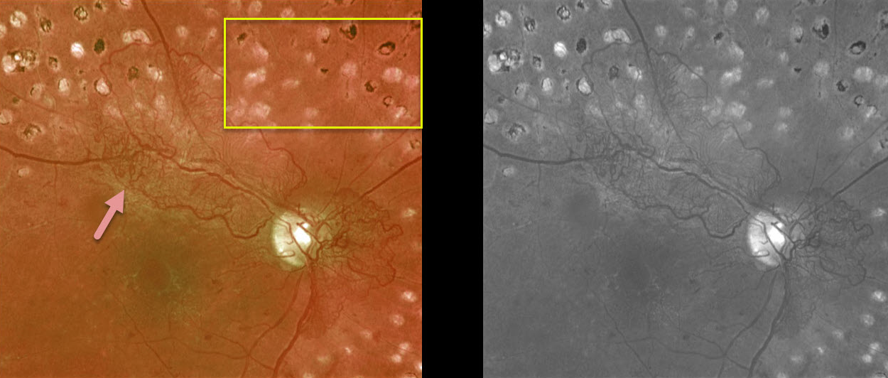
This is a 55YO diabetic patient who had been treated with complete panretinal photocoagulation (PRP). Vision was 20/40. Despite PRP, the patient still had significant residual posterior neovascularization.
Since it is not uncommon for neovascularization to persist following full laser, we are observing for now.
Learning Points:
Panretinal photocoagulation has been around since the 1960s and still remains the mainstay for treating proliferative disease. Often considered a “one and done,” patients can still develop breakthrough hemorrhages. Intravitreal anti-VEGF injections are often helpful, but some will require vitrectomy.
HYPERTENSION + PROLIFERATIVE DIABETIC RETINOPATHY (PDR)
Originally posted on @retina.rocks February 2, 2021
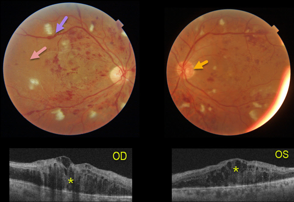
This 41YO male presented after a recent hospitalization for severe hypertension with vision of counting fingers OD and 20/60 OS.
The bilateral nerve fiber layer infarcts (cotton-wool spots) are more characteristic of hypertension than of diabetes.
Severe retinal hemorrhages and disc neovascularization are noted in each eye. The right eye, in particular, is severely ischemic, with Triton imaging showing a featureless, ischemic retina and white vessels, most pronounced in the temporal retina.
The venous sausaging is a clinical sign of severe ischemia, and when present, virtually always shortly precedes or is indicative of concurrent proliferative disease. Triton swept-source OCT shows severe bilateral center-involving diabetic macular edema.
He subsequently received multiple bilateral anti-VEGF injections followed by panretinal photocoagulation, with marked improvement of the central edema.
OPTIC NERVE PSEUDO-DUPLICATION WITH NVD ELSEWHERE
Originally posted on @retina.rocks December 18, 2020
Is that another optic nerve?
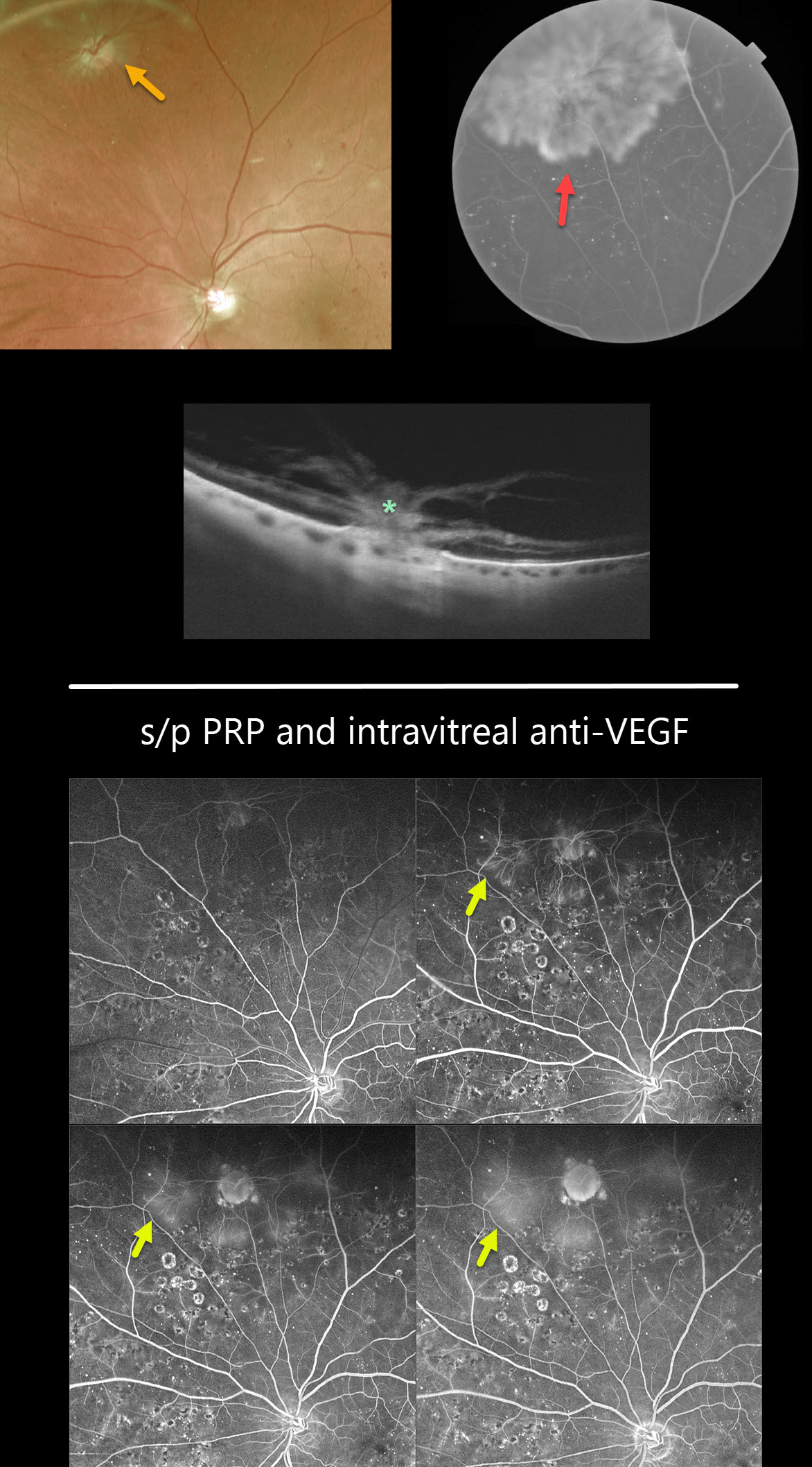
This is a pseudo-duplicated optic disc from peripheral neovascularization emanating from a disc-sized mid-peripheral chorioretinal scar.
The OCT reveals the area of choriovitreal neovascularization.
Learning Points:
The neovascularization regressed following panretinal photocoagulation (PRP) supplemented with intravitreal Eylea injections.
Fluorescein angiography following PRP and anti-VEGF injections shows virtually no leakage from the neovascularization.
PROLIFERATIVE DIABETIC RETINOPATHY (PDR)
Originally posted on @retina.rocks December 11, 2020
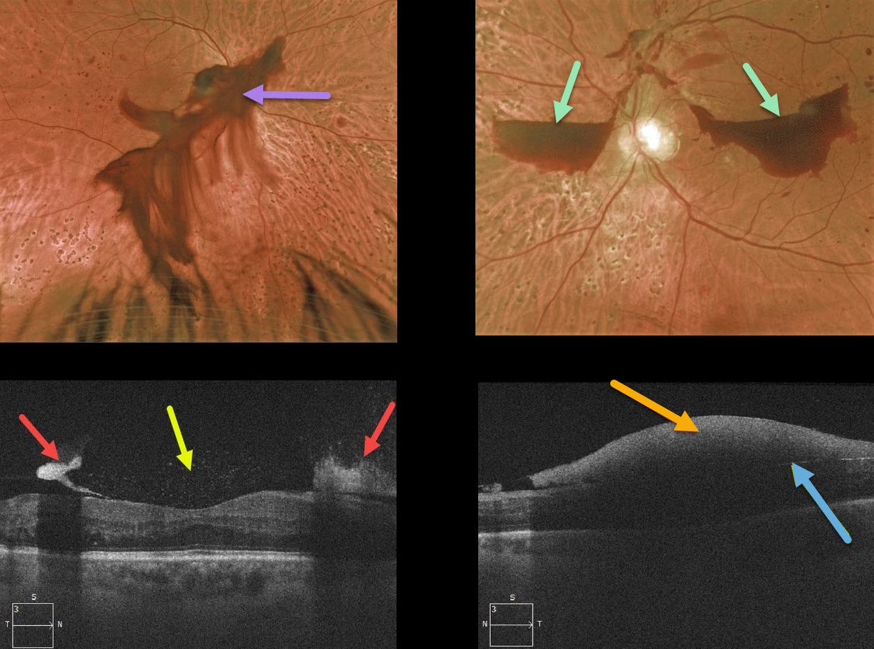
This patient presented with a posterior pole vitreous hemorrhage OD and localized subhyaloid hemorrhages OS.
The OCT of the right eye highlights a thickened hyaloid on either side of the fovea with specks of hyperreflective pre-retinal blood. The left eye shows the subhyaloid blood blocking most of the underlying retinal detail.
This patient has already received partial panretinal photocoagulation bilaterally and will receive additional laser therapy.
PROLIFERATIVE DIABETIC RETINOPATHY (PDR)
Originally posted on @retina.rocks November 10, 2020
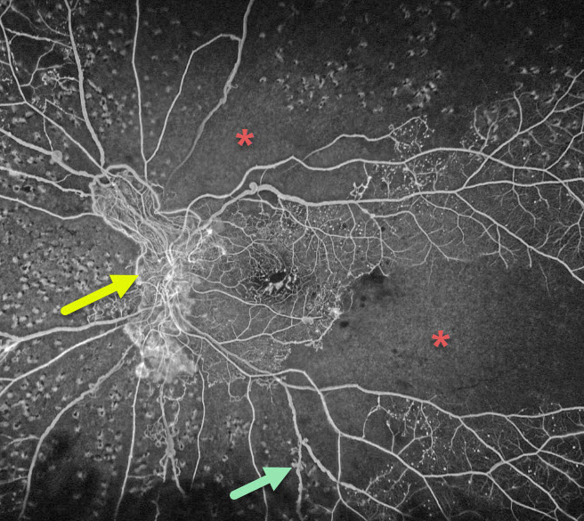
This is an Optos ultra-widefield fluorescein angiogram of a patient with proliferative diabetic retinopathy. Despite recent panretinal photocoagulation (PRP), there is persistent large disc neovascularization. Extensive capillary nonperfusion is noted superotemporal to the disc and temporarily. Prominent venous beading is also noted inferotemporally. Additional PRP was added.
Learning Points:
Panretinal photocoagulation has been around since the 1960s and still remains the mainstay for treating proliferative disease. Often considered a “one and done,” patients can still develop breakthrough hemorrhages. Intravitreal anti-VEGF injections are often helpful, but some will require vitrectomy.
RETINAL VENOUS MALFORMATION
Originally posted on @retina.rocks October 12, 2020
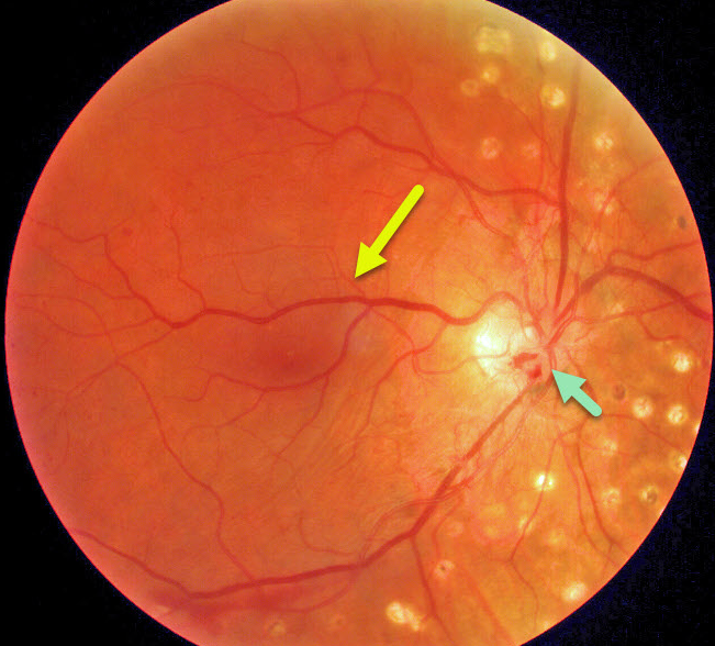
This patient has a retinal venous malformation (formerly known as retinal macrovessel), in addition to full panretinal photocoagulation (PRP) for proliferative diabetic retinopathy. Residual flat disc neovascularization remains despite full PRP.
Learning Points:
A retinal venous malformation is an anomalous retinal vessel that crosses the horizontal raphe (remember that normal retinal vessels respect the horizontal meridian).
These vessels are technically arterio-venous malformations (AVM). Since patients with AVMs can have additional venous anomalies in the brain (24% compared to up to 6% of the population), a brain MRI is recommended to rule out any intracranial abnormalities.
PROLIFERATIVE DIABETIC RETINOPATHY (PDR)
Originally posted on @retina.rocks August 12, 2020
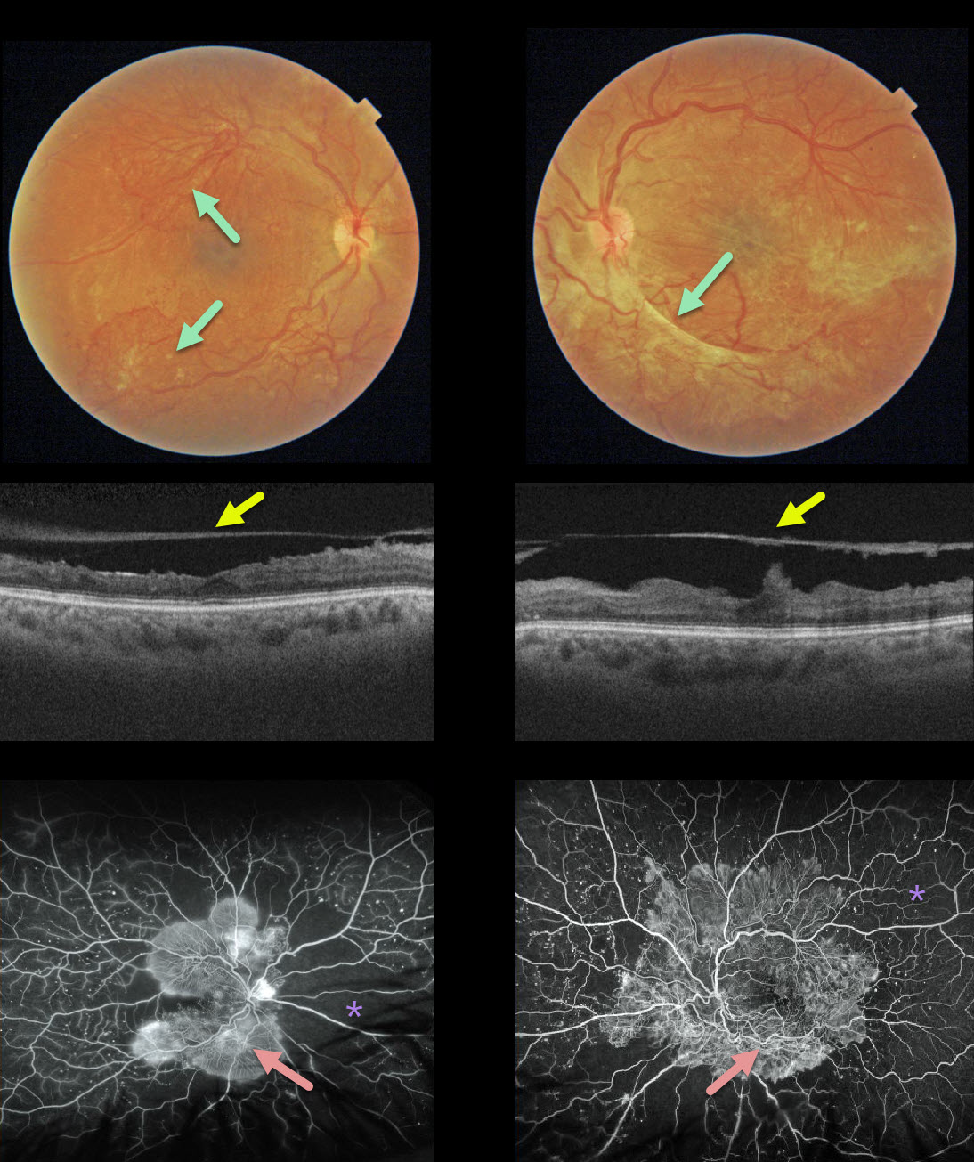
This 32YO female presented with 20/400 vision bilaterally, a hemoglobin A1C over 12%, and a blood glucose level over 300. Fibrosed neovascularization variably covers each posterior pole.
OCT shows that the posterior hyaloid has spontaneously detached from each central macula, which has a rippled contour. This appearance is also often seen following vitrectomy for tractional detachment with peeling of the hyaloid.
Fluorescein angiography shows extensive posterior pole neovascularization with peripheral ischemia.
ASTEROID HYALOSIS
Originally posted on @retina.rocks July 29, 2020
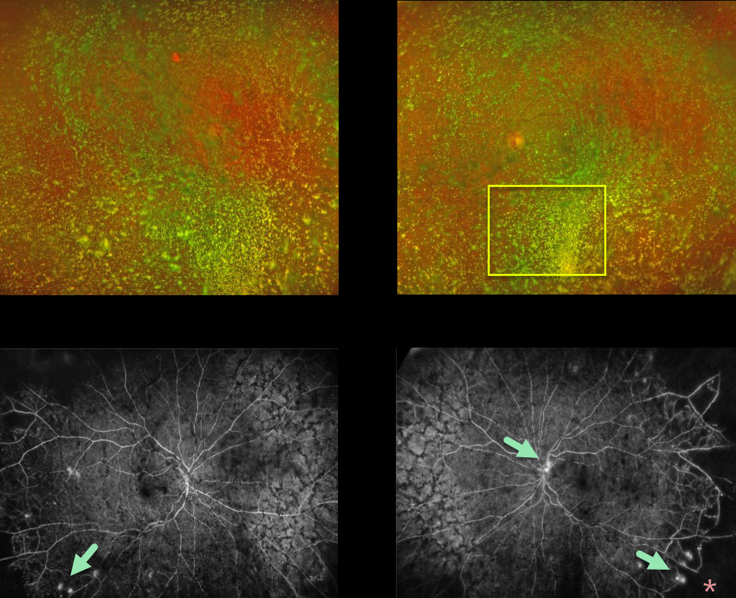
Although this diabetic patient had normal vision with absolutely no symptoms, what we saw on clinical examination and Optos color imaging was nothing but asteroid hyalosis.
Fluorescein angiography (FA) showed bilateral proliferative diabetic retinopathy with areas of leakage and peripheral capillary dropout. In-office panretinal photocoagulation (PRP) was not possible due to the dense asteroid.
We discussed ongoing anti-VEGF injections versus vitrectomy + PRP to clear the visual axis. She decided to proceed with vitrectomy in both eyes.
Learning Points:
Asteroid hyalosis is caused by the accumulation of benign calcium-lipid particles suspended in the vitreous.
Fluoroscein angiography and OCT can image through asteroid hyalosis.
PROLIFERATIVE DIABETIC RETINOPATHY (PDR)
Originally posted on @retina.rocks July 2, 2020
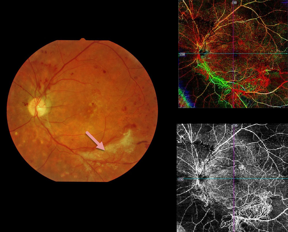
This patient has proliferative diabetic retinopathy. The fundus photo shows scattered red and fibrosed neovascularization.
The color OCTA image is a composite showing all layers of the retinal vasculature: red is the inner retina, green is the mid-retinal layer, and blue is the outer retina (photoreceptors, which are usually devoid of intrinsic vasculature). The bottom right image isolates the inner retina, showing regions of retinal ischemia with neovascularization.
Learning Points:
OCTA is a relatively new and non-invasive modality able to quickly identify diabetic retinal vascular abnormalities. Its role in screening for retinal ischemia and proliferative disease is still being defined.
PROLIFERATIVE DIABETIC RETINOPATHY (PDR)
Originally posted on @retina.rocks June 16, 2020
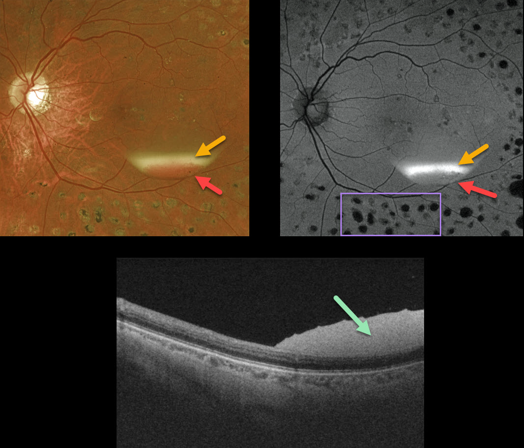
This patient, previously treated for proliferative diabetic retinopathy with panretinal photocoagulation (PRP), presented with an asymptomatic resolving two-toned subhyaloid hemorrhage. The devitalized yellow blood indicates chronicity, although some red blood remains.
The OCT confirms the subhyaloid location of the hyperreflective blood.
Red blood is dark on fundus autofluorescence (FAF), although yellow blood is extremely hyperautofluorescent. Lipofuscin within the RPE is the main fluorophore in FAF. The laser PRP scars are hypoautofluorescent due to loss of the RPE.
PROLIFERATIVE DIABETIC RETINOPATHY (PDR)
Originally posted on @retina.rocks June 3, 2020
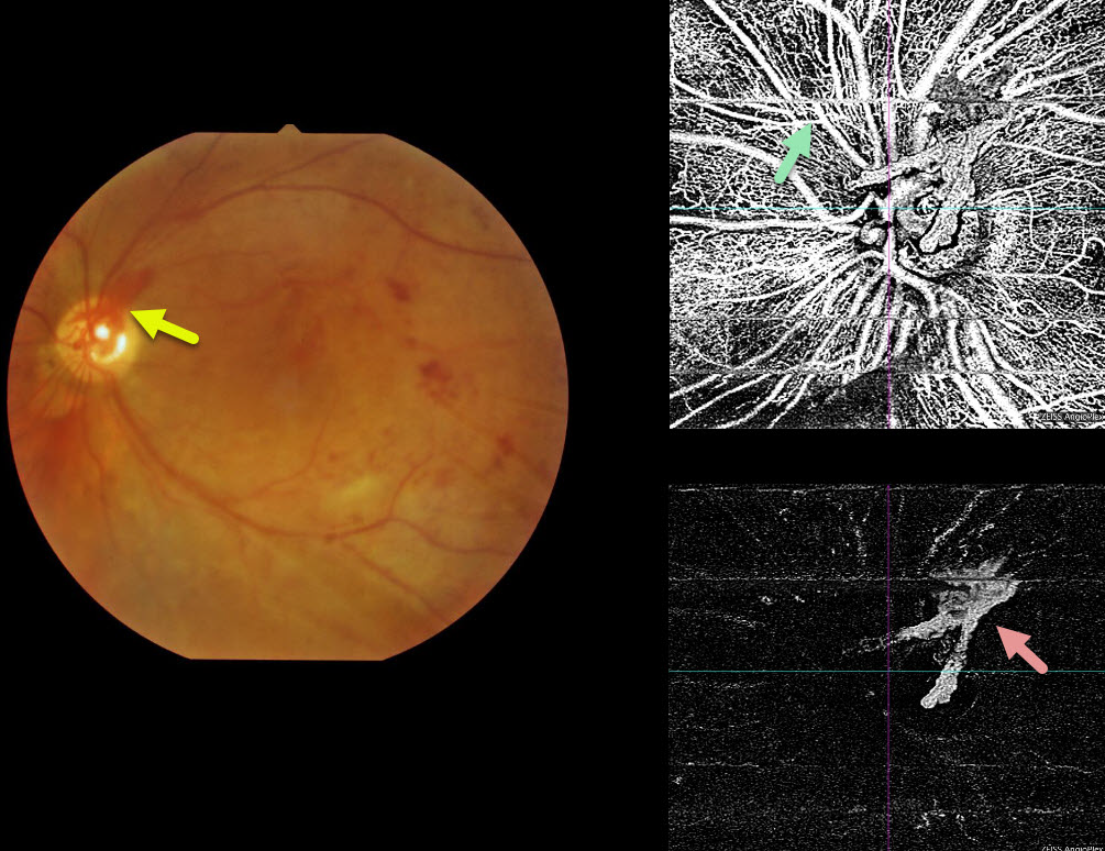
This patient has PDR and prominent neovascularization of the disc (NVD), which is well-captured by the OCT angiogram.
Movement artifacts are noted throughout the scan. The bottom-right image isolates the vitreoretinal interface, showing only the NVD.
Learning Points:
The OCT angiogram captures blood vessels from the full-thickness retina, including the superficial radial peripapillary capillary network, which is not imaged with traditional fluorescein angiography.
PROLIFERATIVE DIABETIC RETINOPATHY (PDR)
Originally posted on @retina.rocks May 4, 2020
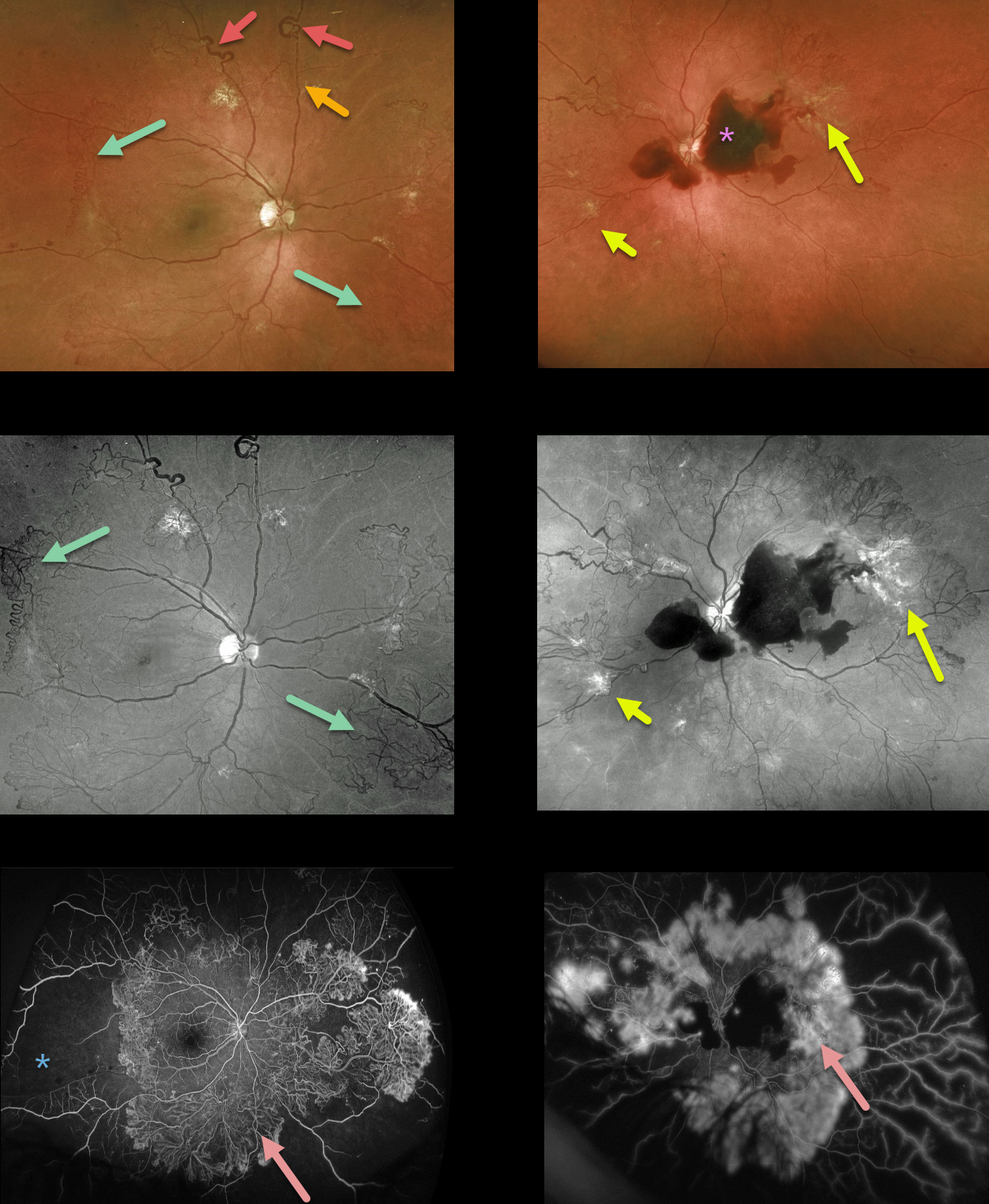
This 39YO presented with severe PDR in both eyes, highlighting nearly all of the key features of proliferative disease. Surprisingly, vision was 20/30 OD and 20/50 OS.
The right eye shows great examples of venous loops and venous beading and extensive scattered NVE, especially highlighted by the green channel Optos.
The left eye has a central subhyaloid hemorrhage along with venous beading and scattered NVE.
Fluorescein angiography shows dramatic and extensive neovascularization encircling both posterior poles with marked anterior ischemia.
Learning Points:
This case further emphasizes the importance of regular diabetic dilated examinations with peripheral retinal examination to unveil the true extent of the retinopathy.
ASTEROID HYALOSIS OBSCURING OCCULT PROLIFERATIVE DIABETIC RETINOPATHY (PDR)
Originally posted on @retina.rocks April 17, 2020
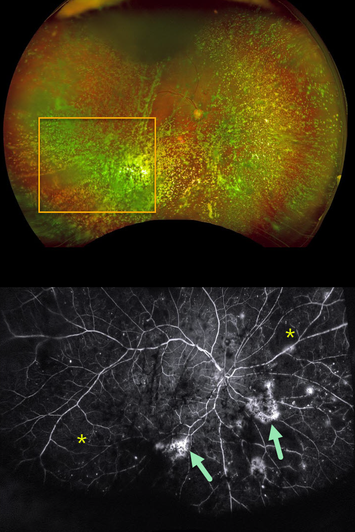
Examining the fundus with dense asteroid hyalosis, as in this diabetic patient, can be extremely challenging.
Fluorescein angiography (FA) and OCT are great modalities since they’re both able to visualize the retina when the clinical exam can’t.
FA was critical in diagnosing proliferative retinopathy with peripheral ischemia in this patient.
Learning Point:
Asteroid hyalosis is caused by calcium-lipid complexes suspended throughout the vitreous. These white opacities resemble stars on a night sky – hence the name.
PROLIFERATIVE DIABETIC RETINOPATHY (PDR)
Originally posted on @retina.rocks April 13, 2020
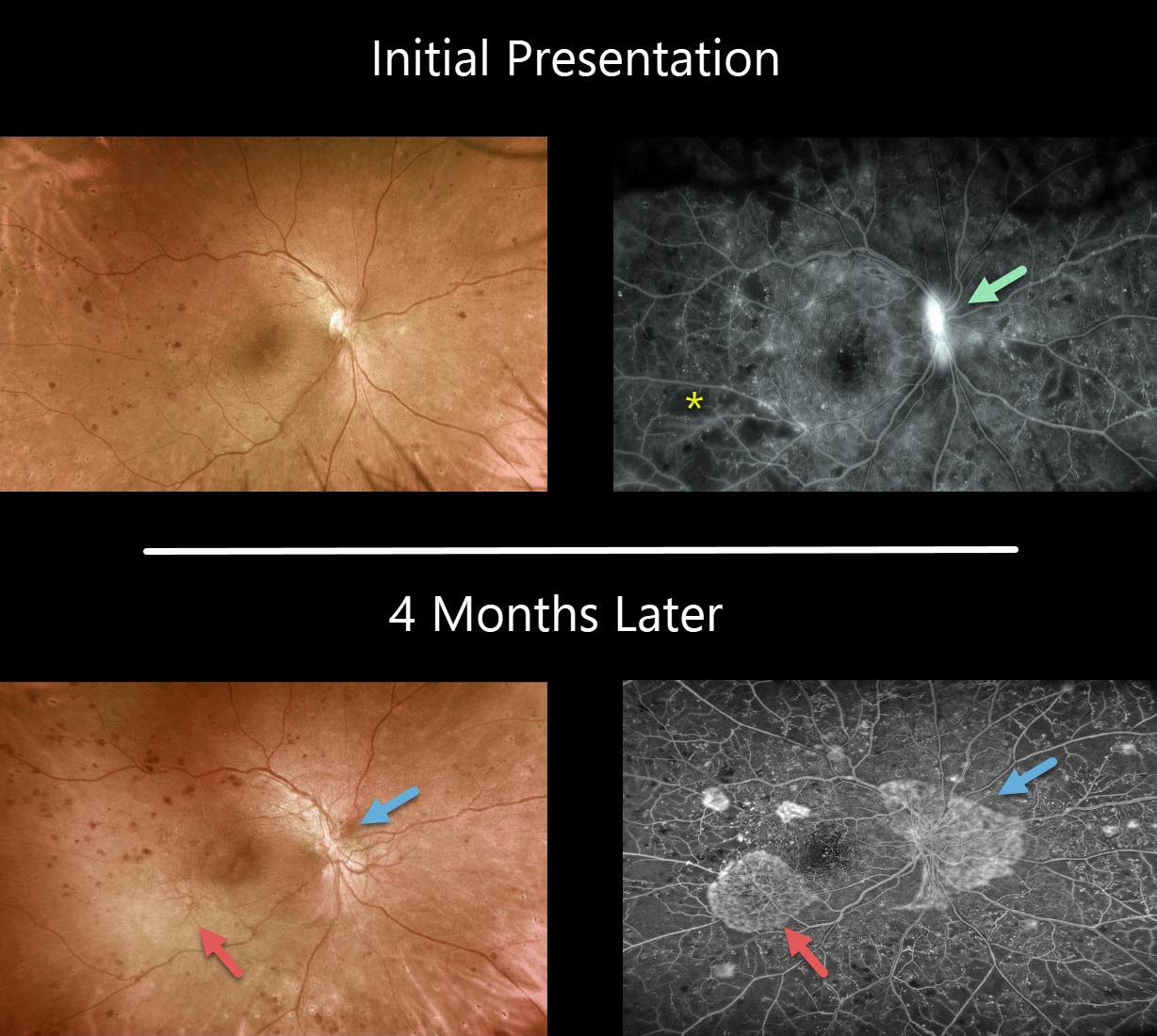
This patient, who was in the process of undergoing panretinal photocoagulation for proliferative diabetic retinopathy, was originally seen with NVD and severe nonperfusion.
Unfortunately, she was lost to follow-up for 4 months and presented again with increased NVD and new NVE. The Optos fluorscein angiogram shows the complex of fine neovascular vessels and capillary dropout in exquisite detail.
PROLIFERATIVE DIABETIC RETINOPATHY (PDR)
Originally posted on @retina.rocks January 31, 2020
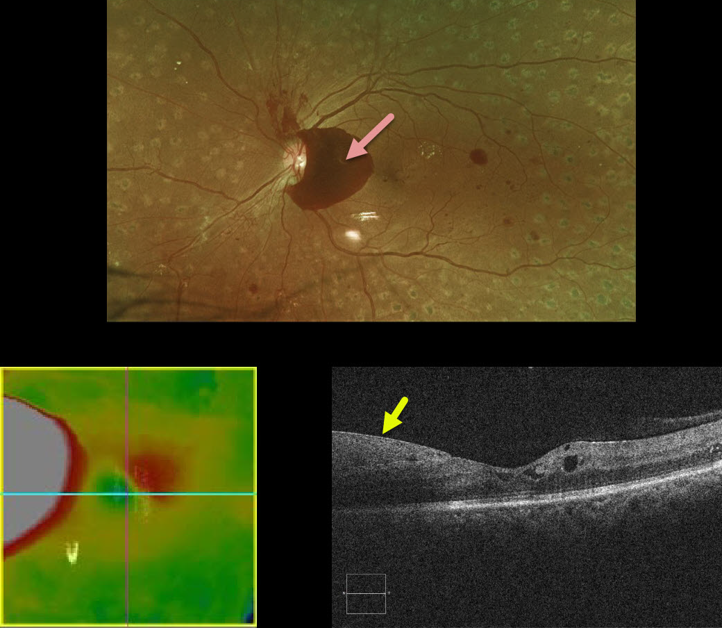
This patient with a history of involuted proliferative diabetic retinopathy following complete panretinal photocoagulation presented with what appeared on OCT to be new diabetic macular edema.
A closer inspection of the B-scan shows the retina to be normal with the pseudo-edema representing subhyaloid blood.
PROLIFERATIVE DIABETIC RETINOPATHY
Originally posted on @retina.rocks January 10, 2020
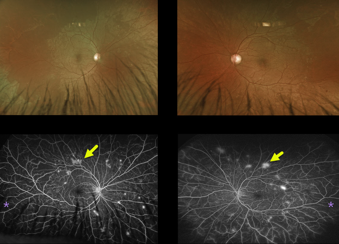
This patient had relatively normal posterior poles with extensive peripheral ischemia and neovascularization on ultra-widefield fluorescein angiography.
Learning Points:
We see this fairly frequently. Since it is so easy to miss proliferative disease, it is not unreasonable for patients with apparent moderate NDPR to have a baseline and periodic ultra-widefield angiogram to rule out subclinical PDR.


