

Purtscher + Purtscher-Like Retinopathy
<< Back to Cases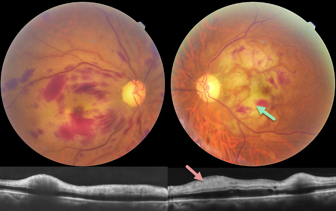
PURTSCHER RETINOPATHY
Abhishek Karra and Ravindra Karra
Originally posted on @retina.rocks July 28, 2026
This 85YO male lost vision in each eye following a fall around 1 week back with multiple rib fractures. Vision was hand motion OU.
Color photography shows bilateral macular retinal hemorrhages with patches of white ischemic retina, most prominent in the left macula. OCT scanning shows a thickened hyperreflective inner retina bilaterally.
Learning Points:
Purtscher retinopathy was first described by Otmar Purtscher in 1910. Although originally described in a man who fell from a tree with cranial trauma, these white patches of retinal ischemia and hemorrhages are found in numerous other etiologies, including pancreatitis, chest trauma, and collagen vascular diseases. The findings are called Purtscher retinopathy when due to trauma (as in our patient), and Purtscher-like retinopathy when seen from other causes.
The underlying cause is unknown, but the findings are thought to result from leukoembolization due to complement activation. The areas of ischemia include more superficial nerve fiber layer infarcts (cotton wool spots) and deeper ischemia involving the middle retinal layers.
PURTSCHER RETINOPATHY
Ayushi Gupta and Vishal Agrawal
Originally posted on @retina.rocks June 1, 2026
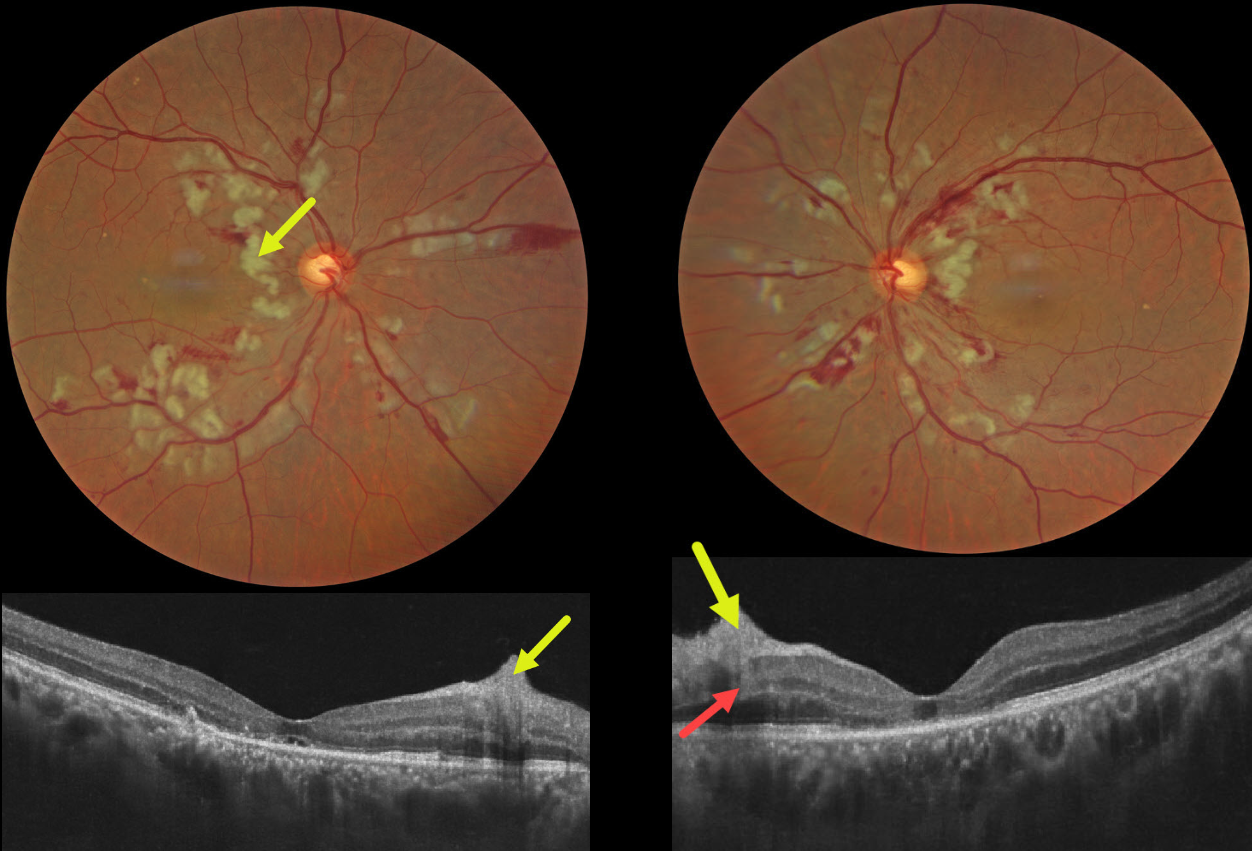
This 55YO male presented with sudden bilateral vision loss that developed immediately following a motor vehicle accident, which included blunt thoracoabdominal trauma and transient loss of consciousness. Vision was 20/200 OU.
Color photography shows bilateral inner and mid-retinal patches of white ischemic retina. The nerve fiber layer lesions appear as inner retinal hyperreflective thickening on OCT. Mid-retinal hyperreflectivity involving the outer plexiform/inner nuclear/outer plexiform layers is noted nasally OS. The outer photoreceptor bands are variably disorganized OU, with a small amount of subfoveal subretinal fluid OD and a foveal cyst OS.
Learning Points:
Purtscher retinopathy was first described by Otmar Purtscher in 1910. Although originally described in a man who fell from a tree with cranial trauma, these white patches of retinal ischemia and hemorrhages are found in numerous other etiologies, including pancreatitis, chest trauma, and collagen vascular diseases. The findings are called Purtscher retinopathy when due to trauma, and Purtscher-like retinopathy when seen from other causes.
The underlying cause of Purtscher is unknown, but it is thought to represent leukoembolization by complement activation. The areas of ischemia include more superficial nerve fiber layer infarcts (cotton wool spots) and deeper ischemia involving the middle retinal layers. This recently described retinal ischemic phenotype, paracentral middle maculopathy (PAMM), occurs as an idiopathic entity or in other ocular disorders, including Purtscher retinopathy, diabetic retinopathy, retinal artery and retinal vein occlusions, and acute macular neuroretinopathy (see Retina 2015;35:1921-1930).
PURTSCHER-LIKE RETINOPATHY
Keissy Sousa, Gil Calvão-Santos, and Rita Gentil
Originally posted on @retina.rocks January 10, 2024
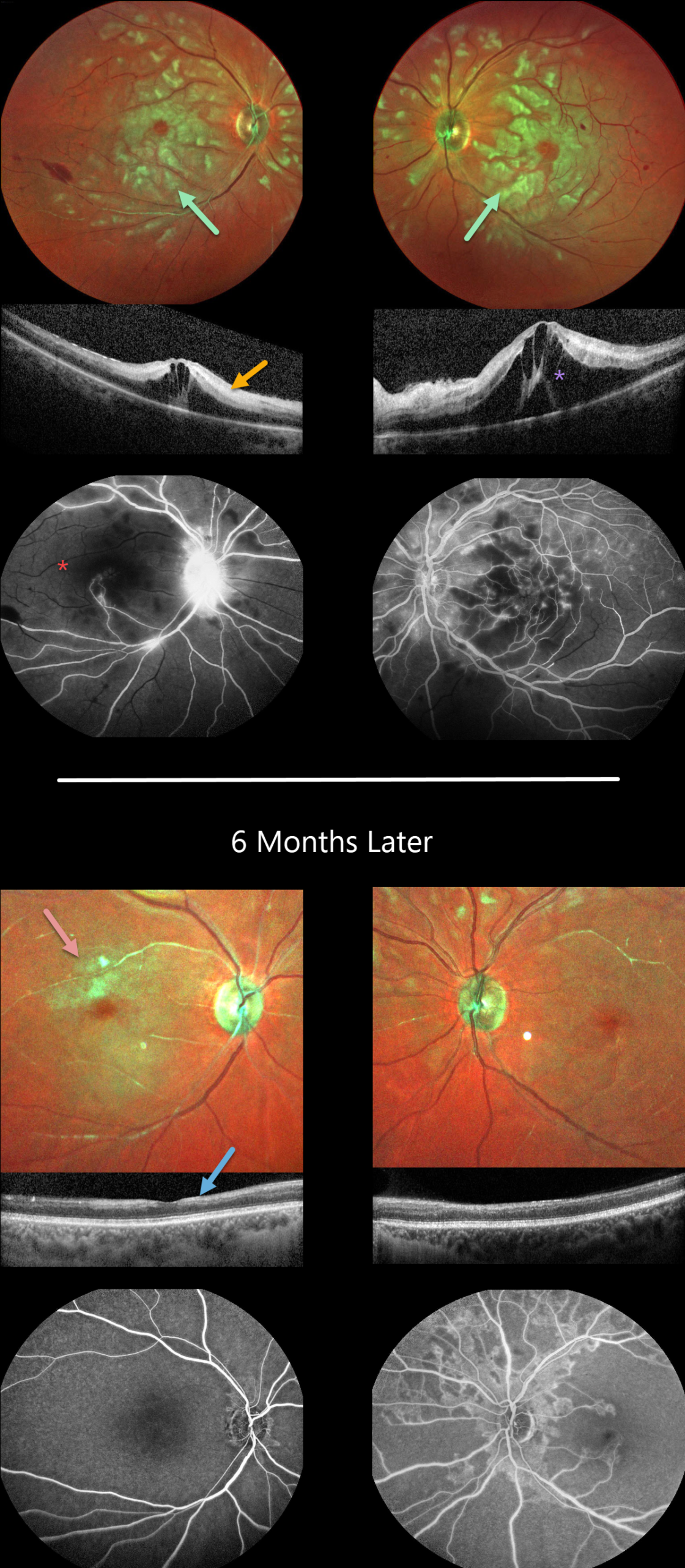
This previously healthy 29YO female presented to the emergency room with acute bilateral vision loss. Vision was counting fingers OU.
Color imaging shows bilateral white patches of inner and mid-retinal ischemia, white narrowed arterioles, and a few inner retinal hemorrhages.
OCT scanning shows variable hyperreflectivity within the inner and mid-retinal layers and cystoid edema. Fluorescein angiography confirms profound retinal ischemia, particularly in the right eye.
Extensive medical workup resulted in a diagnosis of systemic lupus erythematosus (SLE). Rheumatology started systemic rituximab. Bilateral intravitreal dexamethasone followed by intravitreal Eylea was given for the macular edema.
Six months later, although her vision remained at counting fingers OU, she was functioning better and learning to adapt to a new way of living. She was on prednisone 10mg daily and Eylea OU q12 weeks.
Although the acute patches of white retinal ischemia resolved, each fundus is clinically and angiographically markedly ischemic. OCT scanning shows no edema, although there is relative disorganization and thinning of the inner retinal layers.
Learning Points:
Purtscher retinopathy was first described by Otmar Purtscher in 1910. Although originally described in a man who fell from a tree with cranial trauma, these white patches of retinal ischemia and hemorrhages are found in numerous other etiologies, including pancreatitis, chest trauma, and collagen vascular diseases.
The findings are called Purtscher retinopathy when due to trauma, and Purtscher-like retinopathy when seen from other causes. SLE-associated retinopathy more typically appears as hypertensive retinopathy (bilateral nerve fiber layer infarcts and retinal hemorrhages), often in patients with cerebral vasculitis. However, our patient’s blood pressure was not elevated, and there were no signs of central nervous system involvement.
The underlying cause of Purtscher retinopathy is unknown, but it is thought to result from leukoembolization mediated by complement activation. The areas of ischemia include more superficial nerve fiber layer infarcts (cotton wool spots) and deeper ischemia involving the middle retinal layers.
This middle retinal ischemia, paracentral middle maculopathy (PAMM), is a recently-described phenotype found as an idiopathic entity or in other ocular disorders, including Purtscher, diabetic retinopathy, retinal artery and retinal vein occlusions, and acute macular neuroretinopathy (see Retina 2015;35:1921-1930).
CRAO
Asma Samsudeen and Ashish Sharma
Originally posted on @retina.rocks August 10, 2023
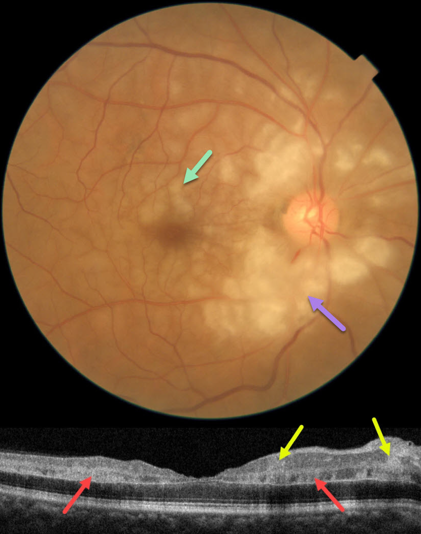
This 60YO male presented with an abrupt, painless loss of vision in his right eye 1 week earlier. There was no prior ocular history or recent history of trauma. Vision was 20/30 in his right eye, and 20/20 in his normal left eye.
Fundus photography shows patches of mostly inner-peripapillary ischemia, with deeper ischemia throughout the macula. This is shown on OCT as patches of inner and mid-retinal hyperreflectivity. Fluorescein angiography showed a markedly delayed transit time (not shown).
We immediately referred him to his cardiologist for an emergency stroke protocol evaluation.
Learning Points:
Purtscher retinopathy was first described by Otmar Purtscher in 1910. Although originally described in a man who fell from a tree with cranial trauma, these white patches of retinal ischemia and hemorrhages are found in numerous other etiologies, including pancreatitis, chest trauma, and collagen vascular diseases.
The findings are called Purtscher retinopathy when due to trauma, and Purtscher-like retinopathy when seen from other causes.
Our patient’s fundus appearance most resembles Purtscher-like retinopathy, with patches of inner- and middle-retinal ischemia. However, given the acute symptoms and delayed angiographic filling time, we believe our patient most likely has an incomplete central retinal artery occlusion (CRAO).
See Mangla et al for the OCT findings in CRAO of varying severities (Int J Retina Vitreous 2023;9;37).
PURTSCHER-LIKE RETINOPATHY
Originally posted on @retina.rocks July 28, 2022
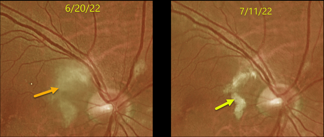
This 65YO male developed inferior field loss in his right eye immediately following cardiac valve replacement on 6/10/22. Vision was 20/30 when he presented to our office 10 days later.
Optos imaging shows patches of inner retinal opacification in the nasal macula and around the nerve, consistent with Purtscher ’s-like retinopathy. Several weeks later, the opacification was resolving with improving symptoms.
Learning Points:
Purtscher’s retinopathy was first described by Otmar Purtscher in 1910. Although originally described in a man who fell from a tree with cranial trauma, these white patches of retinal ischemia and hemorrhages are found in numerous other etiologies, including pancreatitis, chest trauma, and collagen vascular diseases.
The findings are called Purtscher’s retinopathy when due to trauma, and Purtscher-like retinopathy when seen from other causes. The underlying cause of Purtscher’s is unknown, but it is thought to represent leukoembolization by complement activation. The areas of ischemia include more superficial nerve fiber layer infarcts (cotton wool spots) and deeper ischemia involving the middle retinal layers.
This middle retinal ischemia, paracentral middle maculopathy (PAMM), is a recently-described phenotype found as an idiopathic entity or in other ocular disorders, including Purtscher’s, diabetic retinopathy, retinal artery and retinal vein occlusions, and acute macular neuroretinopathy (see Retina 2015;35:1921-1930).
PURTSCHER-LIKE RETINOPATHY
Originally posted on @retina.rocks December 23, 2020
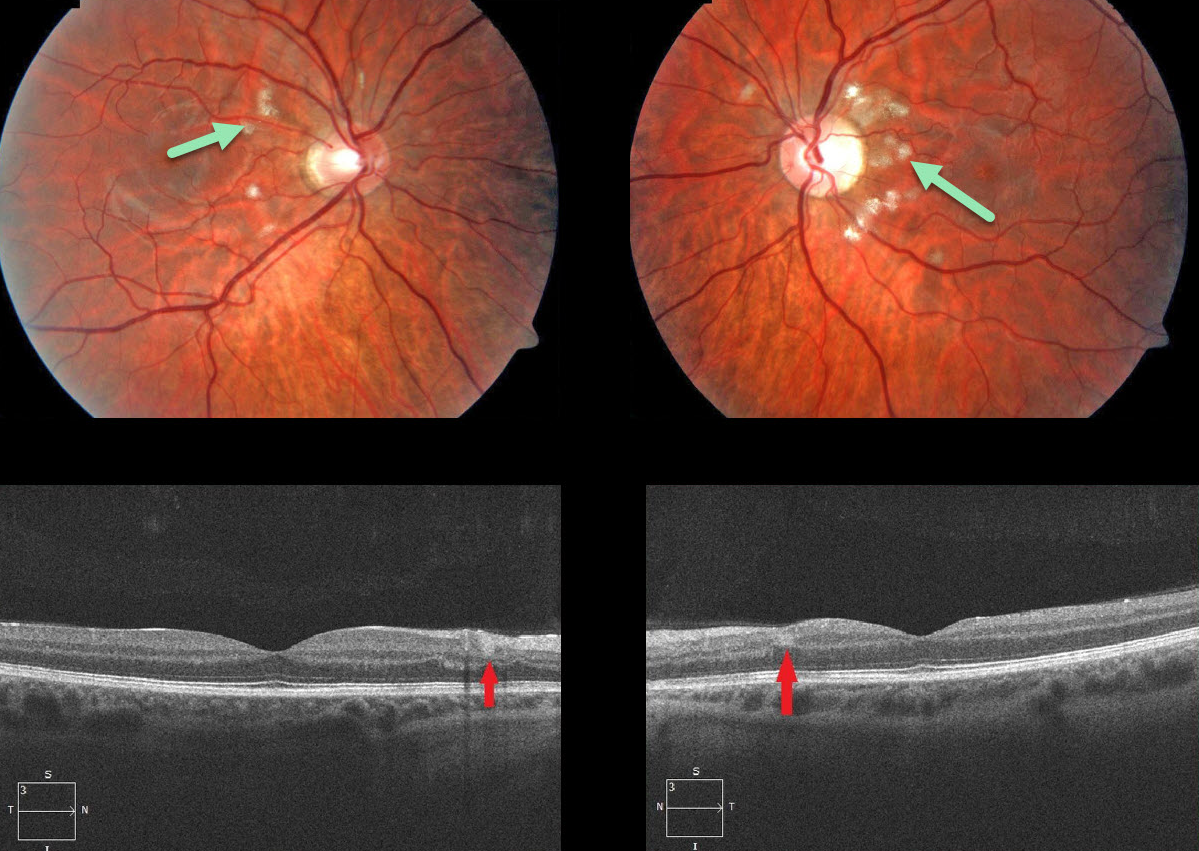
This 24YO male had a history of acute alcohol-related pancreatitis one month earlier and presented with bilateral multifocal areas of superficial peripapillary retinal whitening.
OCT scanning showed small hyperreflective areas spanning the inner plexiform, inner nuclear, and outer plexiform layers.
Learning Points:
Purtscher’s retinopathy was first described by Otmar Purtscher in 1910. Although originally described in a man who fell from a tree with cranial trauma, these white patches of retinal ischemia and hemorrhages are found in numerous other etiologies, including pancreatitis, chest trauma, and collagen vascular diseases.
The findings are called Purtscher’s retinopathy when due to trauma, and Purtscher ‘s-like retinopathy when seen from other causes, as in this case.
The underlying cause of Purtscher’s is unknown, but it is thought to involve leukoembolization due to complement activation. The areas of ischemia include more superficial nerve fiber layer infarcts (cotton wool spots) and deeper ischemia involving the middle retinal layers (paracentral middle maculopathy).


