

Radiation Retinopathy
<< Back to Cases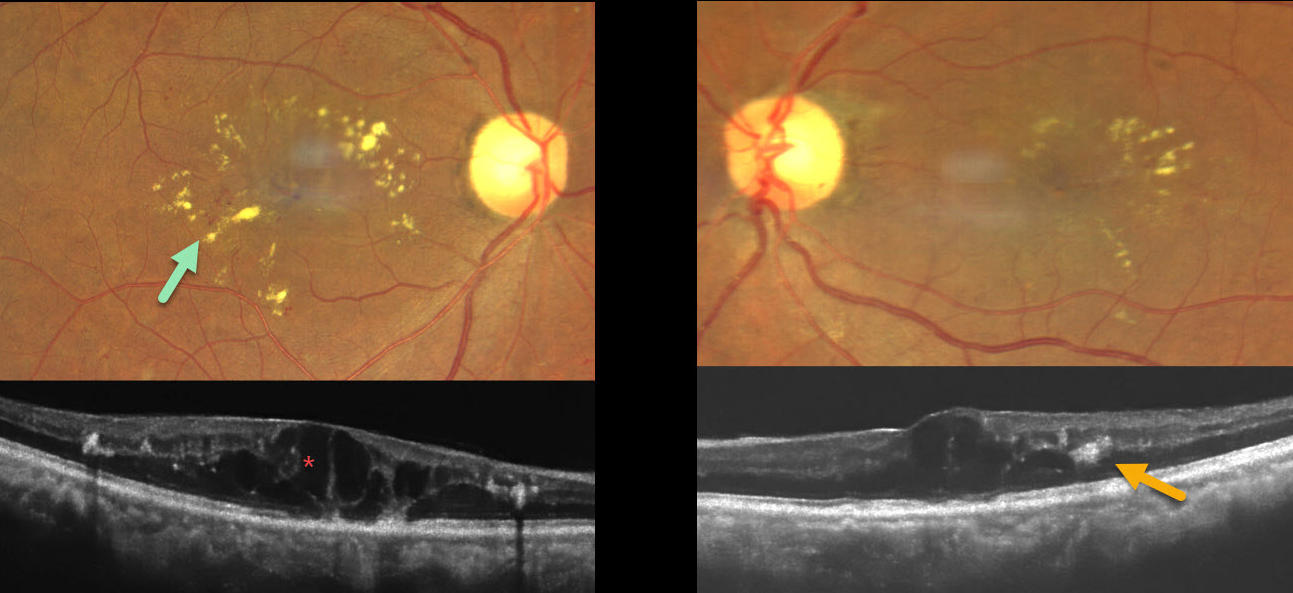
RADIATION RETINOPATHY
Ayushi Gupta and Vishal Agrawal
Originally posted on @retina.rocks April 21, 2026
This 53YO female presented with gradual bilateral vision loss. Three years earlier, she was treated elsewhere for metastatic breast carcinoma, which included a mastectomy followed by radiotherapy to the supraclavicular region and brain. She denied having diabetes. Vision was 20/60 OD and 20/30 OS.
Fundus photography shows bilateral macular telangiectasia with radiating macular lipid flecks. OCT scanning shows diffuse cystic retinal thickening with hyperreflective lipid centered around the outer plexiform layer. Anti-VEGF therapy was recommended.
Learning Points:
Radiation retinopathy usually occurs 3 months to 3 years after external-beam radiation or plaque radiotherapy. The ocular findings and pathogenesis closely resemble those seen with diabetic retinopathy. Radiation optic neuropathy can also develop (Carey et al, Br J Ophthalmol 2023;107:743-749).
RADIATION RETINOPATHY
The European VitreoRetinal Society (EVRS) and Abhishek Upadhyaya
Originally posted on @retina.rocks April 3, 2026
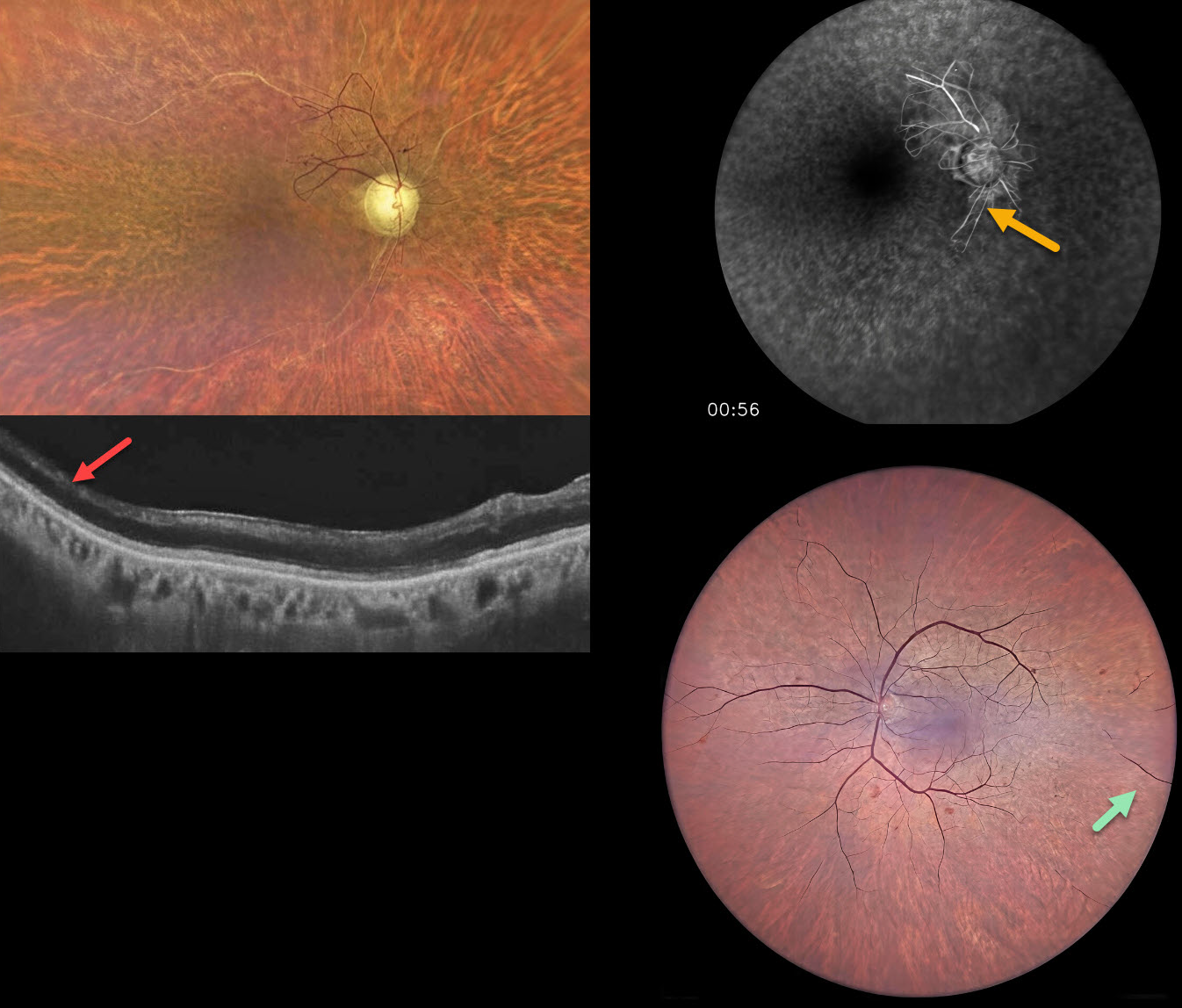
This 65YO male presented for evaluation for cataract surgery. He underwent external beam radiation for throat cancer, predominantly on the right side, 20 years earlier. Vision was hand motion OD and 20/200 OS.
Color photography OD shows severe panretinal ischemia with the major vessels showing absent blood columns except for some abnormal preserved vessels with anastomoses just superotemporal to the nerve. OCT shows temporal inner retinal atrophy. The left posterior pole appears more normal with severe more peripheral ischemia. Fluorescein angiography confirms the profound ischemia noted funduscopically.
Learning Points:
Radiation retinopathy usually occurs 3 months to 3 years following external beam radiation or plaque radiotherapy. The ocular findings closely resemble those seen with diabetic retinopathy. Radiation optic neuropathy can also develop (Carey et al, Br J Ophthalmol 2023;107:743-749) .
Panretinal photocoagulation was recommended for the right eye.
RADIATION RETINOPATHY
Originally posted on @retina.rocks June 15, 2022
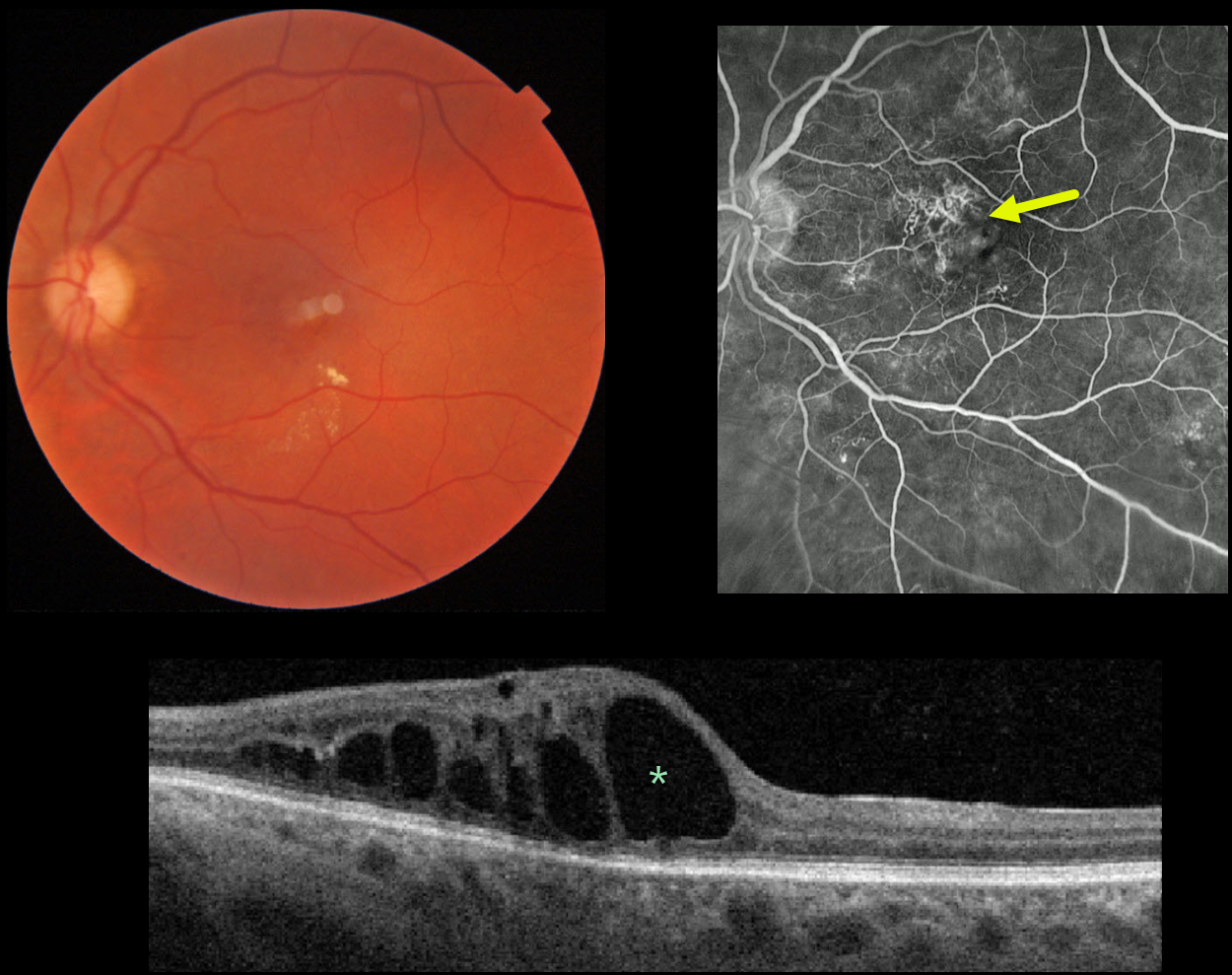
This 60YO male presented with 20/400 vision in his left eye due to radiation retinopathy caused by prior photon radiation treatment for brain cancer 10 years earlier.
Fundus photography shows some small scattered inner retinal hemorrhages and exudates. Fluoroscein angiography shows superonasal foveal retinal telangiectasia, with mild scattered inferior microvascular changes. OCT shows central and nasal thickening from large outer retinal cysts. Anti-VEGF therapy was started.
Learning Points:
Radiation retinopathy usually occurs 3 months to 3 years after external beam radiation (e.g., for facial or orbital cancers) or plaque radiotherapy. The ocular findings closely resemble those seen with diabetic retinopathy.
RADIATION PAPILLOPATHY
Originally posted on @retina.rocks July 2, 2021
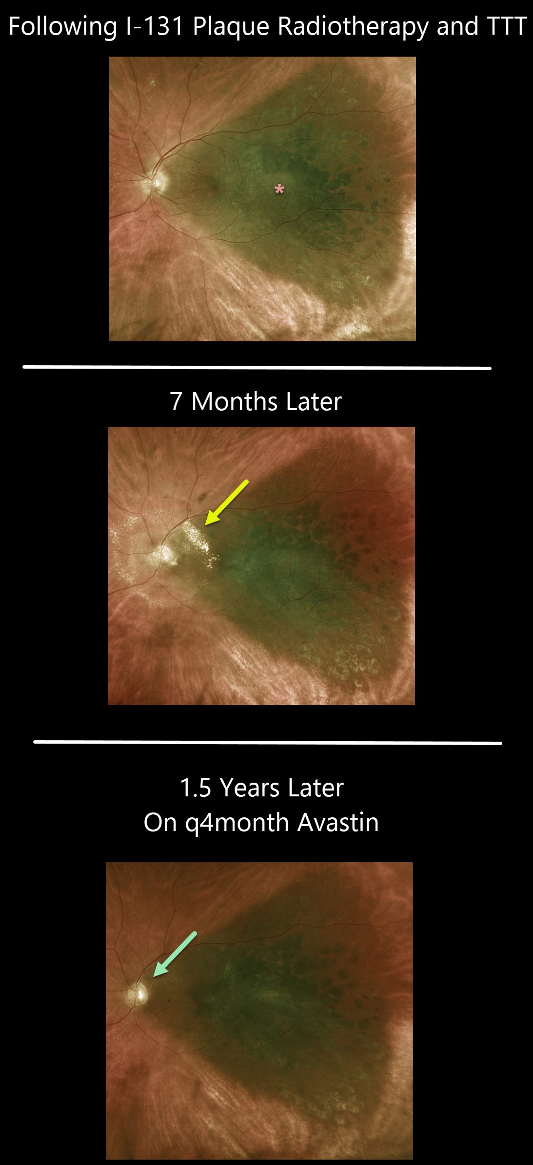
This 66YO male was examined following I-131 plaque radiotherapy and transpupillary thermotherapy for a type 1A, PRAME+ uveal malignant melanoma.
Vision was 20/80, and the tumor was regressing compared to its preoperative appearance (not shown). He was getting intravitreal injections every 4 months as prophylaxis against radiation retinopathy.
The patient returned 8 months later for an overdue examination with new peripapillary lipid from radiation papillopathy. The intravitreal Avastin injections were restarted, and the papillopathy gradually resolved.
Learning Points:
The incidence of radiation retinopathy is relatively high following plaque therapy for uveal malignant melanoma. Ongoing intravitreal injections greatly reduce this complication (see Fallico et al, Surv Ophthalmology 2021;66;441-460).


