

Relentless Placoid (Ampiginous) Chorioretinitis
<< Back to Cases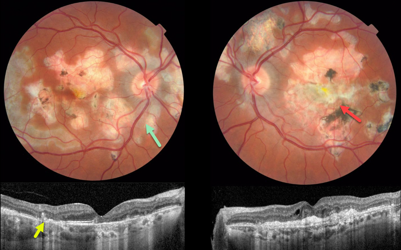
RELENTLESS PLACOID CHORIORETINITIS
Ryan Kern
Originally posted on @retina.rocks April 5, 2023
This 34YO male presented with a one-day history of severe vision loss in his left eye. Vision was 20/30 OD and 20/400 OS.
Color imaging shows bilateral areas of centrifugal pseudopod-like projections of atrophic scarring. The peripapillary scarring in the right eye seems to radiate outward from the optic nerve. There is a large, mostly atrophic disciform scar in the left eye, along with bilateral patches of variably pigmented scarring.
OCT scanning shows hyperreflective subfoveal lesions likely representing spontaneously involuted type 2 macular neovascularization (MNV) with overlying disorganized outer retinal layers.
Our patient’s vision in his left eye is remarkably good, given the macular and OCT appearance. It is unclear why he presented with acute vision loss, given the chronic appearance of his findings.
Learning Points:
Relentless placoid chorioretinitis has clinical and angiographic features resembling both serpiginous and APMPPE. Relentless placoid demonstrates the multifocal lesions of APMPPE, with more meandering, less patchy, serpiginous lesions.
Additionally, these patients will have recurrent inflammation typical of serpiginous but not APMPPE. They may also develop MNV that is more typical of the serpiginous form.
RELENTLESS PLACOID CHORIORETINITIS
Originally posted on @retina.rocks April 20, 2021
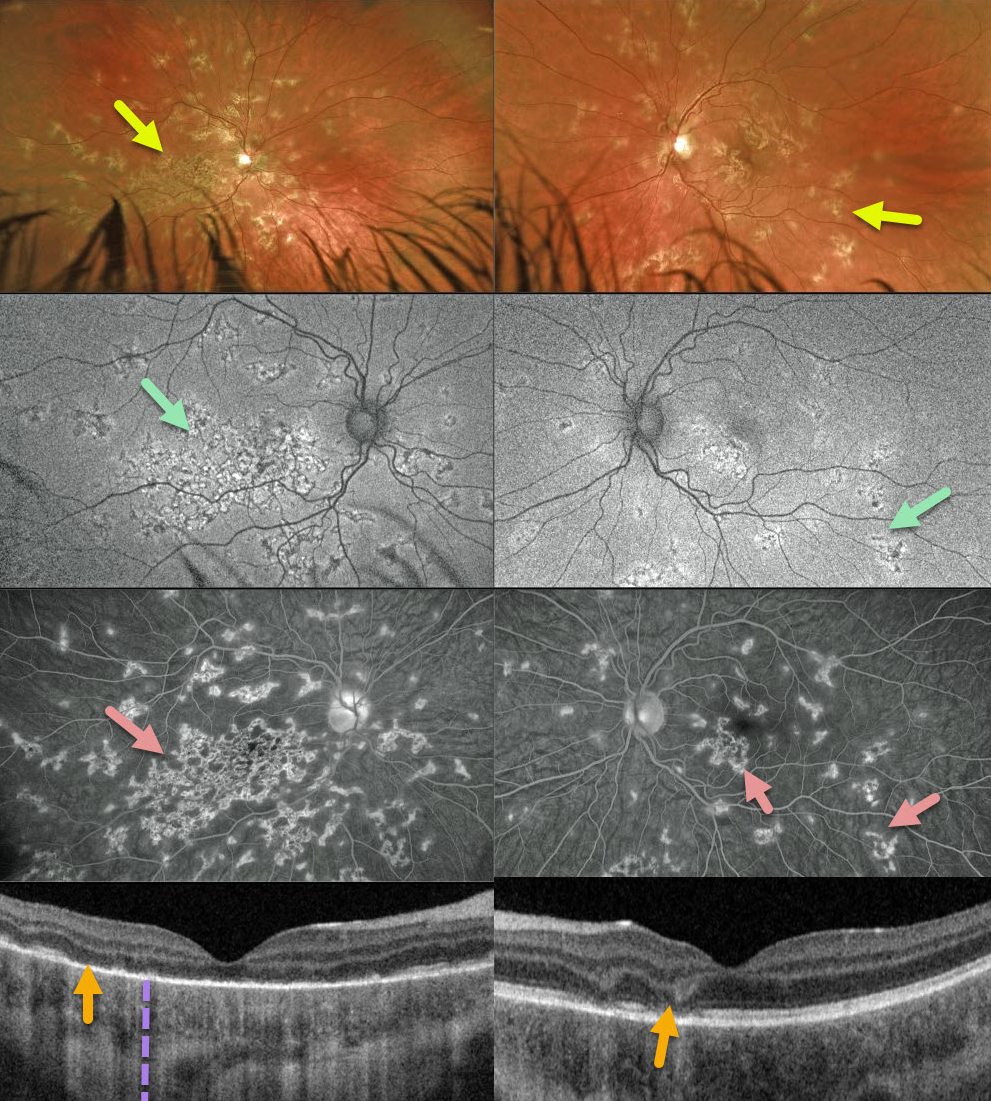
This 12YO boy was referred for asymptomatic retinal changes. Vision was counting fingers OD and 20/25 OS.
Optos imaging shows patchy inactive chorioretinal scarring extending from each macula to the mid-periphery, especially confluent in the right macula.
There is variable hyper- and hypo-autofluorescence, and fluorescein angiography shows areas of staining. Triton swept-source OCT shows variable outer retinal loss with fairly intact RPE associated with increased choroidal transmission.
Our presumed diagnosis was relentless placoid chorioretinitis. Since all the funduscopic findings appeared inactive, we are currently observing him without treatment.
Learning Points:
Relentless placoid chorioretinitis has clinical and angiographic features resembling both serpiginous choroiditis and acute posterior multifocal placoid pigment epitheliopathy (APMPPE).
Relentless placoid demonstrates the multifocal lesions of APMPPE, with more meandering, less patchy, serpiginous lesions.
Additionally, these patients will have recurrent inflammation typical of serpiginous but not APMPPE. They may also develop macular neovascularization, which is more typical of serpiginous.
RELENTLESS PLACOID CHORIORETINITIS
Originally posted on @retina.rocks August 17, 2020
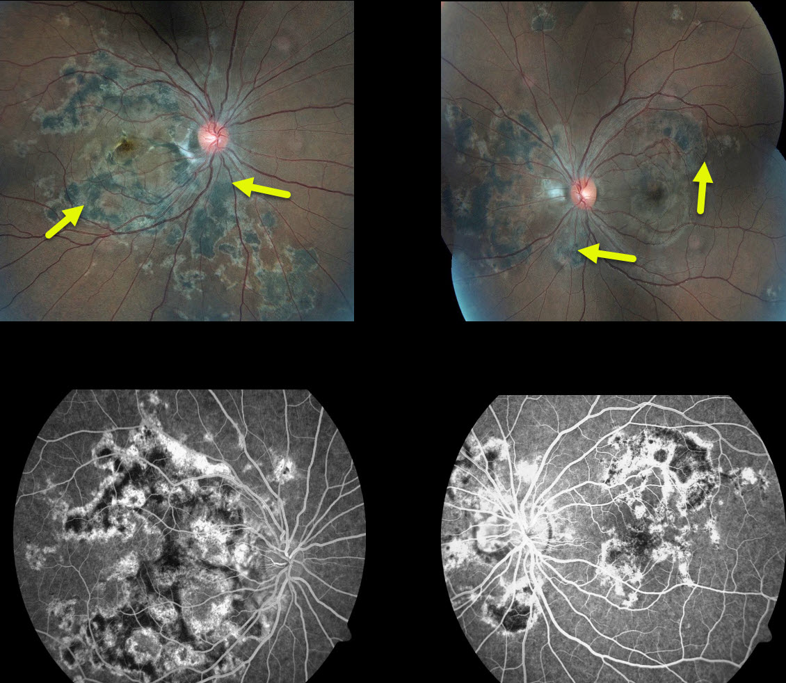
Our patient has relentless placoid chorioretinitis with the multifocal lesions of acute posterior multifocal placoid pigment epitheliopathy (APMPPE) and the peripapillary changes of serpiginous choroiditis.
This patient demonstrates chorioretinal scars surrounding the optic nerve but also throughout the posterior pole. Additionally, he has recurrent inflammation typical of serpiginous but not APMPPE.
His vision is 20/20 OU, which worsens when steroid or anti-inflammatory treatment is tapered. Infectious and inflammatory blood work was negative. He is currently treated and stable with methotrexate.
Learning Points:
Relentless placoid chorioretinitis has clinical and angiographic features resembling both serpiginous chorioretinopathy and acute posterior multifocal placoid pigment epitheliopathy (APMPPE).
Patients with relentless placoid choroiditis can develop recurrent inflammation and macular neovascularization that is more typical of serpiginous than APMPPE.
APMPPE is a bilateral inflammatory vs ischemic disorder of the inner choroid, RPE, and outer retina. These patients present with blurred vision and paracentral/central scotomas often following a viral illness.
Acutely, there are multifocal creamy yellow or grayish-white subretinal placoid lesions scattered throughout the posterior pole.
The lesions and visual symptoms usually resolve within 4 to 8 weeks with secondary chorioretinal changes. Recurrent lesions are rare, and vision is usually fairly good.


