

Retinal Arterial Macroaneurysm (RAM)
<< Back to Cases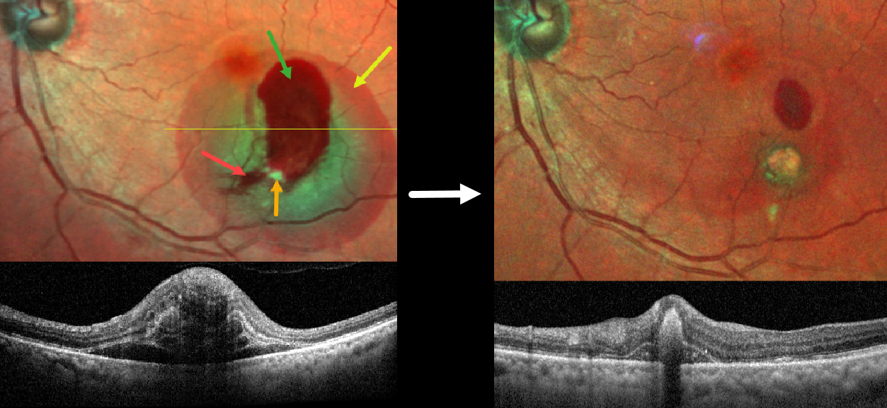
RETINAL ARTERIAL MACROANEURYSM (RAM)
Rohit Agrawal
Originally posted on @retina.rocks May 22, 2026
This 78YO female presented with sudden blurred vision in her left eye. Vision was 20/40.
MultiColor SLO imaging shows subretinal, intraretinal and preretinal blood centered in the inferotemporal macula. A fibrosed retinal arterial macroaneurysm is seen along the inferior edge of the premacular blood. OCT shows a complex laminated pattern of subretinal and intraretinal blood.
A RAM is thought to occur in a weakened arterial wall from arteriosclerosis and appears as a yellow-white dilated lesion along a retinal artery within the first 3 bifurcations from the optic nerve. There are 2 distinct presentations, hemorrhagic or exudative. The hemorrhagic type often presents, as in this case, with pathognomonic multiple layers of blood. Acute macular exudation can be treated with observation, anti-VEGF injections, or thermal laser.
Our patient was treated with a combination of an anti-VEGF injection followed by thermal laser. Five weeks later, vision improved to 20/30 with marked improvement in the blood and macular thickening.
RETINAL ARTERIAL MACROANEURYSM (RAM)
Shilpi Kapoor, Abhishek Sachdeva and Sanjay Sachdeva
Originally posted on @retina.rocks May 11, 2026
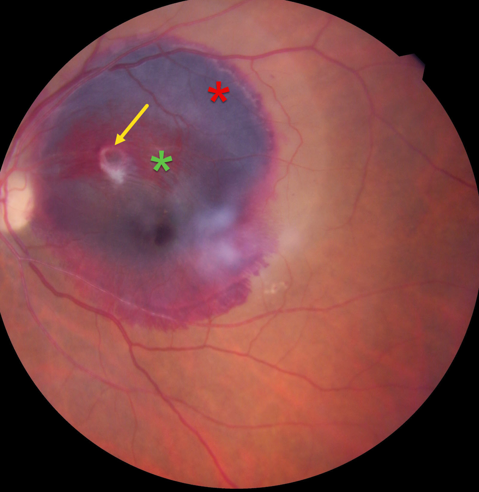
This 65YO female presented with 5 days of acute vision loss in her left eye. Vision was 20/20 in her normal OD and 20/400 OS.
Color photography shows a large subretinal hemorrhage occupying the entire macula, along with a smaller preretinal hemorrhage. A somewhat fibrosed retinal arterial macroaneurysm (RAM) is noted within the preretinal blood. An intravitreal ranibizumab injection was given, but the patient was unfortunately immediately lost to follow-up.
Learning Points:
A RAM is thought to occur in a weakened arterial wall from arteriosclerosis and appears as a yellow-white dilated lesion along a retinal artery within the first 3 bifurcations from the optic nerve. There are 2 distinct presentations, hemorrhagic or exudative. The hemorrhagic type often presents, as in this case, with pathognomonic multiple layers of blood. Acute macular exudation can be treated with observation, anti-VEGF injections, or thermal laser.
RETINAL ARTERIAL MACROANEURYSM
Originally posted on @retina.rocks September 22, 2025
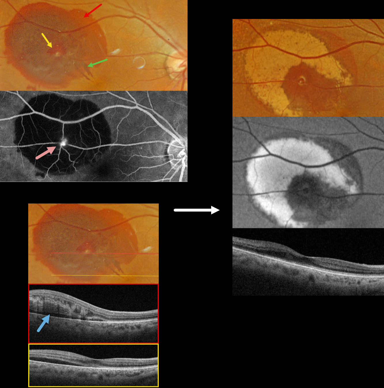
This 83YO female presented with recent blurred vision in her right eye. Vision was 20/40.
Optos color RGB imaging shows subretinal blood in the superotemporal macula. A small amount of inner vs preretinal blood surrounds a small yellow-white retinal arterial macroaneurysm (RAM). In addition, some radiating outer plexiform blood radiates towards the macular center. Fluorescein angiography shows a hyperreflective, leaking RAM with blockage by surrounding subretinal blood. Swept source OCT shows variable outer retinal edema, hyperreflective blood, and subretinal fluid.
Observation was recommended. Six weeks later, the blood is absorbing and turning yellow superiorly and temporally. This devitalized blood is markedly hyper-autofluorescent. The foveal fluid resolved, and vision improved to 20/30.
Learning Points:
A RAM is thought to occur in a weakened arterial wall from arteriosclerosis and appears as a yellow-white dilated lesion along a retinal arterial within the first 3 bifurcations from the optic nerve. There are 2 distinct presentations, hemorrhagic or exudative. The hemorrhagic type often presents, as in this case, with pathognomonic multiple layers of blood. Acute macular exudation can be treated with observation, anti-VEGF injections, or thermal laser
RETINAL ARTERIAL MACROANEURYSM (RAM) WITH MACULAR HOLE
Will Gibson
Originally posted on @retina.rocks January 13, 2025
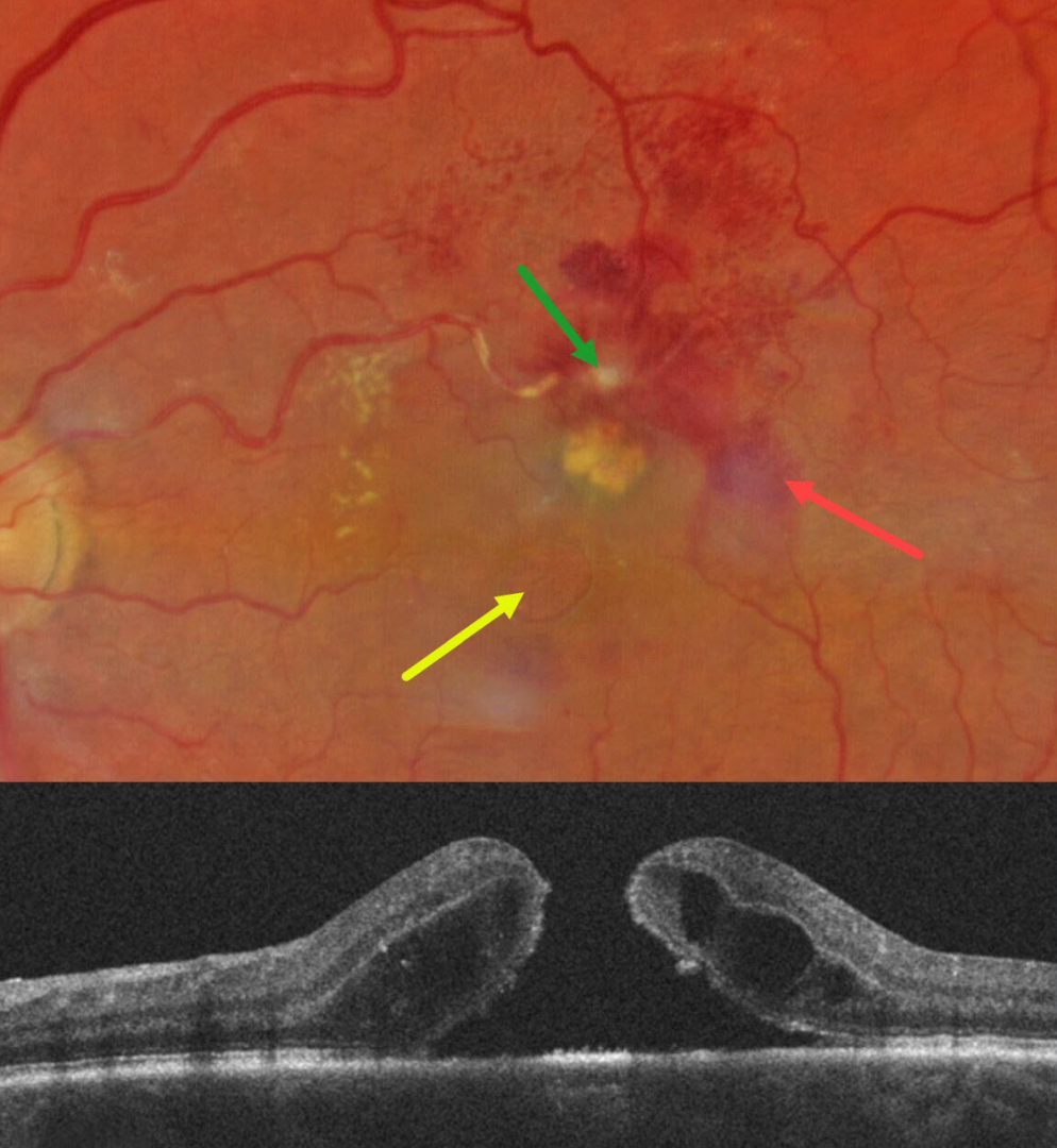
This 67YO white female presented for her six-month diabetic follow-up with a few weeks of vision loss in her left eye. Vision was 20/80 OS and 20/20 in her normal OD.
Color photography shows a fibrosed retinal arterial macroaneurysm (RAM), in the superior macula. Faint subretinal blood is noted temporally, with mostly inner retinal blood superotemporally. The artery is sheathed just proximal to the RAM. A large macular hole with surrounding subretinal fluid is present, accompanied by a lipid clump between the hole and the RAM. Faint lipid is also noted in the nasal macula. She was referred to a retinal specialist for further management.
Learning Points:
Macular hole is a rare finding with retinal arterial macroaneurysms (Sagara et al, Br J Ophthalmol 2009;93:1337-1340). Subretinal and sub-internal limiting membrane blood appears to increase the risk of macular hole.
RETINAL ARTERIAL MACROANEURYSM (RAM) WITH BRAO
Originally posted on @retina.rocks August 13, 2024
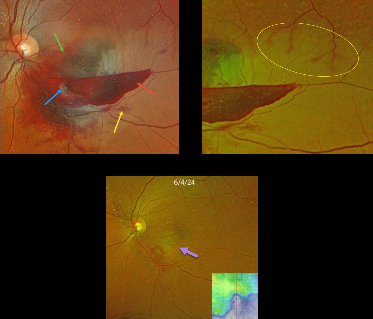
This 59YO female presented with acute vision loss in her left eye. Vision was counting fingers.
Optos color RGB shows a multi-layered macular hemorrhage, including preretinal, intraretinal, and subretinal blood. The causative ruptured retinal arterial macroaneurysm (RAM) is noted within the preretinal blood inferonasally. The surrounding neurosensory retina is somewhat opaque from a secondary inferior branch retinal artery occlusion (BRAO).
Optos RG color imaging shows some periarterial sparing along the superotemporal edge of the BRAO. The perfused superior retinal arteriole, as it enters the region of the occlusion, still has some oxygen that is diffusing into the perivascular tissue.
The patient was treated with several monthly Avastin injections in hopes of more rapidly resolving the surrounding fluid to help relieve arterial compression. Although the blood and fluid resolved, secondary central and inferotemporal macular atrophy resulted from the prior BRAO. Vision was 20/400.
Learning Points:
A BRAO can be a rare complication of direct thermal laser of a RAM (Russell and Folk, AJO 1987;104:186-187), which is one reason it is almost never performed anymore. In this case, we believe the BRAO was secondary to mechanical arterial compression from the surrounding blood.
RUPTURED RETINAL ARTERY MACROANEURYSM (RAM)
Mohammad Abbas
Originally posted on @retina.rocks November 1, 2023
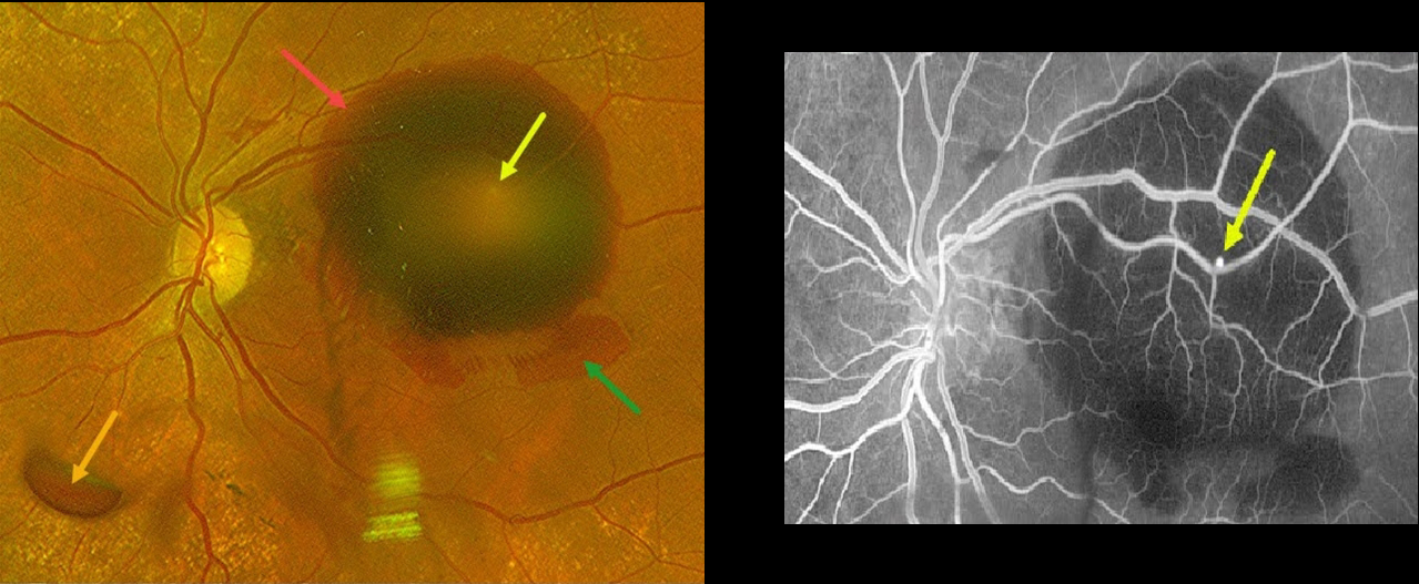
This 78YO hypertensive female presented with sudden vision loss in her left eye. Vision was 20/40 in her normal right eye and counting fingers in her left eye.
Optos color RG imaging shows multiple layers of blood, including a large submacular hemorrhage, intraretinal blood, and preretinal blood.
A tiny retinal arterial macroaneurysm is noted in the superotemporal macula clinically as well as on fluorescein angiography.
This case was submitted by Mohammad Abbas.
Learning Points:
A RAM is thought to occur in a weakened arterial wall from arteriosclerosis, and appears as a yellow-white dilated lesion along a retinal artery within the first 3 bifurcations from the optic nerve.
There are 2 distinct presentations, hemorrhagic and exudative. The hemorrhagic type often presents, as in this case, with pathognomonic multiple layers of blood. Acute macular hemorrhage can be treated with observation, anti-VEGF injections, or thermal laser.
Due to the thick submacular blood, vitrectomy with subretinal TPA was recommended. However, she declined surgery and is being followed for now.
RETINAL ARTERIAL MACROANEURYSM (RAM)
Asma Samsudeen and Ashish Sharma
Originally posted on @retina.rocks October 26, 2023
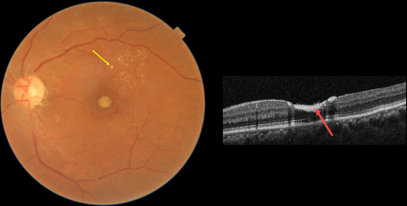
This 54YO female presented with 3 weeks of vision loss in her left eye. Vision was 20/20 in her normal OD and 20/200 OS.
Fundus photography shows devitalized yellow prefoveal blood, some lipid flecks centered in the superotemporal macula, and a tiny fibrosed superior retinal arterial macroaneurysm (RAM).
OCT scanning through the fovea confirms the sub-internal limiting membrane (ILM) location of the hyperreflective blood.
This case was submitted by Asma Samsudeen and Ashish Sharma.
Learning Points:
A RAM is thought to occur in a weakened arterial wall from arteriosclerosis, and appears as a yellow-white dilated lesion along a retinal artery within the first 3 bifurcations from the optic nerve.
There are 2 distinct presentations, hemorrhagic or exudative, and our patient has features of both. Although she reported only a several-week history of vision loss, the retinal findings suggest a much longer course.
The devitalized blood usually takes months to turn yellow, and was likely much larger in extent originally. The blood elsewhere has largely resolved. There was also likely macular thickening and possibly subretinal fluid, which spontaneously resolved, leaving behind residual peripheral macular lipid.
Observation was recommended, and we expect her vision to significantly improve, as the retina appears structurally fairly normal on OCT deep to the blood. Subretinal blood, especially devitalized, is toxic to the outer retina, which is why removal and/or displacement of thick subretinal blood is often recommended. Blood in the sub-ILM or preretinal space does not seem to damage the retina, so it can more safely be observed (see Bloom and Spaide, Retinal Cases 2022;16;401-402).
RETINAL ARTERIAL MACROANEURYSM (RAM)
Asma Samsudeen and Ashish Sharma
Originally posted on @retina.rocks September 28, 2023
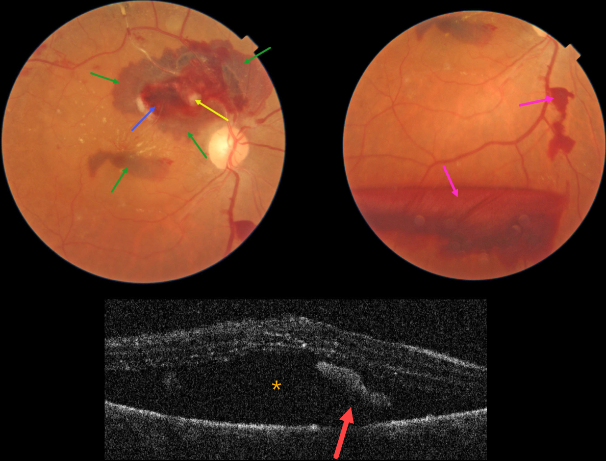
This 76YO female presented with blurred vision in her right eye for one month. Vision was 20/60 in her right eye and 20/30 in her left eye.
Color photography shows a ruptured retinal arterial macroaneurysm (RAM) causing multiple layers of blood. The blood is subretinal, intraretinal, and preretinal. Some scattered lipid flecks are noted centrally and temporally, and the arterial distal to the RAM is sclerosed.
OCT scanning shows marked subretinal fluid and some hyperreflective subretinal blood.
Learning Points:
A RAM is thought to occur in a weakened arterial wall from arteriosclerosis, and appears as a yellow-white dilated lesion along a retinal artery within the first 3 bifurcations from the optic nerve.
There are 2 distinct presentations, hemorrhagic or exudative. The hemorrhagic type often presents, as in this case, with pathognomonic multiple layers of blood. Acute macular exudation can be treated with observation, anti-VEGF injections, or thermal laser.
Due to her symptomatic subretinal fluid, a single intravitreal Avastin injection was given. Four months later, vision improved to 20/30, the macular fluid resolved, and most of the blood layers were gone.
RETINAL ARTERIAL MACROANEURYSM (RAM)
Originally posted on @retina.rocks July 24, 2023
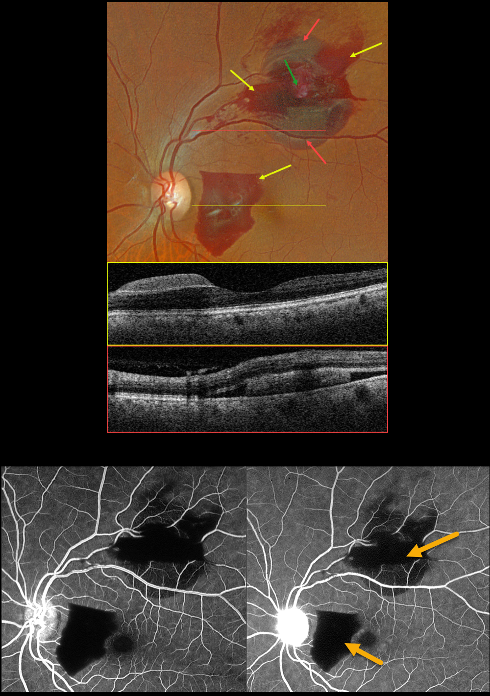
This 65YO female presented with 2 weeks of paracentral vision loss in her left eye. Vision was 20/25.
Optos color RGB imaging shows multilayered hemorrhages in the nasal and superior macula, including preretinal and subretinal blood.
Swept-source OCT through the preretinal and subretinal areas confirms the locations of the blood. Fluorescein angiography shows a blockage in the blood. The retinal arterial macroaneurysm, which is noted as a yellowish mass within the superior blood clinically, is obscured by the blood angiographically.
Learning Points:
A RAM is thought to occur in a weakened arterial wall from arteriosclerosis, and appears as a yellow-white dilated lesion along a retinal artery within the first 3 bifurcations from the optic nerve.
There are 2 distinct presentations, hemorrhagic and exudative. The hemorrhagic type often presents, as in this case, with pathognomonic multiple layers of blood. Acute macular hemorrhage can be treated with observation, anti-VEGF injections, or thermal laser.
RETINAL ARTERIAL MACROANEURYSM WITH SECONDARY BRAO
Originally posted on @retina.rocks December 8, 2022
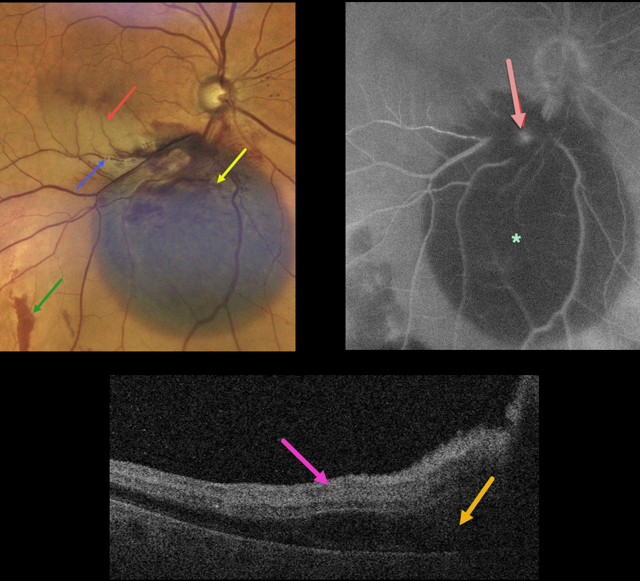
This 79YO female presented with acute inferior paracentral vision loss in her right eye. Vision was 20/50.
A large blister of dark subretinal blood was noted just inferior to the optic nerve. A few small areas of more distal red subretinal blood were seen. An inferior macular branch retinal artery occlusion likely accounted for her symptoms. The blood column is stagnant within the inferior portion of the acute occlusion.
OCT scanning through the BRAO shows hyperreflective inner retina with subretinal fluid.
Fluorescein angiography shows blockage from the subretinal blood, along with a leaking retinal arterial macroaneurysm (RAM).
Learning Points:
A BRAO can be a rare complication from direct thermal laser of a RAM (Russell and Folk, AJO 1987;104:186-187), which is one reason why this is almost never performed anymore. In this case, we believe the BRAO was secondary to mechanical arterial compression from the surrounding blood.
RETINAL ARTERIAL MACROANEURYSM (RAM)
Originally posted on @retina.rocks September 21, 2022
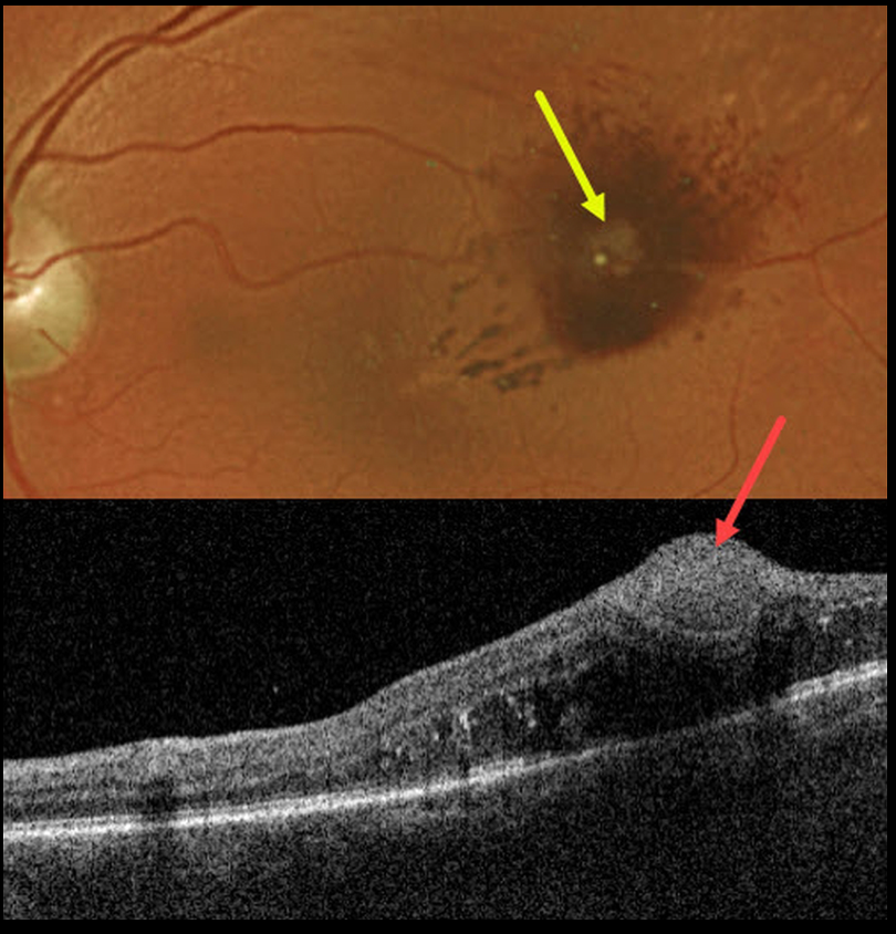
This 70YO female presented with mild vision loss in her left eye. Vision was 20/50.
Optos imaging shows a retinal arterial macroaneurysm (RAM) with surrounding blood. Swept-source OCT shows the hyperreflective RAM in the inner retina. The underlying retina is somewhat thickened.
Learning Points:
A RAM is thought to occur in a weakened arterial wall from arteriosclerosis, and usually appears as a yellow-white dilated lesion along a retinal arteriole within the first 3 bifurcations from the optic nerve.
Although most RAMs spontaneously resolve, chronic central edema or lipid can cause long term vision loss.
Mansour et al, Retina 2019;39;1133-1141, showed that anti-VEGF treatment is effective in reducing macular exudation and improving vision.
RETINAL ARTERIAL MACROANEURYSM (RAM)
Originally posted on @retina.rocks September 1, 2022
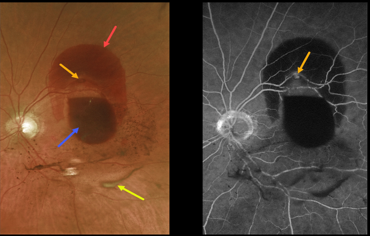
This 85 YO female presented with a retinal arterial macroaneurysm (RAM) with counting fingers vision.
The macroaneurysm has ruptured, causing multiple levels of blood shown on fundus photography and fluorescein angiography. The blood is subretinal superiorly, subhyaloid more centrally, and in the vitreous (both red and devitalized). We are observing this patient.
Learning Points:
A RAM is thought to occur in a weakened arterial wall from arteriosclerosis, and appears as a yellow-white dilated lesion along a retinal arterial within the first 3 bifurcations from the optic nerve.
There are 2 distinct presentations, hemorrhagic or exudative. The hemorrhagic type often presents, as in this case, with pathognomonic multiple layers of blood.
Acute macular hemorrhage can be treated with observation, anti-VEGF injections, or thermal laser.
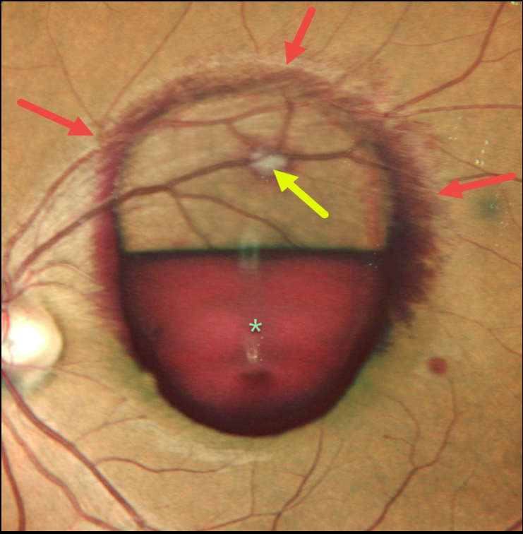
RETINAL ARTERIAL MACROANEURYSM (RAM)
Nivesh Gupta
Originally posted on @retina.rocks June 23, 2022
This 34YO female presented with a symptomatic ruptured retinal arterial macroaneurysm (RAM).
Vision was counting fingers due to the inferior sub-internal limiting membrane blood obscuring the underlying fovea. More peripherally, blood is staining the surrounding nerve fiber layer. The fibrosed RAM is visible superiorly.
Learning Points:
A RAM is thought to occur in a weakened arterial wall from arteriosclerosis, and usually appears as a yellow-white dilated lesion along a retinal arteriole within the first 3 bifurcations from the optic nerve.
There are 2 distinct presentations, hemorrhagic or exudative. The hemorrhagic type often presents with pathognomonic preretinal, intraretinal, and subretinal blood. Acute macular hemorrhage can be treated with observation, anti-VEGF injections, or thermal laser to the RAM.
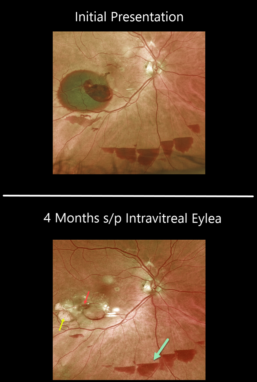
RETINAL ARTERIAL MACROANEURYSM (RAM)
Originally posted on @retina.rocks February 14, 2022
This 85 YO female with a retinal arterial macroaneurysm (RAM) continues to improve with 20/30 vision following a single Eylea injection four months earlier.
The macroaneurysm still appears patent. The old subretinal devitalized blood is devitalized and yellow. There are also multiple patches of resolving inferior subhyaloid blood.
Learning Points:
A RAM is thought to occur in a weakened arterial wall from arteriosclerosis and appears as a yellow-white dilated lesion along a retinal arteriole within the first 3 bifurcations from the optic nerve.
There are 2 distinct presentations: hemorrhagic or exudative. The hemorrhagic type often presents, as in this case, with pathognomonic preretinal, intraretinal, and subretinal blood. Acute macular hemorrhage can be treated with observation, anti-VEGF injections, or thermal laser.
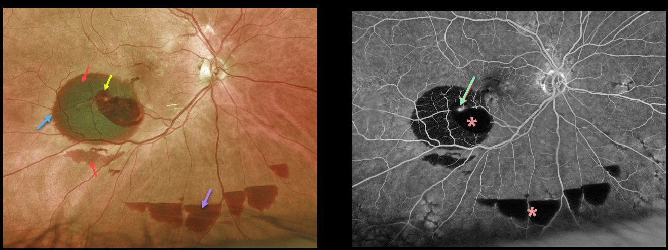
RETINAL ARTERIAL MACROANEURYSM (RAM)
Originally posted on @retina.rocks September 6, 2021
This 85YO female presented with some peripheral vision loss in her right eye. Vision was 20/40.
Optos color imaging shows areas of subretinal blood in the inferotemporal macula and below the inferotemporal arcade, intraretinal blood, and multiple areas of inferior preretinal blood. A rim of radiating outer plexiform blood surrounds the large subretinal blood.
Fluorescein angiography shows blockage from the various layers of blood, along with leakage from the causative macroaneurysm.
We recommended initial observation due to the excellent vision and ectopic blood.
Learning Points:
A retinal arterial macroaneurysm (RAM) is thought to result from a weakened arterial wall due to arteriosclerosis. A RAM appears as a yellow-white dilated lesion along a retinal arterial within the first 3 bifurcations from the optic nerve.
There are 2 distinct presentations, hemorrhagic or exudative. The hemorrhagic type often presents, as in this case, with pathognomonic preretinal, intraretinal, and subretinal blood.
Acute macular hemorrhage can be treated with observation, anti-VEGF injections, or thermal laser.
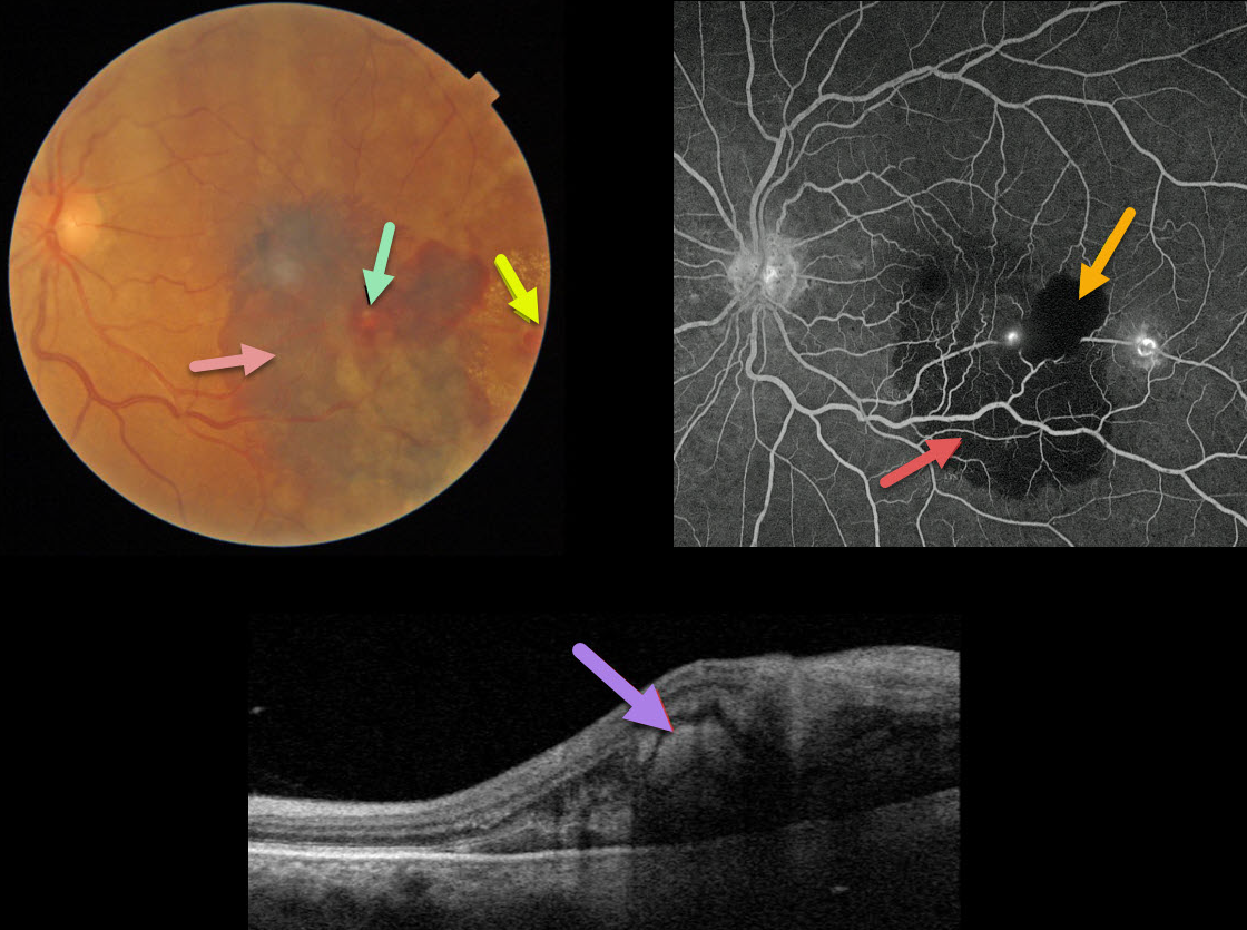
RETINAL ARTERIAL MACROANEURYSM (RAM)
Originally posted on @retina.rocks April 8, 2021
This 68YO female presented with acute vision loss in her left eye. Vision was hand motion from submacular blood.
A retinal arterial macroaneurysm (RAM) is noted overlying the blood, with an additional RAM with lipid more temporally.
Triton swept-source OCT scanning shows subretinal blood beneath the RAM. Fluorescein angiography shows blockage from the subretinal and intraretinal blood, with leakage from both lesions. These multiple layers of blood (subretinal and intraretinal) are classic for RAMs.
In this case, the patient elected for treatment with anti-VEGF injections.
Learning Points:
RAMs present with either acute blood or with chronic exudation, and this case is unique in that we can see both presentations in the same eye.
Acute macular hemorrhage can be treated with observation, anti-VEGF injections, or thermal laser. Macular edema with lipid exudate that threatens or involves the macular center usually requires either thermal laser treatment or anti-VEGF injections.
RETINAL ARTERIAL MACROANEURYSM (RAM)
Originally posted on @retina.rocks October 9, 2020
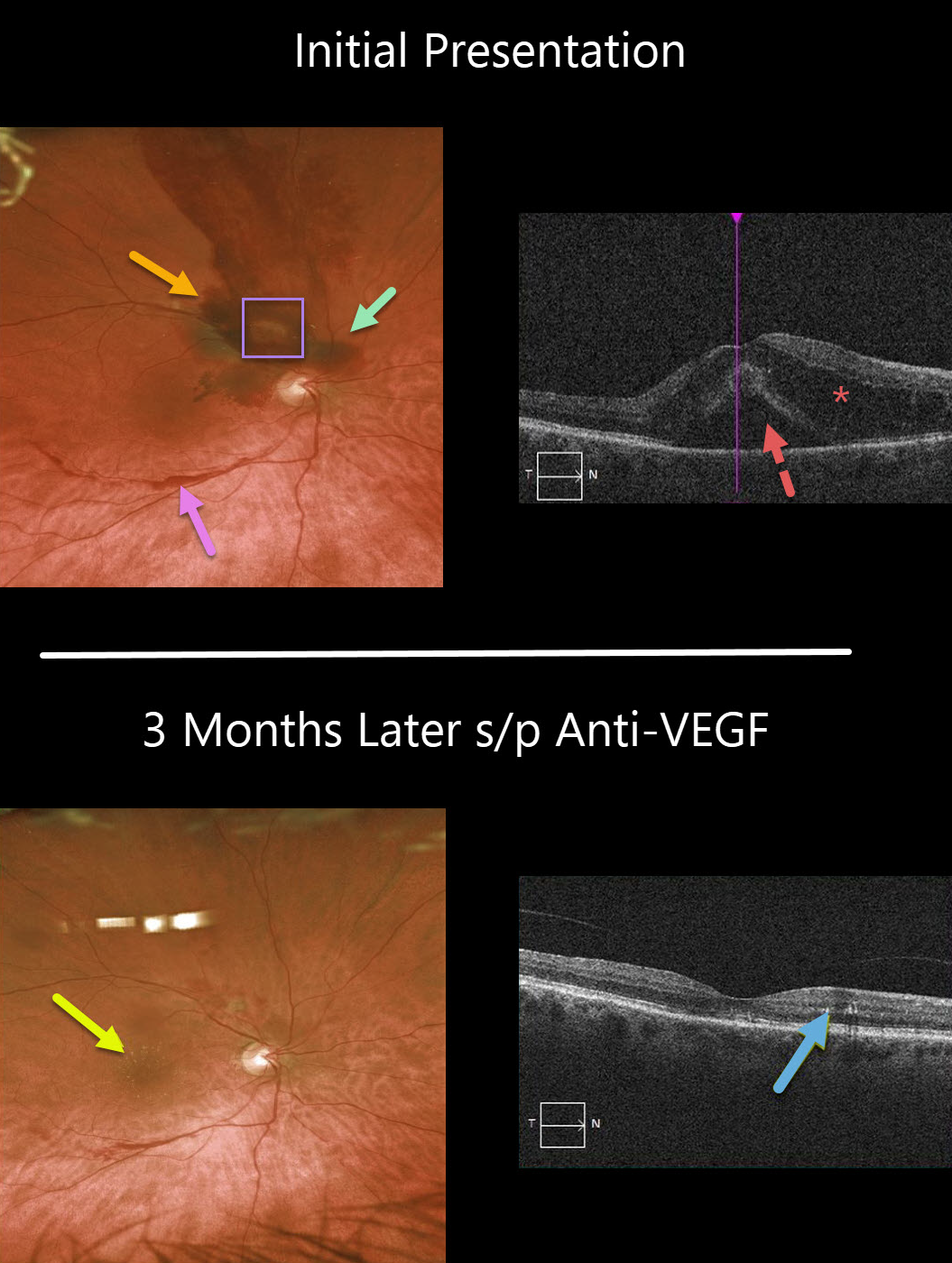
This 71YO female presented with counting-finger vision in her right eye. Clinically, there are multiple layers of blood, including preretinal, intraretinal, and subretinal. These multiple layers are virtually pathognomonic for a ruptured retinal arterial macroaneurysm (RAM).
Despite the causative RAM being just superotemporal to the optic nerve, significant intra- and subretinal fluid extends through the macular center, which is seen on OCT.
Following three monthly anti-VEGF injections, the fluid resolved completely, and vision returned to 20/30. Some residual precipitated lipid is seen in Henle’s layer as a faint lipid star in the color image and as hyperreflective dots in the outer plexiform layer.
Learning Points:
A retinal arterial macroaneurysm (RAM) is thought to result from a weakened arterial wall due to arteriosclerosis.
A RAM appears as a yellow-white dilated lesion along a retinal artery within the first 3 bifurcations from the optic nerve.
There are 2 distinct presentations, hemorrhagic or exudative. The hemorrhagic type often presents as a pathognomonic multilayer macular hemorrhage, with preretinal, intraretinal, and subretinal blood.
RETINAL ARTERIAL MACROANEURYSM (RAM)
Originally posted on @retina.rocks August 20, 2020
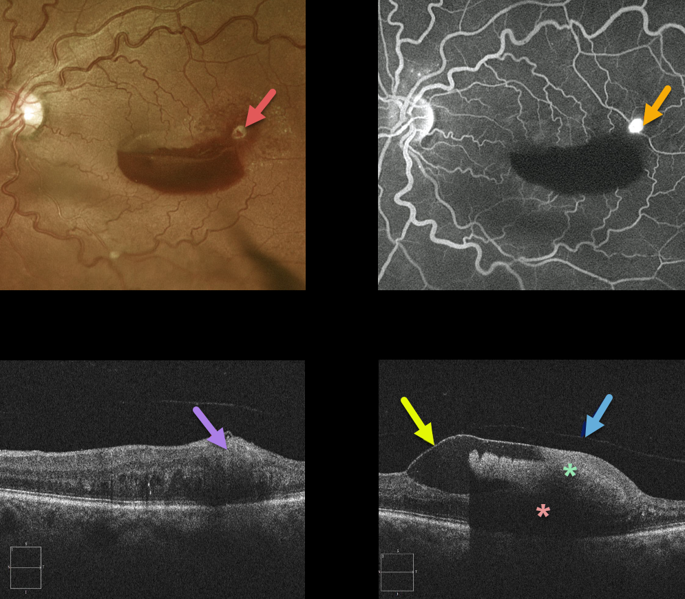
This retinal arterial macroaneurysm (RAM) appears as a yellow-white inner retinal lesion along the superotemporal edge of a macular hemorrhage. Angiography confirms the presence of the hyperfluorescent RAM.
An OCT line scan through the RAM shows its location in the inner retina.
OCT scanning shows that the blood is almost all above the retina and below the detached internal limiting membrane. The detached hyaloid can be seen anterior to the ILM. The dense sub-ILM blood blocks almost all retinal and subretinal detail more posteriorly.
Learning Points:
A retinal arterial macroaneurysm (RAM) is thought to result from a weakened arterial wall due to arteriosclerosis.
A RAM appears as a yellow-white dilated lesion along a retinal artery within the first 3 bifurcations from the optic nerve.
There are 2 distinct presentations, hemorrhagic or exudative. The hemorrhagic type often presents as a pathognomonic multilayer macular hemorrhage, with preretinal, intraretinal, and subretinal blood.
Retinal Arterial Macroaneurysm (RAM)
Originally posted on @retina.rocks March 10, 2020
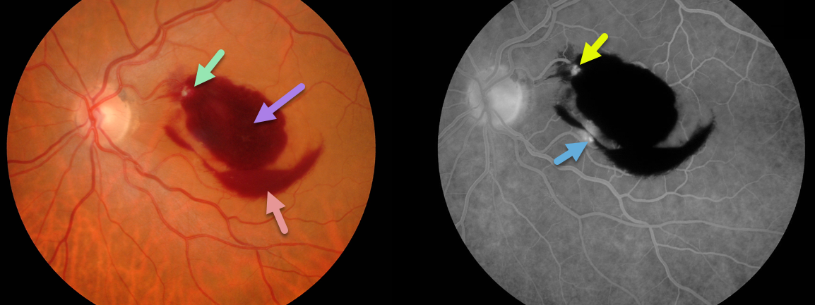
Our patient’s retinal arterial macroaneurysm (RAM) is seen within the superonasal portion of the macular blood. Preretinal and dense intraretinal blood are the clues that a RAM is the cause.
The RAM is confirmed on fluorescein angiography. Although usually singular, our patient may also have a second RAM inferiorly.
Learning Points:
A retinal arterial macroaneurysm (RAM) is thought to occur in a weakened arterial wall from arteriosclerosis.
A RAM appears as a yellow-white dilated lesion along a retinal arterial within the first 3 bifurcations from the optic nerve.
There are 2 distinct presentations, hemorrhagic or exudative. The hemorrhagic type often presents as a pathognomonic multilayer macular hemorrhage, with preretinal, intraretinal and subretinal blood.


